.webp&w=384&q=75)

Loading, please wait...
Loading, please wait...
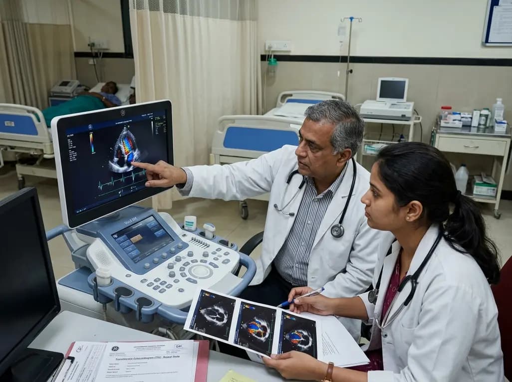
Evaluating patients with suspected infective endocarditis presents a major diagnostic challenge for clinicians. Although guidelines recommend repeat transthoracic echocardiography when clinical suspicion persists despite negative baseline findings, real-world comparative data regarding its true diagnostic value remain limited. A study published in the Journal of the American Heart Association evaluated the real-world diagnostic performance of repeat transthoracic echocardiography within a multimodality imaging approach. Conducted at Lausanne University Hospital, this study evaluated whether a second transthoracic ultrasound significantly improves diagnostic clarity or reclassifies clinical suspicion under updated international diagnostic criteria.
Infective endocarditis requires prompt identification and multidisciplinary management. Echocardiographic examination serves as the initial diagnostic tool, enabling clinicians to visualize endocardial lesions including vegetations and paravalvular complications. However, initial transthoracic examinations frequently yield inconclusive results due to poor acoustic windows or subtle early structural changes. While international guidelines recommend repeating non-invasive cardiac imaging when suspicion persists despite negative baseline scans, the true diagnostic yield of routine repeat scanning required empirical evaluation. The Lausanne study addressed this gap by analyzing adult patients undergoing sequential transthoracic evaluations within twenty-one days. Utilizing the 2023 Duke-International Society for Cardiovascular Infectious Diseases criteria, researchers quantified how often a secondary ultrasound identified new pathological findings or altered patient classification. Understanding these empirical parameters is essential for refining hospital diagnostic pathways and optimizing resource utilization in clinical practice. Clinicians frequently encounter scenarios where initial imaging fails to explain persistent fever or bacteremia, creating uncertainty regarding whether to repeat transthoracic scans or escalate directly to advanced modalities.
The study evaluated five hundred and three patient episodes, with infective endocarditis confirmed in fifty percent of cases. The second repeat transthoracic echocardiography was performed at a median interval of eight days post-baseline. Results demonstrated that repeating the scan identified new cardiac vegetations in seven percent of episodes and uncovered new paravalvular complications in three percent of cases. Diagnostic sensitivity for vegetation detection increased from forty-seven percent on the initial scan to fifty-seven percent on the secondary scan. Excluding non-transthoracic imaging modalities, overall diagnostic sensitivity rose from thirty-four percent to forty-five percent. However, the secondary transthoracic examination led to diagnostic reclassification from possible to definite endocarditis in only one percent of cases. These findings indicate that while repeat imaging modestly enhances lesion detection, its capability to independently alter diagnostic decision-making remains limited. Consequently, physicians must weigh these modest gains against potential delays in definitive diagnosis when relying solely on sequential transthoracic imaging without incorporating complementary modalities.
Modern management of cardiac infections relies on a comprehensive multimodality imaging approach rather than exclusive reliance on standard ultrasound. Transesophageal echocardiography, cardiac computed tomography angiography, and nuclear imaging play critical roles in detecting perivalvular extension and prosthetic valve involvement. The study highlighted that within a multimodality diagnostic strategy, the incremental contribution of repeat transthoracic echocardiography is limited. Relying solely on repeated transthoracic procedures may delay definitive diagnostic confirmation and necessary therapeutic escalations. Transesophageal echocardiography offers superior spatial resolution, making it the preferred second-line modality when clinical suspicion remains elevated. Similarly, cardiac computed tomography excels in identifying paravalvular abscesses and pseudoaneurysms prior to surgical intervention. Nuclear imaging provides crucial metabolic data regarding graft infections and embolic phenomena. Clinicians must recognize that while repeat transthoracic scans are safe and accessible, they should not replace advanced imaging when suspicion persists.
For medical practitioners in India, these findings offer clear guidance for optimizing diagnostic workflows. Infective endocarditis presents significant clinical challenges across Indian tertiary centers, where rheumatic heart disease and healthcare-associated infections remain common. Given high patient volume and variable access to advanced imaging, clinicians often debate whether to perform repeat transthoracic echocardiography or transfer patients for transesophageal or nuclear studies. The evidence confirms that while repeat transthoracic scans capture evolving lesions in a minority of patients, they rarely alter overall patient classification independently. Indian physicians should avoid relying solely on serial transthoracic examinations in high-risk patients with persistent bacteremia, unexplained fever, or prosthetic valves. Instead, clinical pathways should emphasize early escalation to transesophageal echocardiography or cardiac computed tomography whenever baseline findings are equivocal despite high clinical suspicion. This strategy reduces diagnostic delays, avoids unnecessary antibiotic extensions, and improves surgical timing.
The Lausanne study findings support a strategic shift toward early, comprehensive diagnostic pathways in suspected cardiac infections. Future practice guidelines must continue refining indications for serial imaging to avoid low-yield repetitive testing. Rather than protocolizing routine repeat transthoracic scans for all patients, clinical algorithms should advocate risk-stratified imaging cascades. Patients exhibiting high-risk features, such as persistent Staphylococcus aureus bacteremia or new heart block, warrant prompt escalation to transesophageal echocardiography or nuclear imaging rather than awaiting a second transthoracic assessment. Conversely, repeat transthoracic imaging remains valuable when baseline visualization was technically limited or to track dynamic hemodynamic changes in confirmed cases. Medical education must emphasize integrated interpretation combining clinical risk scores, biomarkers, and advanced imaging. By adopting structured multimodality imaging pathways, healthcare teams can improve diagnostic precision, accelerate early surgical consultation, and reduce morbidity and mortality associated with endocardial infections.
Repeat transthoracic echocardiography modestly improves the detection of typical cardiac vegetations and paravalvular complications in patients with suspected infective endocarditis. However, its isolated contribution to reclassifying patients from possible to definite endocarditis is remarkably low, occurring in only one percent of overall cases. Therefore, it should be utilized selectively when advanced complementary imaging modalities are unavailable or when tracking specific hemodynamic changes in confirmed clinical cases.
Clinicians should escalate to transesophageal echocardiography immediately when clinical suspicion of infective endocarditis remains moderate to high despite a negative or non-diagnostic initial transthoracic scan. Transesophageal imaging provides far superior spatial resolution and sensitivity, making it substantially more effective for detecting small vegetations, prosthetic valve dysfunction, perivalvular abscesses, and other subtle anatomical structural complications that require urgent medical or surgical management.
Multimodality imaging combines transthoracic ultrasound, transesophageal echocardiography, cardiac computed tomography, and nuclear imaging to comprehensively evaluate anatomical, structural, and metabolic features of cardiovascular infections. This integrated clinical approach overcomes the technical limitations of isolated non-invasive tests, significantly boosting diagnostic sensitivity, detecting early perivalvular extension, uncovering silent embolic phenomena, and facilitating timely, informed multidisciplinary clinical decision-making for complex cardiac infection management.
Disclaimer: This content is for informational and educational purposes only and does not constitute medical advice. Refer to the latest local and national guidelines for clinical practice.
References
1. Tzimas G et al. Real-World Diagnostic Performance of Repeat Transthoracic Echocardiography Within a Multimodality Imaging Approach in Patients With Suspected Infective Endocarditis. J Am Heart Assoc. 2026 Aug 07. doi: 10.1161/JAHA.126.050020. PMID: 42568077.
2. Habib G et al. 2023 ESC Guidelines for the management of infective endocarditis. European Heart Journal. 2023; 44(39): 3948–4042.

Read summarized clinical updates, watch expert medical content, and earn CME certifications right from your smartphone.
Open your smartphone camera to scan and install.





A study evaluates the diagnostic performance of repeat transthoracic echocardiography in suspected infective endocarditis, showing modest improvements in lesion detection but limited incremental value within a multimodality imaging approach.
last month

A breakthrough study demonstrates that BAP31 protects against acute lung injury by inhibiting ferroptosis in alveolar epithelial cells. Through activating the AKT/GSK3β/Nrf2 pathway and upregulating GPX4 and HMOX1, BAP31 preserves the alveolar-capillary barrier, offering novel avenues for critical care management.
Today

A recent longitudinal study evaluated the link between prenatal loss of control eating and cardiovascular health using Life's Essential 8 frameworks. While direct associations across pregnancy were non-significant, the high prevalence of dysregulated eating highlights critical implications for obstetric practice.
Today

Explore modern perioperative management protocols for clinical xenotransplantation. Learn how costimulation blockade, anti-inflammatory perfusion, and coagulopathy rescue overcome interspecies hurdles.
Today
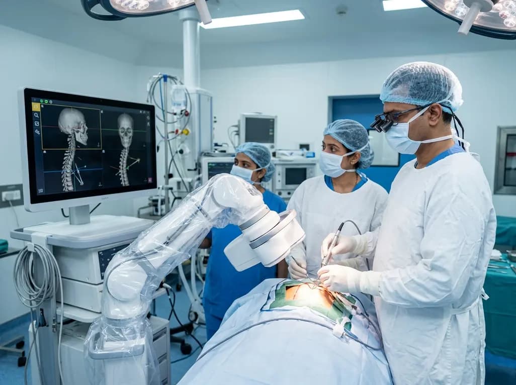
North India's first Centre of Excellence for Brain and Spine at Amrita Hospital Faridabad integrates Brainlab's Loop-X mobile robot. This landmark facility combines cone-beam computed tomography with surgical navigation to elevate precision, optimize operative workflows, and enhance specialized clinician training.
Today

At Homa CME 2026 at Hyderabad's T-Hub, healthcare leaders emphasized moving beyond routine metrics through digital calculators and precision stratification. Discover why characterizing individual insulin secretion, resistance patterns, and vascular risks transforms chronic metabolic care in India.
Today
