.webp&w=384&q=75)

Loading, please wait...
Loading, please wait...
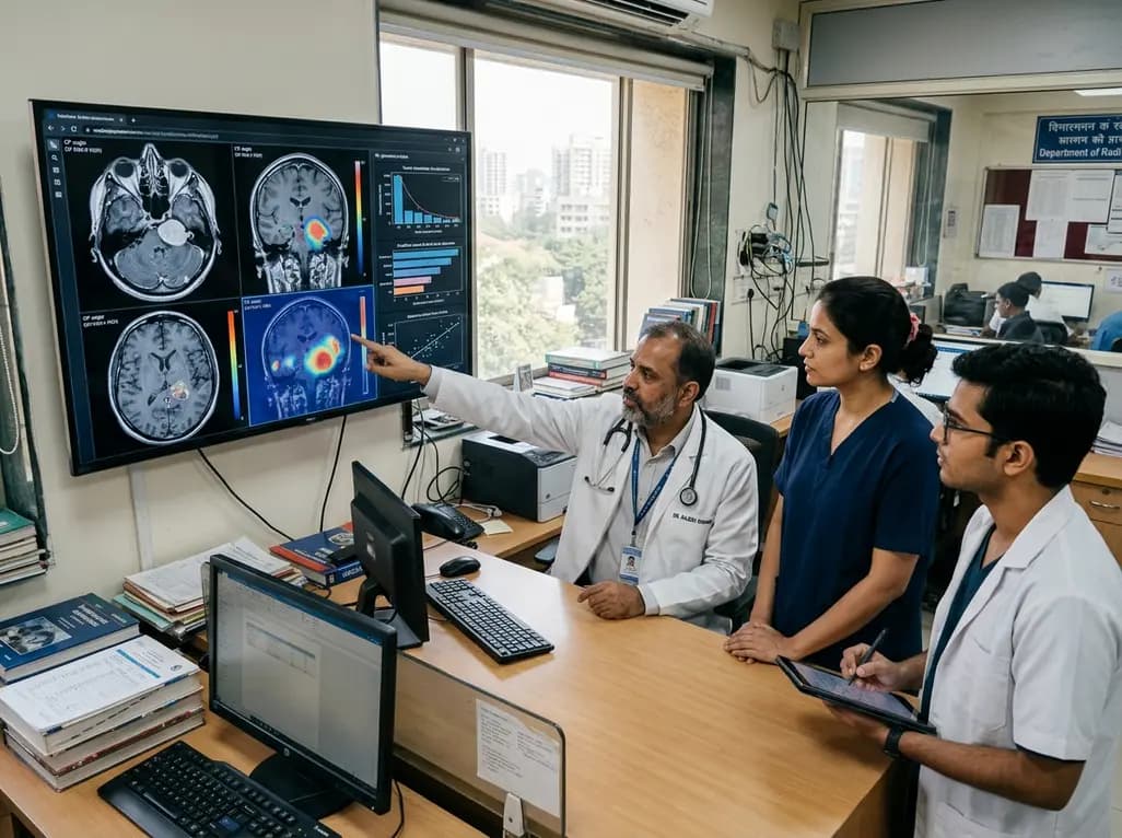
Vestibular schwannomas represent benign yet clinically formidable skull base neoplasms that arise from the eighth cranial nerve sheath. Managing these lesions requires delicate multidisciplinary decision-making because their anatomical proximity to critical neurovascular structures creates substantial functional risks. Clinicians traditionally rely on qualitative magnetic resonance imaging to monitor tumor progression and select appropriate therapeutic interventions. However, the emerging field of radiomics in vestibular schwannoma care is transforming conventional paradigms by converting medical scans into high-dimensional mineable datasets. Through advanced computational algorithms, radiomics extracts imperceptible quantitative imaging metrics that illuminate underlying tumor biology and behavior.
Vestibular schwannomas exhibit highly unpredictable natural growth patterns, creating dilemmas for skull base surgeons and oncologists. Consequently, clinicians must constantly choose between active surveillance, microsurgical resection, and stereotactic radiosurgery. Conventional magnetic resonance imaging provides structural dimensions and anatomical relationships, but it fails to capture subtle intra-tumoral heterogeneity. Moreover, standard surveillance protocols cannot reliably forecast rapid enlargement or sudden indolence in newly diagnosed patients. Therefore, healthcare teams need robust non-invasive biomarkers to guide tailored management pathways. Radiomic analysis addresses this unmet clinical need by evaluating texture distributions, spatial intensity gradients, and shape descriptors. By uncovering microscopic tissue patterns beyond human visual perception, computational extraction delivers crucial prognostic insights. Ultimately, these quantitative signatures empower clinicians to stratify patient risk before functional deficits emerge.
Pre-treatment risk stratification plays a pivotal role in deciding whether to pursue conservative observation or prompt intervention. Recent systematic evaluations indicate that machine learning models trained on baseline magnetic resonance images achieve moderate predictive accuracy. Specifically, pre-procedural models demonstrate area under the receiver operating characteristic curve values ranging between 0.66 and 0.70 for determining tumor behavior. These computational workflows analyze baseline contrast-enhanced T1-weighted and high-resolution T2-weighted sequences to quantify tissue architectural heterogeneity. Furthermore, non-machine learning texture analyses demonstrate significant correlations between gray-level spatial variations and longitudinal volumetric enlargement. Although baseline models currently exhibit moderate accuracy, they still offer objective adjunct information for multidisciplinary tumor boards. Consequently, integrating quantitative imaging metrics with routine clinical parameters strengthens baseline surveillance frameworks.
While pre-treatment modeling offers helpful guidance, post-procedural radiomic applications demonstrate even greater diagnostic and prognostic strength. Recent systematic findings show that post-intervention radiomics models achieve area under the curve metrics spanning from 0.75 to 1.00. These algorithms successfully predict long-term tumor control, radiosurgical response, and secondary treatment necessity. For instance, serial texture parameter alterations after stereotactic radiosurgery indicate therapeutic efficacy long before macroscopic volumetric regression becomes visually apparent. Additionally, machine learning algorithms process multi-parametric MRI sequences to identify radiation-induced changes and early recurrence patterns. Therefore, post-treatment radiomic profiling provides clinicians with an objective surveillance mechanism to evaluate therapeutic success. By detecting treatment failure early, medical teams can modify management strategies before severe neurological symptoms manifest.
Preserving cranial nerve function remains a primary surgical goal during vestibular schwannoma management. Postoperative facial nerve palsy severely compromises patient quality of life, yet traditional visual assessment cannot reliably predict post-surgical facial nerve outcomes. Fortunately, deep learning architectures offer transformative capabilities in this neurosurgical domain. Specifically, a recent convolutional neural network model achieved an outstanding area under the curve of 0.89 in predicting postoperative facial nerve function. Notably, this neural network significantly outperformed conventional machine learning classifiers, which achieved performance scores between 0.64 and 0.85. Deep learning architectures independently analyze spatial voxel hierarchies and subtle tissue interfaces along the cerebellopontine angle. As a result, artificial intelligence delivers actionable preoperative risk assessments, enabling neurosurgeons to customize surgical dissection strategies and enhance patient counseling.
Despite encouraging predictive performance, the translation of radiomics in vestibular schwannoma workflows into routine clinical practice faces substantial hurdles. Most available studies rely on small, single-center retrospective cohorts, which increases susceptibility to overfitting and selection bias. Furthermore, significant heterogeneity exists across imaging acquisition protocols, magnetic field strengths, and reconstruction parameters. Another major bottleneck involves tumor segmentation, as current workflows depend almost entirely on labor-intensive manual or semi-automated boundary delineations. In addition, the lack of external validation across multi-institutional datasets limits model generalizability and algorithmic reproducibility. To overcome these barriers, future investigators must adopt standardized reporting frameworks, such as the Image Biomarker Standardisation Initiative. Establishing shared multi-institutional data repositories will also prove indispensable for robust validation.
Integrating artificial intelligence into neuro-oncology workflows requires seamless collaboration between neurosurgeons, otolaryngologists, neuroradiologists, and data scientists. Prospective clinical trials represent the critical next step to validate whether radiomic predictions truly improve functional outcomes. Furthermore, combining radiomics with molecular markers, clinical indices, and advanced perfusion metrics could create comprehensive multi-modal decision support tools. Standardized automated segmentation tools will also streamline clinical deployment by eliminating observer variability in lesion contouring. In clinical settings, these tools could help clinicians pinpoint which asymptomatic tumors require immediate radiosurgery versus conservative observation. In addition, automated decision support can enhance individualized counseling regarding hearing preservation and facial nerve risk. Ultimately, rigorous prospective validation will enable radiomics to evolve from an experimental tool into everyday neuro-oncological practice.
Radiomics extracts hundreds of imperceptible quantitative features from routine magnetic resonance imaging scans. By converting imaging data into mineable metrics, computational algorithms predict tumor growth trajectories, assess treatment responsiveness, and forecast post-procedural cranial nerve outcomes, thereby assisting clinicians in tailoring individualized vestibular schwannoma treatment strategies.
Post-procedural radiomics models demonstrate moderate to strong predictive performance, with area under the curve metrics ranging from 0.75 to 1.00. These models effectively evaluate response to stereotactic radiosurgery and forecast tumor control, outperforming pre-treatment baseline models that currently achieve area under the curve scores between 0.66 and 0.70.
Yes, advanced artificial intelligence algorithms accurately assess postoperative facial nerve risk. Specifically, convolutional neural networks have achieved predictive accuracy with an area under the curve of 0.89, outperforming traditional machine learning classifiers. These models analyze complex spatial relationships to help neurosurgeons anticipate nerve vulnerability before surgical intervention.
Disclaimer: This content is for informational and educational purposes only and does not constitute medical advice or establish a doctor-patient relationship. Treatment decisions should always be made in consultation with qualified healthcare professionals based on individual patient circumstances. While we strive to present accurate and up-to-date information, medical knowledge evolves rapidly. Healthcare providers should verify details and exercise clinical judgment independently. Refer to the latest local and national guidelines for clinical practice.
References

Read summarized clinical updates, watch expert medical content, and earn CME certifications right from your smartphone.
Open your smartphone camera to scan and install.





A systematic review evaluates MRI radiomics and machine learning models in vestibular schwannoma, highlighting high predictive capacity for post-procedural outcomes and facial nerve preservation while outlining current validation challenges.
last month

A systematic review of 41 preclinical studies reveals that baicalin dampens systemic inflammation, inhibits NF-κB and NLRP3 pathways, and improves organ preservation in sepsis models. However, substantial study heterogeneity and a complete lack of human clinical trials highlight the need for further translational research.
Today
Discover the crucial distinctions between mesonephric and mesonephric-like proliferations of the female genital tract. Learn the key histomorphologic, immunohistochemical, and molecular differences essential for accurate diagnosis and clinical management.
Today

A large UK Biobank study reveals that plasma proteomic signatures capture preclinical organ damage and improve multiorgan risk prediction across early CKM syndrome stages 0 to 2 beyond traditional PREVENT clinical models.
Today
A narrative review investigates whether the articularis genus muscle functions as an independent anatomical entity or blends with the vastus intermedius. We evaluate its morphology, role in retracting the suprapatellar bursa, and direct clinical significance in anterior knee pain and arthroplasty.
Today

A Bayesian multilevel meta-analysis reveals that aerobic training combined with moderate carbohydrate restriction modestly lowers HbA1c in type 2 diabetes. However, sparse data and very low certainty leave incremental benefits over exercise or diet alone unproven, highlighting the need for individualized care.
Today
