

Loading, please wait...
Loading, please wait...

Glioblastoma and solitary brain metastases represent the vast majority of malignant intracranial neoplasms in adults. Although these distinct entities possess fundamentally different biological origins, they frequently present with identical imaging characteristics on routine magnetic resonance scans. Clinicians frequently encounter solitary ring-enhancing lesions with extensive vasogenic edema, creating diagnostic dilemmas. Consequently, the clinical implementation of radiomics in brain tumors provides a transformative computational approach. By converting conventional images into mineable quantitative data, machine learning algorithms discern intricate intratumoral textures imperceptible to the human eye. This objective assessment delivers vital noninvasive guidance for personalized neuro-oncological management.
Accurate preoperative discrimination between glioblastoma and solitary metastatic lesions remains critical for therapeutic planning. Glioblastoma typically demands aggressive maximal safe surgical resection followed by concurrent chemoradiotherapy. In contrast, systemic brain metastases often require stereotactic radiosurgery, whole-brain radiation, or targeted systemic therapies. Furthermore, an incorrect provisional diagnosis can subject patients to unnecessary surgical morbidity, procedural risks, and treatment delays.
Conventional magnetic resonance imaging relies upon contrast enhancement patterns, central necrosis, and peritumoral fluid collection. Unfortunately, solitary metastases frequently mimic the classic ring-enhancing characteristics, irregular borders, and microvascular proliferation typical of high-grade gliomas. Moreover, the non-enhancing perilesional T2/FLAIR hyperintensity contains infiltrative neoplastic cells in glioblastoma, whereas it reflects pure vasogenic edema in metastatic tumors. Standard structural imaging cannot reliably distinguish these subtle cellular differences. Therefore, physicians often resort to stereotactic biopsy, which introduces risks of intracranial hemorrhage, neurological deficits, and sampling errors. Noninvasive digital biomarkers help bridge this clinical diagnostic gap effectively.
Radiomic analysis transforms standard radiological imaging into multidimensional quantitative arrays. Specifically, advanced neuroimaging protocols evaluate contrast-enhanced T1-weighted, T2-weighted, and diffusion-weighted sequences. Computational algorithms extract hundreds of mathematical features from segmented regions of interest. These features capture first-order voxel intensity distributions, spatial shapes, and complex higher-order textural relationships.
Consequently, radiomics in brain tumors deciphers hidden phenotypic spatial heterogeneity and microstructural architecture beyond human visual perception. Machine learning classifiers, including support vector machines, random forest models, and gradient boosting algorithms, identify subtle patterns from these multi-parametric features. In particular, the peritumoral boundary between the enhancing margin and surrounding edema demonstrates distinct radiomic signatures. Infiltrating neoplastic cells in glioblastoma disrupt local tissue architecture, altering spatial entropy and contrast gradients. Conversely, brain metastases exhibit mechanical displacement of adjacent parenchyma without infiltrative neoplastic seeding. Thus, machine learning classifiers leverage these subtle textural gradients to distinguish tumor histopathology reliably without requiring invasive tissue sampling.
A rigorous systematic review and meta-analysis synthesized diagnostic evidence across 39 international studies involving 11,558 patients. The investigation evaluated 31 binary studies distinguishing glioblastoma from brain metastases and 8 complex multiclass cohorts. For binary classification during internal validation, radiomics models demonstrated an impressive pooled sensitivity of 88.9% and a pooled specificity of 83.0%. Importantly, independent external validation cohorts retained strong diagnostic accuracy, achieving a pooled sensitivity of 83.8% and a pooled specificity of 80.7%.
These robust external validation metrics confirm that extracted computational features reflect genuine biological distinctions rather than dataset overfitting. Furthermore, the researchers evaluated multiclass models differentiating glioblastoma, brain metastases, and primary central nervous system lymphoma. In these demanding multi-disease cohorts, pooled sensitivity reached 84.4% for glioblastoma, 70.6% for brain metastases, and 75.0% for lymphoma. The corresponding specificities remained high at 82.5%, 88.8%, and 94.4%, respectively. Therefore, computational radiomics demonstrates substantial diagnostic value across complex differential scenarios in neuro-oncology.
Evaluating artificial intelligence against clinical human experts provides essential perspective for real-world medical practice. Interestingly, the systematic review demonstrated that radiomics algorithms achieved diagnostic performance comparable or superior to experienced neuroradiologists. While clinicians evaluate qualitative morphological characteristics, human perception cannot systematically quantify voxel-level spatial gradients, directional textural coherence, or fine mathematical asymmetry.
For example, microstructural cellular infiltration within the non-enhancing peritumoral T2 hyperintense zone routinely eludes visual inspection but generates distinct computational texture signatures. Consequently, machine learning models detect these microscopic tumor alterations with remarkable consistency. However, computational models should assist rather than replace clinical experts. When neuroradiologists interpret complex neuroimaging scans alongside algorithmic probability outputs, diagnostic accuracy and clinical confidence improve considerably. Additionally, artificial intelligence tools can reduce diagnostic discrepancies between specialized academic medical centers and regional hospitals. As a result, community neurosurgeons and general radiologists gain immediate access to standardized diagnostic support, improving care coordination.
Despite remarkable diagnostic accuracy, several critical methodological challenges currently prevent routine clinical deployment. Quality evaluations utilizing the Radiomics Quality Score 2.0 revealed substantial deficits in study design and standardized reporting. The majority of included studies relied upon retrospective single-center data, creating susceptibility to selection bias and institutional imaging idiosyncrasies.
Furthermore, technical variability across scanner vendors, magnetic field strengths, acquisition sequences, and reconstruction algorithms introduces feature instability. Manual tumor segmentation remains labor-intensive and subjective, whereas automated deep learning segmentation models require broader prospective testing across diverse patient populations. Consequently, regulatory approval demands large-scale prospective multi-center studies with consecutive patient enrollment. In particular, researchers must standardize image pre-processing protocols and publish reproducible algorithmic code. Moreover, integrating physiological imaging sequences, such as perfusion MRI and magnetic resonance spectroscopy, with clinical parameters will likely enhance precision. Thus, rigorous prospective validation remains essential to translate these promising computational algorithms into daily clinical practice.
Radiomics cannot replace definitive histopathology or molecular genetic testing in clinical neuro-oncology. Histopathological examination, immunohistochemical analysis, and molecular profiling of biomarkers such as IDH mutation and MGMT promoter methylation remain essential standards for therapeutic decision-making. However, radiomics serves as an invaluable noninvasive diagnostic adjunct. It offers critical preoperative diagnostic guidance when surgical biopsy poses substantial procedural morbidity, in deep surgically inaccessible brain regions, or when rapid clinical intervention is urgently required.
Differentiating these intracranial neoplasms on standard magnetic resonance imaging is notoriously difficult because both tumors frequently appear as solitary, thick, ring-enhancing lesions with central necrotic cores and extensive perilesional edema. Furthermore, conventional visual inspection cannot reliably determine whether surrounding T2/FLAIR hyperintensity represents pure vasogenic edema typical of metastasis or infiltrative non-enhancing neoplastic cells characteristic of glioblastoma. This morphological overlap creates diagnostic uncertainty that frequently necessitates invasive stereotactic biopsy.
Routine clinical translation requires large-scale prospective multi-center clinical trials evaluating consecutive patients across varied scanner hardware and standardized imaging protocols. Additionally, investigators must validate fully automated segmentation pipelines and adopt transparent, reproducible radiomic feature extraction standards. Finally, software developers must integrate these algorithms into existing hospital picture archiving and communication systems while demonstrating measurable improvements in patient outcomes, diagnostic timeliness, and clinical workflow efficiency.
Disclaimer: This content is for informational and educational purposes only... Refer to the latest local and national guidelines for clinical practice.
References
Hong C et al. Diagnostic Performance of Radiomics-Based Machine Learning in Differentiating Glioblastoma From Brain Metastasis: Systematic Review and Meta-Analysis. J Med Internet Res. 2026 Nov 19. doi: 10.2196/104060. PMID: 43056705.
Na YS et al. Radiomics-Based Machine Learning for Differentiating Glioblastoma From Brain Metastases. Investig Magn Reson Imaging. 2026;30(3):145-154.
Bae S et al. Robust performance of deep learning for distinguishing glioblastoma from single brain metastasis using radiomic features: model development and validation. Sci Rep. 2020;10(1):12110.
Gillies RJ, Kinahan PE, Hricak H. Radiomics: Images Are More than Pictures, They Are Data. Radiology. 2016;278(2):563-577.

Read summarized clinical updates, watch expert medical content, and earn CME certifications right from your smartphone.
Open your smartphone camera to scan and install.





A systematic review of 39 studies and 11,558 patients reveals that radiomics-based machine learning models achieve high diagnostic accuracy in differentiating glioblastoma from brain metastasis, yielding 83.8% sensitivity and 80.7% specificity in external validation cohorts.
Today
A clinical decision-support framework using health checkup data from 231,683 adults predicts incident obesity with high discrimination (AUROC 0.918). The system integrates temporal survival analysis and risk-factor explanations to prioritize individualized preventive interventions.
Today

A large cohort study demonstrates that fetal growth restriction in a first pregnancy raises maternal cardiovascular risk within five years postpartum, including elevated rates of heart failure, stroke, and myocardial infarction. Clinicians must initiate proactive postpartum screening and cardiovascular prevention.
Yesterday
A multicenter observational study evaluated an 8-week digital health monitoring platform using wearable sensors in ambulatory heart failure. While participants achieved high daily wear-time of 11.7 hours and 80% completion, initial recruitment revealed significant technology-related onboarding barriers.
Today
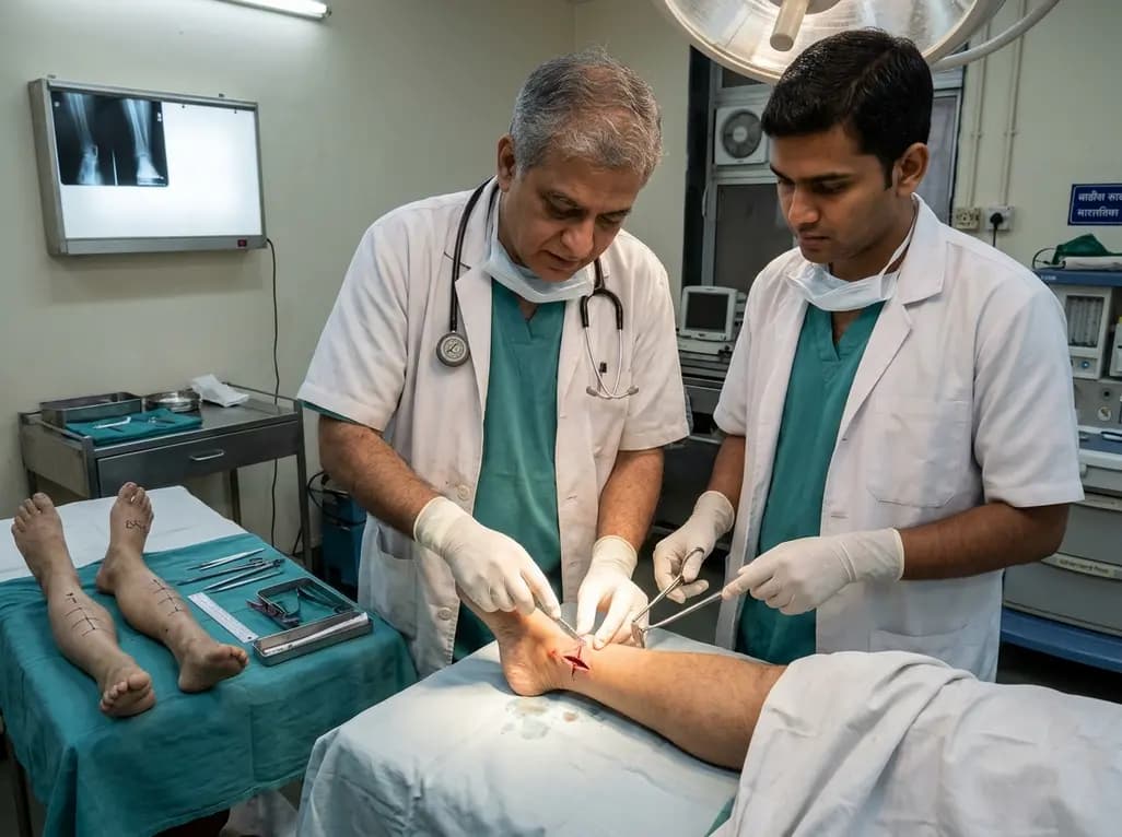
A paired cadaveric study compared triple and quintuple percutaneous Achilles tendon lengthening. While quintuple hemisection showed numerically greater elongation, differences were not statistically significant.
Yesterday
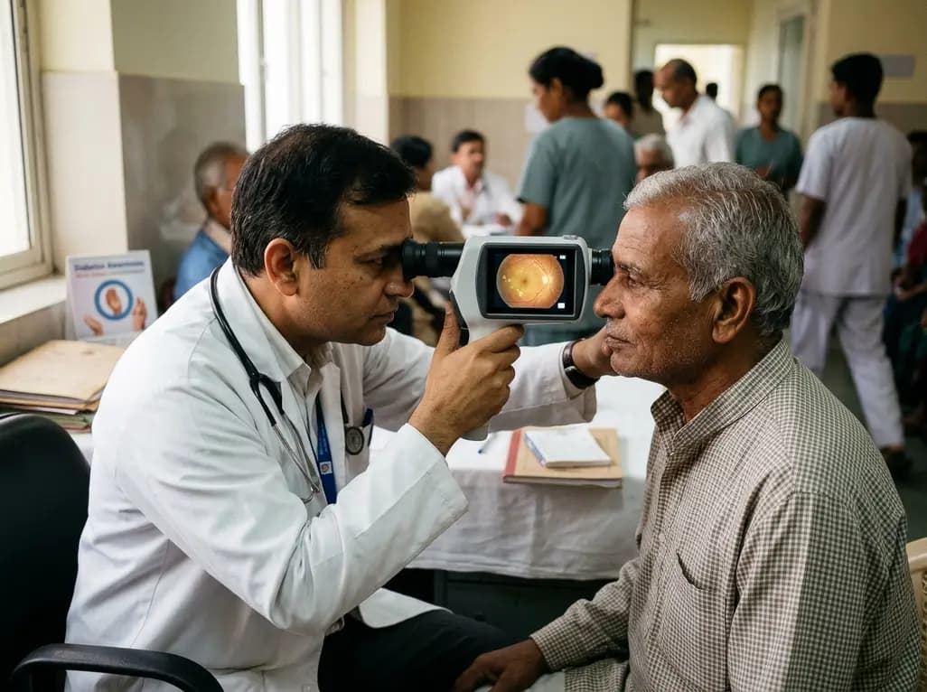
Diabetic retinopathy remains a leading cause of preventable visual impairment. This review outlines key strategies for screening, systemic risk factor control, advanced diagnostic imaging, and timely specialist referrals within primary and multidisciplinary care models.
Today
