

Loading, please wait...
Loading, please wait...

Diabetic retinopathy screening serves as an indispensable tool for preventing irreversible visual impairment in individuals with diabetes mellitus. Consequently, healthcare systems worldwide face mounting pressure to expand early detection protocols. Microvascular damage progresses silently during early disease stages, and patients frequently remain asymptomatic until severe visual loss occurs. Therefore, structured screening initiatives bridge the critical gap between early tissue injury and timely therapeutic intervention. In clinical practice, routine fundus evaluation detects microaneurysms, dot-and-blot hemorrhages, and macular edema long before subjective visual changes arise. Moreover, delayed screening drastically increases the likelihood of catastrophic outcomes such as vitreous hemorrhage or tractional retinal detachment. Healthcare providers must recognize that systematic ocular evaluations preserve functional vision and significantly reduce long-term medical expenditures. Furthermore, socioeconomic disparities and limited geographic access to ophthalmologists frequently restrict patient participation in conventional eye clinics. Primary care teams can eliminate these persistent barriers by integrating point-of-care retinal evaluations into regular chronic disease consultations. Ultimately, timely identification of retinal changes preserves personal independence and enhances overall quality of life for vulnerable patient populations. As a result, regular surveillance remains the cornerstone of modern ophthalmic preventive care.
Persistent hyperglycemia drives the initial molecular cascade underlying diabetic retinopathy. Specifically, elevated blood glucose levels induce oxidative stress, endothelial damage, and chronic retinal capillary occlusion. As pericytes detach and apoptosis accelerates, retinal vessels lose structural stability and leak fluid into surrounding tissues. Consequently, localized tissue ischemia stimulates substantial upregulation of vascular endothelial growth factor. This signaling molecule triggers aberrant neovascularization, which characterizes proliferative diabetic retinopathy. In addition, systemic hypertension accelerates vascular shearing stress and substantially exacerbates retinal microvascular permeability. Dyslipidemia also correlates strongly with the development of hard exudates and central macular edema. Therefore, clinicians must assess metabolic control comprehensively rather than focusing solely on ocular findings. Glycated hemoglobin targets below seven percent significantly retard disease onset and slow progression. Furthermore, strict blood pressure regulation protects vulnerable microvasculature against hydrostatic trauma. Similarly, lipid-lowering therapies stabilize vascular walls and minimize retinal lipid deposition. Clinicians must address nephropathy as well, because microalbuminuria indicates generalized vascular vulnerability. Thus, holistic cardiometabolic control forms the true backbone of effective ocular preservation. Moreover, multidisciplinary teamwork ensures that patients achieve individualized glycemic and cardiovascular targets throughout their lifespan.
Nurse practitioners and primary care physicians occupy a unique vantage point on the frontlines of diabetes care. In fact, these frontline providers routinely interact with patients far more frequently than subspecialty eye surgeons. Therefore, mid-level clinicians can proactively orchestrate early detection workflows during standard wellness visits. Furthermore, advanced practice providers evaluate glycemic stability, adjust insulin regimens, and manage coexisting cardiovascular conditions. By taking proactive responsibility for surveillance, mid-level providers bridge communication gaps between community clinics and tertiary eye centers. Additionally, these clinicians identify social determinants of health that frequently hinder clinic attendance, such as transportation barriers and financial distress. They can prescribe convenient local screenings and establish electronic consultations with retinal experts. Consequently, health centers achieve higher compliance rates when mid-level clinicians oversee screening protocols. Clinicians also reinforce dietary counseling, physical activity targets, and strict medication adherence during every clinical encounter. In doing so, they convert routine diabetes visits into comprehensive organ-preservation checkups. Ultimately, empowering primary care teams establishes a durable safety net that captures disease progression before visual acuity suffers irreparable damage. Moreover, sustained clinical engagement fosters deep mutual trust, which dramatically enhances long-term adherence to therapeutic recommendations.
Modern diagnostic technology has transformed diabetic eye evaluations into streamlined, accessible clinical procedures. Digital non-mydriatic fundus cameras allow trained medical staff to capture sharp retinal images without requiring chemical pupillary dilation. In addition, validated artificial intelligence algorithms can now analyze fundus photographs with exceptional accuracy within minutes. These automated systems rapidly differentiate normal retinas from vision-threatening retinopathy, thereby streamlining clinical decision-making. However, artificial intelligence tools serve to augment rather than replace experienced ophthalmic specialists. When autonomous systems or human evaluators identify moderate non-proliferative changes, clinicians must generate an expedited referral. Specifically, patients displaying severe non-proliferative retinopathy, macular edema, or neovascularization require urgent vitreoretinal evaluation. Optical coherence tomography provides microscopic cross-sectional images that precisely quantify retinal thickening and intraretinal fluid. Furthermore, telemedicine networks allow remote retina specialists to review ambiguous images and guide local management strategies. Consequently, structured digital referral pathways prevent unnecessary travel for stable patients while expediting care for critical cases. Standardizing these referral thresholds ensures that sight-threatening lesions receive immediate medical attention. Therefore, primary care clinics that implement telemedicine networks significantly expand access and eliminate diagnostic backlogs across regional communities.
Empowering individuals through structured health education forms an indispensable component of successful retinopathy management. Many patients erroneously assume that good visual acuity guarantees the absence of retinal disease. Consequently, clinicians must proactively explain that microvascular lesions frequently develop without producing noticeable symptoms. Clear communication regarding glycemic control, blood pressure maintenance, and regular examinations dispels widespread misconceptions and drives patient engagement. Furthermore, discussing advanced therapeutic interventions reassures patients that clinical options exist to halt vision loss. Intravitreal anti-vascular endothelial growth factor injections currently serve as the first-line therapy for central-involved diabetic macular edema. In addition, targeted panretinal photocoagulation stabilizes ischemic tissue and induces regression of fragile neovascular vessels. Vitrectomy surgery successfully restores useful vision in advanced situations involving non-clearing vitreous hemorrhage or retinal detachment. However, these advanced surgical and pharmacological therapies achieve the best visual outcomes when initiated before widespread tissue destruction occurs. Therefore, educating patients about urgent warning signs, such as new floaters or sudden blurriness, facilitates timely self-referral. Ultimately, informed patients collaborate more effectively with their healthcare teams, safeguarding their sight over decades of living with diabetes. As a result, longitudinal education remains as vital as pharmacological therapy in preserving lifelong functional vision.
Patients diagnosed with type 2 diabetes must undergo comprehensive retinal screening immediately upon diagnosis, because occult microvascular changes often develop years before clinical detection. Conversely, individuals with type 1 diabetes should initiate annual evaluations five years after diagnosis. Furthermore, clinicians must evaluate pregnant women with pre-existing diabetes during their first trimester, because hormonal fluctuations accelerate microvascular alterations. Therefore, strict adherence to these initial screening timelines prevents irreversible retinal damage across all patient cohorts.
Primary clinicians must immediately refer patients exhibiting sudden vision loss, flashes of light, or a sudden burst of dark floaters. These acute symptoms frequently signal vitreous hemorrhage or imminent retinal detachment. Additionally, objective findings such as extensive retinal neovascularization, preretinal bleeding, or clinically significant macular edema warrant urgent specialist intervention. Therefore, prompt vitreoretinal referral remains essential to arrest irreversible photoreceptor injury, implement targeted intravitreal anti-VEGF therapies, and preserve long-term functional vision.
Nurse practitioners bridge critical healthcare delivery gaps by integrating digital non-mydriatic fundus photography into regular primary care consultations. In addition, these clinicians manage underlying hypertension, dyslipidemia, and hyperglycemia, directly addressing systemic drivers of microvascular destruction. Nurse practitioners also deliver culturally competent health education that clarifies the asymptomatic nature of early retinal lesions. Consequently, their proactive involvement boosts annual screening compliance, facilitates timely ophthalmic referrals, and significantly reduces rates of preventable blindness in underserved communities.
Disclaimer: This content is for informational and educational purposes only... Refer to the latest local and national guidelines for clinical practice.
References

Read summarized clinical updates, watch expert medical content, and earn CME certifications right from your smartphone.
Open your smartphone camera to scan and install.





Diabetic retinopathy remains a leading cause of preventable visual impairment. This review outlines key strategies for screening, systemic risk factor control, advanced diagnostic imaging, and timely specialist referrals within primary and multidisciplinary care models.
Today

A large cohort study demonstrates that fetal growth restriction in a first pregnancy raises maternal cardiovascular risk within five years postpartum, including elevated rates of heart failure, stroke, and myocardial infarction. Clinicians must initiate proactive postpartum screening and cardiovascular prevention.
Today

Cardiovascular surgery is transforming rapidly across India as tertiary hospitals embrace robotic assistance and AI-driven planning. Delhi-NCR now spearheads this shift, demonstrating higher procedural volumes, lower tissue trauma, and accelerated patient recovery compared to conventional sternotomy approaches.
Today
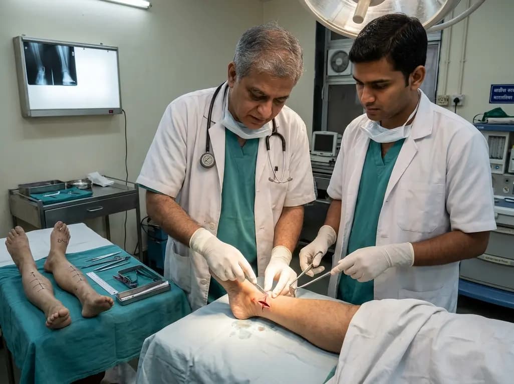
A paired cadaveric study compared triple and quintuple percutaneous Achilles tendon lengthening. While quintuple hemisection showed numerically greater elongation, differences were not statistically significant.
Yesterday
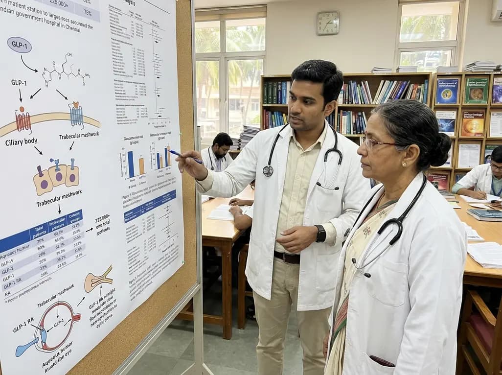
A systematic review and meta-analysis of over 225,000 patients explores whether GLP-1 receptor agonists reduce the risk of glaucoma and ocular hypertension. While pooled data show a 31% lower risk, low certainty and wide prediction intervals urge cautious optimism and further randomized trial evaluation.
Today
