

Loading, please wait...
Loading, please wait...
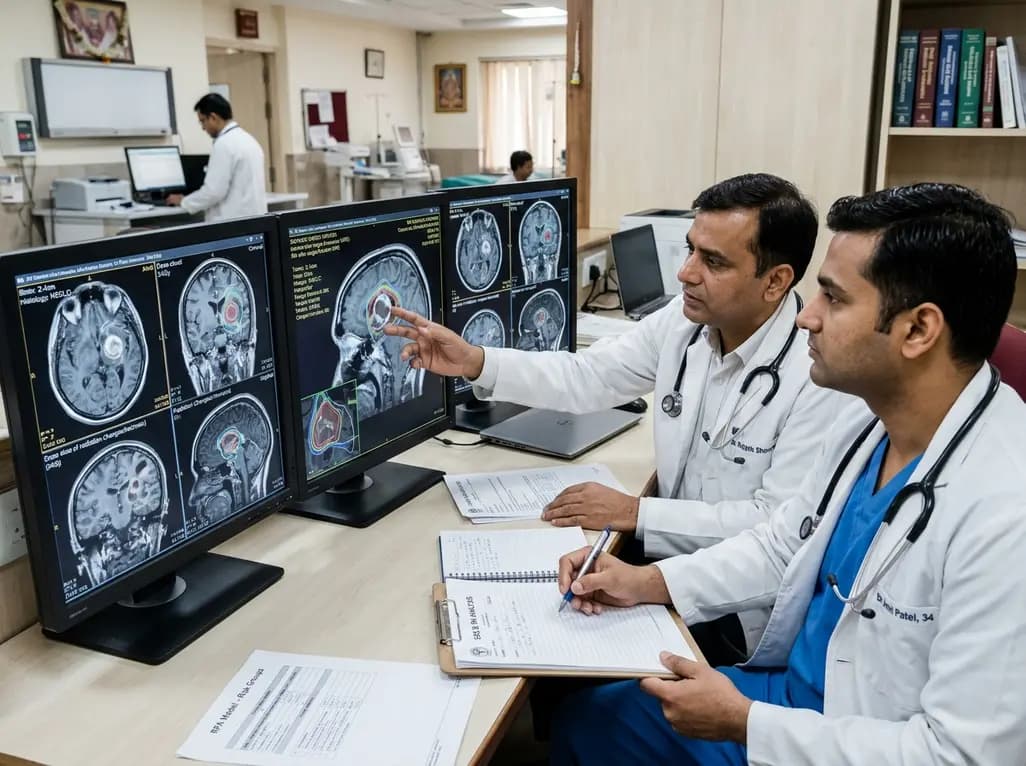
Stereotactic radiosurgery provides exceptional local disease control for secondary intracranial malignancies. However, radiation necrosis after SRS remains a distressing late adverse effect that significantly impairs patient quality of life. As targeted agents and immune checkpoint inhibitors prolong survival in oncology, managing radiosurgery neurotoxicity has become increasingly paramount. Radiation necrosis mimics tumor progression radiologically and clinically, often prompting unwarranted surgical resections or premature cessation of active systemic therapies. Recently, radiation oncologists at the Cleveland Clinic developed a practical decision-tree model using recursive partitioning analysis to predict necrosis risk in intact brain metastases treated with single-fraction radiosurgery. This educational guide explores the clinical evidence, risk stratification classes, and individualized strategies designed to optimize neuro-oncological care.
Cerebral radiation injury reflects a dynamic and complex cascade of parenchymal and vascular damage. Following single-fraction radiosurgery, high focal doses trigger microvascular endothelial cell death and disruption of the blood-brain barrier. Consequently, plasma proteins extravasate into surrounding brain tissue, provoking extensive vasogenic edema. Furthermore, reactive astrocytes and microglia release potent inflammatory mediators, particularly vascular endothelial growth factor (VEGF) and interleukin cytokines. This persistent inflammatory microenvironment exacerbates tissue hypoxia, fibrinoid vascular degeneration, and subsequent coagulative necrosis of white matter. In addition, tumor histology and concurrent pharmacotherapies modulate local tissue radiosensitivity. For instance, histology with inherent vascular vulnerability or enhanced radiosensitization can amplify glial apoptosis. Notably, post-treatment microhemorrhages can also worsen tissue inflammation by releasing toxic free hemoglobin and hemosiderin. Because cerebral tissue has limited regenerative capacity, this persistent oxidative stress perpetuates a vicious neuroinflammatory cycle. Therefore, identifying baseline clinical drivers is critical to prevent debilitating, irreversible intracranial tissue injury.
To refine risk prediction, investigators evaluated 170 patients with 919 intact brain metastases treated between 2017 and 2021 with single-fraction radiosurgery. The patient cohort demonstrated a median follow-up of 9 months, presenting diverse primaries including non-small cell lung cancer (49%), breast cancer (12%), melanoma (11%), and renal cell carcinoma (6%). The median radiosurgery prescription dose was 24 Gy, and the median maximum lesion dimension was 0.7 cm. Overall, radiation necrosis occurred in 110 lesions (12.2%), with 32 lesions (3.5%) presenting with neurological symptoms. The median time to necrosis development was 4.9 months after radiosurgery. Through multivariable Cox regression, investigators identified key predictors with significance thresholds of p < 0.1 for tree construction. In addition to lesion diameter, variables included primary tumor histology, lesion location, dosimetry metrics such as prescription isodose volume, prior radiosurgery exposure, concurrent systemic therapies, and intratumoral hemorrhage. Consequently, these robust data points established the foundational parameters for objective, decision-tree modeling.
The primary output of the study is a clinically intuitive recursive partitioning analysis that divides treated lesions into four distinct prognostic groups. In this model, maximum lesion dimension serves as the primary initial branching node, split cleanly at the 0.8 cm threshold. Lesions measuring 0.8 cm or smaller further branch by primary tumor histology. Specifically, Group 1 comprises lesions 0.8 cm or smaller arising from non-breast, non-lung, and non-renal histologies, achieving an extraordinary two-year necrosis-free survival rate of 99%. Group 2 includes lesions 0.8 cm or smaller with breast cancer, non-small cell lung cancer, or renal cell carcinoma, yielding an 89% two-year necrosis-free survival rate. Conversely, for larger lesions exceeding 0.8 cm, the model identifies post-radiosurgery hemorrhage as the decisive secondary factor. Group 3 consists of lesions greater than 0.8 cm without post-radiosurgery hemorrhage, exhibiting a 70% two-year necrosis-free survival rate. Finally, Group 4 encompasses lesions larger than 0.8 cm complicated by post-treatment hemorrhage, demonstrating a markedly reduced two-year necrosis-free survival rate of only 52%.
Accurate differentiation between radiation-induced necrosis and tumor recurrence represents an everyday clinical challenge for multidisciplinary tumor boards. Both conditions demonstrate rim enhancement, central hypointensity, and surrounding vasogenic edema on standard contrast-enhanced T1 and T2 magnetic resonance imaging. However, misinterpreting radiation injury as recurrence leads to unnecessary repeat irradiation or risky salvage surgeries. Conversely, mistaking disease progression for benign necrosis delays essential oncological interventions. Clinicians can resolve this ambiguity by integrating advanced neuroimaging modalities. For example, dynamic susceptibility contrast magnetic resonance perfusion evaluates relative cerebral blood volume; elevated perfusion indicates active hypervascular neoplasm, whereas depressed perfusion indicates avascular necrotic tissue. Similarly, MR spectroscopy detects elevated lipid and lactate peaks alongside suppressed choline in necrosis. In contrast, viable tumors show marked elevation of choline-to-creatine ratios. Furthermore, positron emission tomography utilizing radiolabeled amino acids, such as fluoroethyltyrosine, provides outstanding diagnostic specificity. Applying the recursive partitioning model allows clinicians to anticipate necrosis probabilities before interpreting these complex neuroimaging findings.
These predictive findings provide concrete opportunities to personalize radiosurgery treatment plans for oncology patients. For lesions designated within Group 1 and Group 2, single-fraction radiosurgery delivers excellent local control with minimal risk of late parenchymal injury. Therefore, radiation oncologists can confidently maintain standard ablative single-fraction dosing schedules in these favorable subsets. In contrast, lesions allocated to Group 3 and Group 4 carry a substantial risk of necrosis, reaching nearly 50% in the presence of hemorrhage. For these high-risk targets, clinicians should actively consider dose de-escalation or transition toward fractionated stereotactic radiotherapy regimens, delivering three to five fractions. Fractionation exploits radiobiological repair mechanisms in normal brain tissue, thereby maintaining durable tumor ablation while substantially reducing endothelial toxicity. Moreover, for patients in high-risk categories who develop post-radiosurgery hemorrhage or increasing perilesional edema, clinicians should establish shorter follow-up imaging intervals. Early identification of asymptomatic swelling enables timely therapeutic interventions, such as conservative corticosteroid tapering or anti-VEGF therapy with bevacizumab, before irreversible neurological deficits occur.
Optimal care for patients with brain metastases requires synchronized coordination among radiation oncologists, medical oncologists, and neurosurgeons. Increasingly, clinicians combine intracranial radiosurgery with powerful modern systemic drugs, including antibody-drug conjugates and immune checkpoint inhibitors. Although these systemic combinations improve intracranial disease response, certain targeted agents exacerbate vascular permeability and heighten tissue susceptibility to necrosis. Therefore, medical teams must carefully coordinate systemic treatment timing relative to radiosurgery delivery. Furthermore, while the Cleveland Clinic decision-tree model provides actionable clinical insights, prospective multi-institutional trials must externally validate its generalizability across varied machine platforms, such as Gamma Knife and linear accelerators. In addition, incorporating artificial intelligence radiomics and liquid biopsy biomarkers into the four-tier schema could enhance predictive precision. Ultimately, adopting structured prognostic frameworks allows multidisciplinary teams to preserve neurological function, minimize iatrogenic harm, and optimize oncological outcomes in cancer patients with intracranial metastases.
Radiation necrosis is a localized, non-malignant inflammatory reaction of brain tissue occurring months or years after radiosurgical treatment. It is characterized by vascular hyalinization, endothelial injury, fibrinoid necrosis, and disruption of the blood-brain barrier. Patients may remain entirely asymptomatic or develop headaches, seizures, focal neurological deficits, and debilitating cognitive impairment.
The recursive partitioning analysis model identifies maximum lesion dimension as the primary determinant, using an 0.8 cm cutoff. Secondary branching factors include primary tumor histology—specifically breast, non-small cell lung, and renal cancer histologies—for smaller lesions, and post-treatment hemorrhage for lesions exceeding 0.8 cm in maximum diameter.
Intratumoral hemorrhage occurring after radiosurgery substantially amplifies parenchymal inflammation and local oxidative damage. In the recursive partitioning model, lesions larger than 0.8 cm that experienced post-treatment hemorrhage exhibited the highest risk, with two-year necrosis-free survival dropping to 52%, compared to 70% in lesions without hemorrhage.
Disclaimer: This content is for informational and educational purposes only and should not be taken as medical advice. Clinical decisions must always be made based on independent evaluation and patient assessment. Refer to the latest local and national guidelines for clinical practice.
References

Read summarized clinical updates, watch expert medical content, and earn CME certifications right from your smartphone.
Open your smartphone camera to scan and install.





A Cleveland Clinic study establishes the first recursive partitioning analysis model predicting radiation necrosis after single-fraction SRS for intact brain metastases, categorizing lesions into four risk tiers based on tumor diameter, histology, and post-treatment hemorrhage to guide clinical management.
Today

A qualitative study investigates whether traditional bedside clinical examinations still mirror contemporary physician practice. Findings support retaining long cases while reforming short cases toward hypothesis-driven assessments.
Today

A pilot study evaluated WearDOXXN, a capillary microfluidic sweat patch, for noninvasive breast cancer monitoring by comparing sweat and serum CEA and PIP/GCDFP-15 levels.
Yesterday

India faces a massive diabetes burden with over 89 million adults affected. Managing blood sugar in isolation fails to prevent severe cardiorenal complications. Clinicians must adopt an integrated metabolic ecosystem that prioritizes early detection, multifactorial risk assessment, and personalized therapies.
Yesterday

The Gross Motor Function Classification System (GMFCS) stops its validated age bands at 18. Recent evidence emphasizes that evaluating adult cerebral palsy requires purpose-sensitive documentation combining adolescent ratings with dynamic profiles of mobility, fatigue, pain, and community participation.
Yesterday

A breakthrough 3D-printed piezoelectric collagen hydrogel activated by ultrasound accelerates diabetic wound repair, demonstrating 98.5% wound closure, robust antibacterial action, and enhanced vascularization without toxic chemical crosslinkers.
Today
