.webp&w=384&q=75)

Loading, please wait...
Loading, please wait...

Neoadjuvant chemotherapy downstages locally advanced breast tumors and provides a direct in vivo assessment of treatment efficacy. When patients achieve a pathological complete response, their long-term event-free survival improves substantially. Therefore, oncologists continually search for noninvasive imaging biomarkers to predict therapeutic outcomes early during treatment. Dynamic contrast-enhanced magnetic resonance imaging effectively evaluates tumor geometry and functional vascularity. Recently, researchers evaluated quantitative background parenchymal enhancement to determine whether normal tissue enhancement predicts chemotherapeutic response. However, clinicians must understand whether automated parenchymal metrics provide meaningful prognostic utility beyond intrinsic tumor biology.
Background parenchymal enhancement describes how normal fibroglandular tissue enhances with gadolinium contrast during breast dynamic contrast-enhanced magnetic resonance imaging. Clinicians historically categorized this enhancement qualitatively using standard BI-RADS descriptions ranging from minimal to marked. However, qualitative assessments frequently suffer from observer variability and inconsistent reproducibility between different institutions. Consequently, investigators developed automated computational techniques to quantify parenchymal enhancement objectively. Because microvascular permeability and circulating hormonal stimuli drive parenchymal contrast uptake, physiological changes during cytotoxic chemotherapy might mirror therapeutic drug delivery. Therefore, many oncologists hypothesized that quantitative background parenchymal enhancement dynamics could independently predict pathological complete response. Furthermore, researchers hoped automated imaging might detect tumor responsiveness before structural tumor shrinkage becomes clinically noticeable. Nonetheless, historical studies produced conflicting results regarding the prognostic value of parenchymal perfusion changes. While several small studies linked declining enhancement to improved tumor eradication, other reports indicated that parenchymal features merely reflect systemic patient factors like age and menopausal status. Thus, standardized clinical investigations remained vital to clarify these contradictory findings.
To resolve existing uncertainties, researchers conducted a comprehensive retrospective investigation involving 142 breast cancer patients receiving neoadjuvant chemotherapy. Every patient completed standardized dynamic contrast-enhanced magnetic resonance imaging scans before and after chemotherapy. The research team implemented the custom-trained nnU-Net deep learning framework to execute automated segmentations of both breast parenchyma and primary malignant lesions. Consequently, this computational approach eliminated subjective human contouring variations and produced standardized volumetric measurements. The investigators subsequently calculated quantitative enhancement parameters across multiple signal intensity thresholds and signal enhancement ratios. In addition, the team recorded essential clinicopathological variables for every participant. These variables included age, menopausal status, body mass index, histological grade, and hormone receptor status. Furthermore, pathologists evaluated human epidermal growth factor receptor 2 status and Ki-67 proliferation indices on baseline biopsy specimens. The authors performed multivariable logistic regression analyses to identify independent predictors of treatment response. Additionally, they conducted pre-specified subgroup analyses using Mann-Whitney U tests while adjusting for body mass index and menopausal status. Through this rigorous methodology, the study isolated the genuine predictive performance of automated imaging biomarkers.
The investigation delivered clear conclusions regarding the predictive capability of parenchymal imaging metrics compared to tumor biology. Notably, none of the evaluated quantitative imaging metrics demonstrated a statistically significant association with pathological complete response across the complete cohort. Neither baseline parenchymal enhancement nor longitudinal enhancement alterations predicted treatment success in unselected patients. In sharp contrast, traditional clinicopathological markers strongly correlated with therapeutic outcomes. Specifically, multivariable logistic regression revealed that human epidermal growth factor receptor 2 positivity tripled the likelihood of achieving complete response, demonstrating an adjusted odds ratio of 2.99. Furthermore, a high Ki-67 proliferation index independently predicted complete response, showing an adjusted odds ratio of 1.03 per unit increase. The multivariable diagnostic model incorporating these clinicopathological determinants achieved an area under the curve of 0.76. Conversely, combining quantitative parenchymal enhancement with clinicopathological variables produced a cross-validated discrimination of merely 0.69. Moreover, intrinsic tumor markers entirely drove this discrimination, while parenchymal metrics added zero incremental predictive accuracy. Therefore, the data confirm that tumor-intrinsic biological properties remain the primary determinants of therapeutic responsiveness.
Although the overall cohort demonstrated neutral results, exploratory subgroup analyses revealed fascinating biological nuances across specific tumor subtypes. In the human epidermal growth factor receptor 2-negative cohort, patients achieving complete response exhibited significantly lower baseline parenchymal enhancement than non-responders. Similarly, patients with hormone receptor-positive disease who attained complete response demonstrated lower baseline enhancement. Furthermore, a relative increase in parenchymal enhancement during chemotherapy correlated with pathological complete response among hormone receptor-positive tumors. Conversely, patients with triple-negative breast cancer showed an entirely different physiological pattern. In this aggressive subgroup, significantly lower post-treatment parenchymal enhancement accompanied complete tumor eradication. Consequently, these distinct patterns suggest that local tissue microenvironments and vascular responses behave differently according to molecular subtype. For example, host parenchymal dynamics in hormone-driven tumors may reflect circulating endocrine fluctuations and anti-estrogenic effects. Meanwhile, triple-negative tumors might induce profound vascular normalization and inflammation suppression following effective cytotoxic therapy. However, the study authors emphasize that these subgroup observations remain purely hypothesis-generating. Because these exploratory signals lack multivariable validation, clinicians must not apply them to clinical decision-making.
These clinical findings provide valuable guidance for surgical oncologists, medical oncologists, and breast radiologists evaluating neoadjuvant therapy. First, clinicians should avoid relying on automated magnetic resonance imaging parenchymal metrics when evaluating chemotherapy efficacy. Although deep learning frameworks achieve standardized and reproducible image segmentation, normal tissue perfusion metrics fail to predict systemic responsiveness independently. Therefore, clinicians must continue prioritizing intrinsic tumor markers, particularly human epidermal growth factor receptor 2 status and Ki-67 indices. Second, practicing oncologists should not alter chemotherapy regimens based solely on parenchymal enhancement changes observed on follow-up imaging. Because normal tissue perfusion reflects multiple systemic factors including patient age and menopausal status, fluctuating enhancement rarely indicates chemoresistance. In addition, prematurely modifying successful therapy could compromise curative treatment intent. Third, radiologists should recognize quantitative parenchymal enhancement as an evolving research metric rather than a proven clinical test. Multidisciplinary teams must maintain focus on validated pathological parameters while researchers conduct prospective multicenter validation trials. Ultimately, anchoring patient care in proven pathological biomarkers ensures optimal oncologic outcomes and prevents clinical misinterpretation.
Current clinical research indicates that quantitative background parenchymal enhancement does not independently predict pathological complete response across unselected breast cancer patients. Although deep learning algorithms generate standardized and reproducible imaging metrics, these parenchymal measurements provide zero incremental predictive value over conventional tumor biomarkers. Therefore, clinicians should not utilize quantitative parenchymal enhancement as a standalone prognostic test to anticipate chemotherapy response or modify ongoing systemic treatments.
The most dependable determinants of complete response remain intrinsic tumor biological characteristics established via pretreatment biopsy. Multivariable analyses confirm that human epidermal growth factor receptor 2 positivity and an elevated Ki-67 proliferation index independently predict pathological eradication following neoadjuvant chemotherapy. Consequently, oncology teams must continue prioritizing standardized histopathological assessments, receptor assays, and tumor proliferation indices rather than experimental host tissue imaging metrics when estimating response probabilities and tailoring patient therapy.
Parenchymal enhancement dynamics vary among biological subtypes because distinct tumors exert diverse hormonal, inflammatory, and microenvironmental influences. In hormone receptor-positive disease, parenchymal perfusion reflects host estrogen sensitivity and systemic endocrine signaling during treatment. Conversely, in triple-negative tumors, rapid pathological clearance often correlates with pronounced vascular shutdown and reduced post-treatment perfusion. While these exploratory subtype associations offer compelling biological insights, they currently lack independent statistical validation to guide routine clinical oncologic practice.
Disclaimer: This content is for informational and educational purposes only... Refer to the latest local and national guidelines for clinical practice.
References

Read summarized clinical updates, watch expert medical content, and earn CME certifications right from your smartphone.
Open your smartphone camera to scan and install.





A new study reveals that deep learning-based quantitative background parenchymal enhancement does not independently predict pathological complete response after neoadjuvant chemotherapy in breast cancer, underscoring that intrinsic tumor biomarkers like Ki-67 and HER2 remain the most reliable predictors.
Today
A study of over 340,000 Medicare beneficiaries shows that hospital artificial intelligence tools, particularly risk prediction algorithms, significantly lower readmissions and acute care utilization in older adults with Alzheimer's disease and dementia without driving up total healthcare spending.
Today
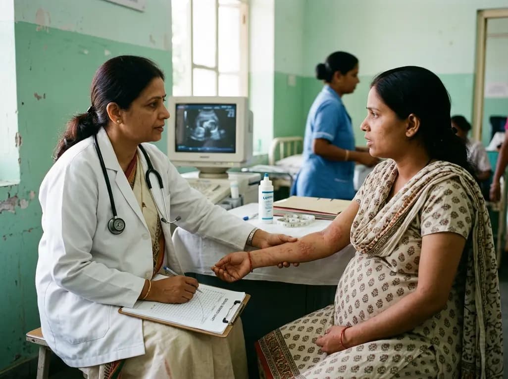
A comprehensive meta-analysis of over 278,000 women demonstrates that maternal atopic dermatitis significantly increases risks of premature rupture of membranes and low birth weight. Discover essential clinical insights on disease mechanisms, serial antenatal growth monitoring, and safe therapeutic interventions.
Today

Over 60 international public health researchers have advised FSSAI to mandate front-of-pack warning labels for any packaged product high in even one critical nutrient: sugar, saturated fat, or sodium. This reform counters phased rollouts to accelerate consumer protection and address India's chronic disease burden.
Today
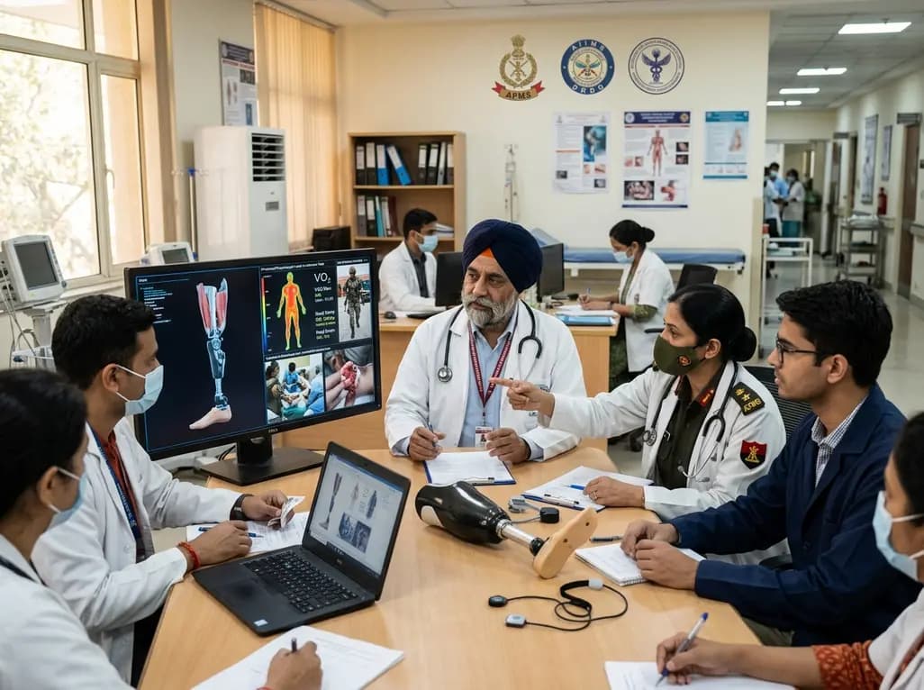
India's premier scientific bodies have joined the Armed Forces Medical Services to solve unique physiological and operational challenges faced by soldiers. The national collaboration covers combat casualty care, artificial intelligence diagnostics, bionic prosthetics, and extreme-environment physiological resilience.
Today

A cross-sectional study reveals that elevated METS-IR is independently associated with higher myopia odds in adolescents, showing biological synergy with obesity and highlighting the need for metabolic screening in pediatric eye care.
Yesterday
