

Loading, please wait...
Loading, please wait...

Achieving an accurate pulmonary embolism diagnosis remains a formidable clinical hurdle across outpatient and urgent care environments. Venous thromboembolic events claim over one hundred thousand lives annually in the United States alone. However, many presentations remain deceptively subtle. Patients frequently arrive with isolated dyspnea, mild pleurisy, or vague fatigue rather than textbook circulatory collapse. Consequently, initial evaluations frequently overlook thromboembolism, exposing vulnerable individuals to preventable complications. Clinicians must maintain a heightened index of suspicion whenever patients report sudden or unexplained breathlessness.
Ambulatory facilities encounter vast numbers of patients complaining of acute respiratory discomfort. Most of these individuals suffer from self-limiting viral bronchitis, asthma exacerbations, or musculoskeletal strain. Therefore, primary care clinicians understandably suspect common respiratory ailments first. Nevertheless, anchoring bias creates serious clinical vulnerabilities. Because thromboembolic occlusions restrict pulmonary arterial perfusion without producing focal lung consolidation, physical examinations often appear completely unremarkable. Normal breath sounds can mislead practitioners into underestimating disease severity. Furthermore, non-specific tachypnea and subtle anxiety might mimic hyperventilation or functional disorders. When clinicians fail to consider acute vascular obstruction, diagnostic delays escalate substantially. Studies confirm that up to one-third of pulmonary vascular occlusions receive erroneous initial assessments during initial outpatient consultations. Thus, evaluating clinicians must systematically correlate patient symptoms with cardiopulmonary physiology. Recognizing unexplained shortness of breath as an alarming herald sign prevents premature discharge. By integrating thorough history-taking with objective vital sign analysis, providers successfully uncover occult cardiopulmonary emergencies before catastrophic hemodynamic instability occurs.
Clinicians historically associate venous thromboembolism with overt risk factors like recent major surgery, prolonged immobilization, active malignancy, or known hypercoagulable states. However, real-world data demonstrate that numerous patients lack classic predisposing elements. Younger adults and physically active individuals can develop acute vascular thrombi following minor trauma, occult thrombophilia, or estrogen-containing oral contraceptive use. Moreover, atypical presentations frequently obscure clinical perception. A patient might describe brief lightheadedness, mild subcostal discomfort, or transient pleuritic irritation without peripheral leg edema. Because unilateral lower extremity swelling occurs in fewer than half of confirmed cases, its absence never excludes the diagnosis. In addition, baseline physiological reserve often conceals significant pulmonary vascular obstruction. Young, healthy individuals typically maintain stable arterial pressures despite elevated right ventricular afterload. Consequently, normal blood pressure readings provide false reassurance during initial triage. Clinicians must therefore avoid discounting thromboembolism solely because a patient looks robust. Careful scrutiny of subtle oxygen desaturations on exertion and resting tachycardia provides indispensable diagnostic warnings.
A standard twelve-lead electrocardiogram provides rapid, invaluable diagnostic insight during urgent care assessments. While sinus tachycardia represents the most frequent electrocardiographic rhythm in acute pulmonary vascular occlusion, clinicians must actively search for signs of acute right ventricular strain. Specifically, the classical S1Q3T3 pattern provides a distinctive marker of acute cor pulmonale. This finding features a deep S wave in lead I, a prominent Q wave in lead III, and T-wave inversion in lead III. Pathophysiologically, sudden pulmonary artery obstruction raises right ventricular pressure, causing acute right heart dilatation and clockwise cardiac rotation. Although the isolated S1Q3T3 sign lacks high sensitivity, its presence in a dyspneic patient strongly indicates elevated right ventricular strain. Furthermore, concurrent findings like T-wave inversions across precordial leads V1 through V4 or new incomplete right bundle branch block amplify diagnostic concern. Because urgent care clinics often lack immediate advanced imaging, electrocardiography serves as a rapid bedside triage tool. Prompt recognition of these electrical anomalies alerts practitioners to transfer patients rapidly to acute care emergency departments.
Navigating acute pulmonary vascular evaluation requires structured clinical decision pathways to prevent diagnostic oversights and avoid unnecessary testing. International consensus guidelines strongly recommend validated clinical prediction scores, including the Wells criteria and the Revised Geneva score. These instruments stratify patients into low, intermediate, or high pretest probability tiers. In low-risk ambulatory settings, clinicians can apply the Pulmonary Embolism Rule-out Criteria to safely exclude the condition without laboratory testing. However, when patients fail any PERC metric, objective testing becomes mandatory. For low and intermediate probability cohorts, quantitative D-dimer testing offers outstanding negative predictive value. High-sensitivity assays permit safe exclusion when plasma levels fall below age-adjusted thresholds. Conversely, elevated biomarker levels necessitate immediate anatomical imaging via computed tomography pulmonary angiography. Clinicians must remember that clinical decision instruments perform reliably only when applied systematically before therapeutic decisions. Unstructured clinical gestalt frequently underestimates disease risk in atypical patients. Consequently, standardized algorithmic protocols safeguard both patients and practitioners across outpatient healthcare settings.
Once clinicians suspect an acute pulmonary vascular event in an urgent care setting, prompt stabilization and emergency transfer become top priorities. Outpatient facilities rarely possess the imaging modalities or critical care monitoring necessary to manage developing cardiovascular collapse. Therefore, medical teams must establish direct transfer protocols to hospital emergency departments. While awaiting transport, providers must administer supplemental oxygen to correct arterial hypoxemia and secure reliable intravenous access. Additionally, continuous cardiac monitoring helps detect emerging right bundle branch block, severe sinus tachycardia, or malignant ventricular arrhythmias. Clinicians should avoid aggressive intravenous fluid boluses, because excessive volume expansion worsens right ventricular wall tension and impairs left ventricular filling. When transfer intervals extend significantly, emergency physicians may consider initiating parenteral anticoagulation after carefully evaluating hemorrhagic risks. Clear, concise inter-facility communication ensures that receiving emergency departments immediately mobilize specialized imaging and pulmonary embolism response teams. Ultimately, organized referral networks bridge the critical gap between initial outpatient presentation and life-saving tertiary intervention.
The S1Q3T3 electrocardiographic pattern reflects acute right ventricular strain caused by sudden pulmonary vascular occlusion. Although this sign occurs in only a minority of patients, its presence provides meaningful evidence of right heart strain. Therefore, when clinicians observe a deep S wave in lead I alongside a Q wave and inverted T wave in lead III in a dyspneic patient, they must immediately suspect severe pulmonary vascular obstruction and initiate rapid diagnostic imaging.
Practitioners should apply the Pulmonary Embolism Rule-out Criteria exclusively to patients whom clinical gestalt or structured scoring categorizes as very low risk. If the patient satisfies all eight criteria—including age under fifty, pulse below one hundred, and normal room-air saturation—clinicians can safely exclude pulmonary embolism without laboratory testing. However, if a patient fails even a single criterion, clinicians must proceed to quantitative D-dimer testing or definitive diagnostic imaging without hesitation.
Normotensive individuals often deceive healthcare providers because preserved systemic arterial blood pressure masks underlying right ventricular microvascular compromise. Young or previously healthy patients effectively compensate for elevated pulmonary arterial vascular resistance through endogenous sympathetic activation. Consequently, clinicians may incorrectly attribute their breathlessness to anxiety, asthma, or mild viral syndromes. Therefore, providers must rigorously evaluate resting tachycardia, subtle exertional hypoxemia, and electrocardiographic strain patterns rather than relying entirely on blood pressure numbers.
Disclaimer: This content is for informational and educational purposes only... Refer to the latest local and national guidelines for clinical practice.
References

Read summarized clinical updates, watch expert medical content, and earn CME certifications right from your smartphone.
Open your smartphone camera to scan and install.





A timely pulmonary embolism diagnosis remains a major clinical challenge in urgent care. Nonspecific symptoms like dyspnea can disguise acute right heart strain. Recognizing subtle ECG signs such as S1Q3T3 and applying structured scoring tools enable early detection and lifesaving emergency management.
Today

A large cohort study demonstrates that fetal growth restriction in a first pregnancy raises maternal cardiovascular risk within five years postpartum, including elevated rates of heart failure, stroke, and myocardial infarction. Clinicians must initiate proactive postpartum screening and cardiovascular prevention.
Today
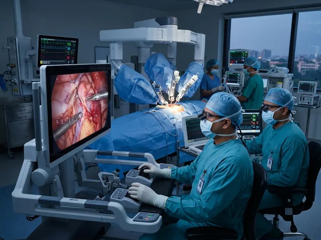
Cardiovascular surgery is transforming rapidly across India as tertiary hospitals embrace robotic assistance and AI-driven planning. Delhi-NCR now spearheads this shift, demonstrating higher procedural volumes, lower tissue trauma, and accelerated patient recovery compared to conventional sternotomy approaches.
Today
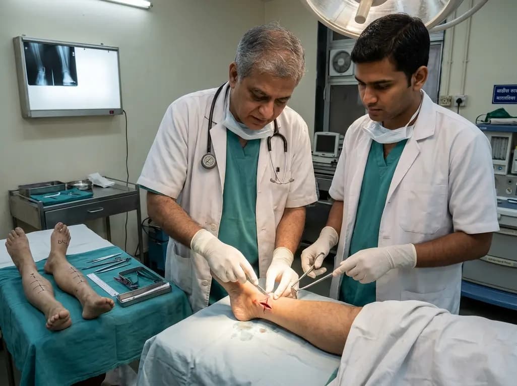
A paired cadaveric study compared triple and quintuple percutaneous Achilles tendon lengthening. While quintuple hemisection showed numerically greater elongation, differences were not statistically significant.
Yesterday
Diabetic retinopathy remains a leading cause of preventable visual impairment. This review outlines key strategies for screening, systemic risk factor control, advanced diagnostic imaging, and timely specialist referrals within primary and multidisciplinary care models.
Today
