.webp&w=384&q=75)

Loading, please wait...
Loading, please wait...

Managing benign breast disorders requires accurate diagnostic differentiation to avoid overtreating harmless lesions or missing rare malignancies. A recent single-centre retrospective study published in September 2026 highlights the complex presentations of pseudoangiomatous stromal hyperplasia (PASH). This benign mesenchymal condition arises from myofibroblastic proliferation within the breast stroma. Consequently, it creates cleft-like anastomosing spaces that frequently mimic vascular formations. Clinicians regularly detect these lesions during routine breast examinations or opportunistic health checks in India. Although many cases remain indolent, aggressive forms can cause clinical uncertainty. Therefore, physicians must maintain high diagnostic precision when assessing these patients.
The recent cohort evaluated thirty-one female patients with histopathologically confirmed PASH. Notably, the patients presented at a mean age of approximately thirty-four years. Premenopausal women comprised 94 percent of the study group, demonstrating a marked hormonal predilection. Circulating estrogens and progesterone strongly stimulate myofibroblastic activation in breast parenchymal tissue. Consequently, younger females demonstrate the highest prevalence of these lesions. Clinicians also occasionally identify similar proliferation in postmenopausal women receiving hormone replacement therapy. Most individuals present with a discrete, non-tender, mobile breast nodule that closely resembles a fibroadenoma. Alternatively, screening modalities detect the lesion as an unexpected incidental finding. Understanding this demographic profile prevents unwarranted invasive workups in younger female demographics. Nevertheless, physicians must thoroughly evaluate rapidly changing physical masses to rule out aggressive stromal lesions promptly.
Sonography serves as the primary diagnostic imaging modality for evaluating palpable breast nodules in young patients. On ultrasound, PASH most frequently presents as an oval, circumscribed, hypoechoic mass with parallel orientation. However, radiologists also observe suspicious features, such as indistinct margins and non-parallel alignment. Mammographic examinations demonstrate wide variability, ranging from entirely negative assessments to dense mass-like tissue with localized architectural alterations. In addition, magnetic resonance imaging displays significant structural heterogeneity. Radiologists regularly identify both circumscribed mass lesions and non-mass enhancement patterns. Dynamic contrast curves predominantly reveal persistent kinetic patterns, but rapid uptake curves also occur occasionally. Thus, imaging features overlap significantly with fibroadenomas, benign phyllodes tumors, and soft-tissue sarcomas. Because of these overlapping radiologic findings, imaging alone cannot establish a definitive diagnosis. Radiologists must always suggest tissue confirmation for suspicious or expanding nodules.
Definite diagnosis relies upon core needle biopsy and expert histopathologic assessment. Microscopic examination demonstrates dense collagenous mammary stroma containing slender, elongated spindle cells. These cells border empty slit-like clefts that deceptively resemble true blood vessels. However, these spaces lack genuine red blood cells and do not demonstrate endothelial linings. Immunohistochemical analysis serves a pivotal role in confirming the myofibroblastic phenotype. The lining spindle cells stain positive for CD34 and vimentin while remaining consistently negative for cytokeratins. Crucially, they lack vascular markers such as CD31 and Factor VIII-related antigen. This specific immunophenotypic profile decisively distinguishes the lesion from angiosarcoma and phyllodes tumors. In the single-centre cohort, 58 percent of lesions occurred alongside fibroadenoma-spectrum changes. Pathologists must therefore assess core samples thoroughly to verify that focal stromal changes do not obscure coexisting disease.
While most PASH lesions remain microscopic or incidental, dominant nodular or diffuse subtypes can present dramatically. The study documented two young patients who developed rapidly growing, dominant breast masses. Contrast-enhanced MRI demonstrated striking diffusion restriction, raising strong clinical concern for low-grade angiosarcoma. In such challenging cases, high mitotic activity or prominent cellularity can mislead the multidisciplinary team. Rapidly enlarging lesions distort the breast architecture and generate immense anxiety for both patient and clinician. Therefore, practitioners must not dismiss rapidly progressing lumps as simple fibroadenomas. Prompt surgical intervention with wide local excision becomes mandatory when tissue biopsies cannot exclude high-grade vascular pathology. Total excision provides complete specimen evaluation, ensuring clear margins and definitive diagnostic resolution. Multidisciplinary tumor boards provide indispensable guidance for these ambiguous, rapidly advancing breast presentations.
Formulating an appropriate treatment strategy requires comprehensive radiologic-pathologic correlation after core needle biopsy. When histology confirms uncomplicated PASH concordant with benign imaging, conservative observational surveillance remains the preferred standard. Patients can safely resume regular clinical follow-up without undergoing invasive surgical intervention. Conversely, surgical excision becomes strictly necessary when radiologic-pathologic discordance occurs, or when the mass grows rapidly. Complete surgical removal also relieves symptoms in symptomatic patients suffering from significant pain or asymmetric deformity. When performing excision, surgeons aim for clear tissue margins to minimize recurrence. Historical evidence indicates that post-excision recurrence remains relatively low, occurring in approximately ten to fifteen percent of cases. Hormonal modulation, such as discontinuing exogenous oral contraceptives or hormone replacement therapies, may also attenuate lesion enlargement. Overall, close collaboration among surgeons, radiologists, and pathologists ensures optimal, evidence-based management.
The exact cause of this condition remains uncertain, but substantial clinical evidence links it to abnormal myofibroblastic responses to reproductive hormones. Circulating estrogen and progesterone stimulate the mammary stromal fibroblasts to proliferate excessively. Consequently, the condition occurs most commonly in premenopausal women and in postmenopausal females undergoing hormone replacement therapy. Discontinuing exogenous hormonal agents often stabilizes lesion dimensions over time.
Pseudoangiomatous stromal hyperplasia is a completely benign condition and does not increase a patient's risk of developing breast carcinoma. However, because its clinical and radiological appearances frequently mimic complex fibroadenomas, phyllodes tumors, or low-grade angiosarcomas, clinicians must perform a thorough core needle biopsy. Once pathologists confirm radiologic-pathologic concordance, patients can confidently continue routine breast cancer surveillance without additional cancer risk.
Surgeons typically recommend operative excision when a breast mass demonstrates rapid expansion, causes substantial pain, or produces severe breast asymmetry. Furthermore, surgery becomes essential whenever core needle biopsy yields radiologic-pathologic discordance or fails to exclude a malignant vascular neoplasm conclusively. In contrast, asymptomatic patients with stable, biopsy-proven lesions can safely avoid surgical intervention under routine clinical surveillance.
Disclaimer: This content is for informational and educational purposes only. It is not intended to provide medical advice, diagnosis, or treatment. Healthcare professionals must use their clinical judgment and consider each patient’s unique circumstances. Refer to the latest local and national guidelines for clinical practice.
References
Karaböcek SM et al. Pseudoangiomatous Stromal Hyperplasia of the Breast: Clinical, Radiologic and Histopathologic Spectrum in a Retrospective Single-Centre Series. J Med Imaging Radiat Oncol. 2026 Sep 14. doi: 10.1111/1754-9485.70186. PMID: 42734059.
Estes A, Cao L, Miller ME. Pseudoangiomatous stromal hyperplasia: overview and clinical management. Ann Breast Surg. 2021;5:1-9.
Jones KN, Glazebrook KN, Reynolds C. Pseudoangiomatous stromal hyperplasia: imaging findings with pathologic and clinical correlation. AJR Am J Roentgenol. 2010;195(4):1036-1042.
Alikhassi A, Skarpathiotakis M, Lu FL, Curpen B. Pseudoangiomatous stromal hyperplasia of the breast, imaging and clinical perspective: A review. Breast Dis. 2023;42(1):147-153.

Read summarized clinical updates, watch expert medical content, and earn CME certifications right from your smartphone.
Open your smartphone camera to scan and install.





Discover key clinical, radiologic, and histopathologic findings of pseudoangiomatous stromal hyperplasia (PASH) of the breast, highlighting its differentiation from vascular malignancies and management approaches.
Today
A study of over 340,000 Medicare beneficiaries shows that hospital artificial intelligence tools, particularly risk prediction algorithms, significantly lower readmissions and acute care utilization in older adults with Alzheimer's disease and dementia without driving up total healthcare spending.
Today
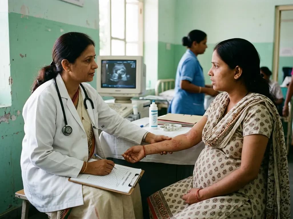
A comprehensive meta-analysis of over 278,000 women demonstrates that maternal atopic dermatitis significantly increases risks of premature rupture of membranes and low birth weight. Discover essential clinical insights on disease mechanisms, serial antenatal growth monitoring, and safe therapeutic interventions.
Today

Over 60 international public health researchers have advised FSSAI to mandate front-of-pack warning labels for any packaged product high in even one critical nutrient: sugar, saturated fat, or sodium. This reform counters phased rollouts to accelerate consumer protection and address India's chronic disease burden.
Today
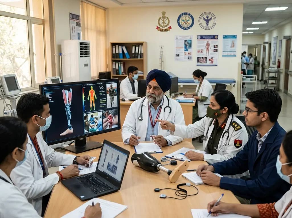
India's premier scientific bodies have joined the Armed Forces Medical Services to solve unique physiological and operational challenges faced by soldiers. The national collaboration covers combat casualty care, artificial intelligence diagnostics, bionic prosthetics, and extreme-environment physiological resilience.
Today

A cross-sectional study reveals that elevated METS-IR is independently associated with higher myopia odds in adolescents, showing biological synergy with obesity and highlighting the need for metabolic screening in pediatric eye care.
Yesterday
