.webp&w=384&q=75)

Loading, please wait...
Loading, please wait...

Primary small cell neuroendocrine carcinoma of the breast represents an exceedingly rare and aggressive malignancy. Historically, mammary neuroendocrine neoplasms account for less than one percent of all breast cancers. Because of their rarity, pathologists and oncologists frequently encounter substantial diagnostic dilemmas. Clinicians must first distinguish primary mammary tumors from metastatic disease arising in the lungs or digestive tract. Therefore, comprehensive clinical assessment and detailed systemic imaging remain paramount for establishing the correct diagnosis.
In addition, recent clinicopathological analyses shed critical light on this unique tumor type. Most documented cases occur in middle-aged and older postmenopausal women who present with palpable breast masses. Moreover, these tumors frequently attain significant dimensions prior to clinical detection. Consequently, healthcare providers must identify these tumors promptly to prevent therapeutic delays. Furthermore, multidisciplinary collaboration between surgical oncologists, medical oncologists, and pathologists ensures optimal clinical pathways. As a result, clinicians can tailor aggressive multimodal treatment strategies to individual patient profiles. Ultimately, recognizing this uncommon variant improves diagnostic precision and patient survival.
Histologically, primary small cell neuroendocrine carcinoma of the breast closely mirrors small cell carcinoma of pulmonary origin. The neoplastic cells display round to ovoid shapes, scant cytoplasm, finely stippled nuclear chromatin, and absent nucleoli. In addition, these tumors demonstrate high nuclear molding and extensive crushed chromatin artifacts on biopsy specimens. Therefore, pathologists must carefully inspect architectural patterns under light microscopy. Tumor sheets typically show nested, insular, or diffuse infiltrative arrangements devoid of glandular formation.
Moreover, tissue sections frequently exhibit widespread geographic necrosis and striking mitotic activity. In published case series, tumor diameters range widely, occasionally surpassing ten centimeters at initial diagnosis. Furthermore, pathologists frequently observe intravascular tumor thrombi and perineural invasion during surgical evaluation. Some cases also demonstrate localized invasion into overlying dermis or nipple epidermis. Thus, these histological findings highlight the profoundly aggressive nature of the disease. Consequently, clinicians must recognize these morphological markers to gauge locoregional recurrence risks and systemic progression potential.
Immunohistochemistry provides indispensable support for confirming neuroendocrine differentiation in breast tissue. Specifically, these tumors display diffuse and strong positivity for pan-cytokeratin, synaptophysin, and chromogranin A. Additionally, a substantial proportion of cases demonstrates positive immunoreactivity for CD56. However, pathologists must evaluate organ-specific markers to exclude extramammary metastases. Mammary lineage markers like GATA3 and TRPS1 show variable, often diminished expression in poorly differentiated tumors.
In contrast, typical invasive ductal carcinomas reliably maintain strong nuclear GATA3 expression. Furthermore, hormone receptor profiling reveals that only a minority of cases expresses estrogen receptor or progesterone receptor. Human epidermal growth factor receptor 2 remains predominantly negative across most cohorts. Crucially, the Ki-67 proliferation index is markedly elevated, typically ranging between sixty and ninety percent. Mutant-type p53 protein expression and complete loss of RB1 protein expression occur uniformly. Therefore, this distinct immunophenotype helps pathologists establish a definitive diagnosis while ruling out alternative malignancies.
Recent advances in next-generation sequencing illuminate the complex genomic architecture of these aggressive mammary tumors. Molecular profiling demonstrates that TP53 is the most frequently altered gene in this cohort. Specifically, pathogenic missense mutations in TP53 occur in the vast majority of tested cases. Furthermore, genomic disruption of the RB1 pathway frequently accompanies TP53 inactivation. This dual TP53 and RB1 loss mirrors the classic molecular hallmark of pulmonary small cell carcinoma.
Nevertheless, breast neuroendocrine carcinomas display distinct secondary genetic features. For example, some tumors harbor concurrent mutations in the PI3K and AKT signaling pathways. In addition, evaluation of tumor mutational burden generally shows low to moderate mutation counts. Microsatellite instability remains exceptionally uncommon in these malignancies. Consequently, immune checkpoint inhibitors may offer limited monotherapy benefit without targeted combination regimens. Understanding these molecular alterations provides valuable biological insights into tumor pathogenesis. Therefore, genomic testing serves as an essential tool for identifying future therapeutic vulnerabilities in affected patients.
Managing primary small cell neuroendocrine carcinoma of the breast requires an aggressive, multidisciplinary therapeutic approach. Because standardized clinical guidelines remain limited, oncologists often adapt systemic regimens from small cell lung cancer protocols. Specifically, platinum-based chemotherapy paired with etoposide serves as the frontline systemic foundation. In addition, some clinicians utilize traditional breast cancer regimens containing anthracyclines and taxanes. Neoadjuvant chemotherapy frequently induces substantial initial tumor shrinkage prior to planned surgical resection.
However, pathological complete response rates remain disappointingly low despite notable radiographic improvements. Therefore, definitive surgery involving modified radical mastectomy or breast-conserving therapy remains necessary for locoregional control. Furthermore, post-mastectomy radiation therapy significantly reduces local recurrence rates in locally advanced presentations. Clinicians also consider adjuvant endocrine therapy for rare patients whose tumors retain hormone receptor expression. Nevertheless, systemic relapses occur rapidly, with early dissemination to liver, bone, and brain. Consequently, vigilant postoperative monitoring and proactive restaging protocols remain vital components of ongoing patient management.
Pathologists differentiate primary breast lesions from metastatic lung tumors by combining clinical imaging, histological context, and immunohistochemical panels. Systemic positron emission tomography or chest computed tomography must first exclude a primary pulmonary mass. Histopathologically, finding in situ ductal carcinoma definitively confirms mammary origin. In addition, immunohistochemical panels assess TTF-1 and breast markers like TRPS1 or GATA3. Correlating systemic radiological findings with lineage-specific biomarkers ensures an accurate, conclusive diagnostic determination.
Oncologists typically select platinum-based chemotherapy regimens combined with etoposide, mirroring established protocols for small cell lung cancer. This cytotoxic combination produces rapid objective responses in poorly differentiated, high-grade neuroendocrine tumors. Alternatively, clinicians may utilize anthracycline and taxane regimens commonly prescribed for aggressive invasive breast carcinomas. Because high-grade neuroendocrine carcinomas exhibit rapid cellular turnover, aggressive platinum-based systemic therapy remains the preferred frontline approach to control micrometastatic dissemination and reduce tumor burden.
Molecular testing provides critical diagnostic confirmation and reveals therapeutic targets in high-grade neuroendocrine breast cancer. Next-generation sequencing detects characteristic alterations, including concurrent TP53 mutations and RB1 loss, distinguishing true neuroendocrine carcinomas from conventional ductal variants. Furthermore, comprehensive profiling identifies potentially actionable mutations in PI3K or FGFR signaling pathways. Molecular assessment also calculates tumor mutational burden to evaluate future immunotherapy potential, thereby guiding individualized precision oncology regimens for high-risk patients facing limited conventional therapeutic options.
Disclaimer: This content is for informational and educational purposes only... Refer to the latest local and national guidelines for clinical practice.
References

Read summarized clinical updates, watch expert medical content, and earn CME certifications right from your smartphone.
Open your smartphone camera to scan and install.





A comprehensive analysis of primary small cell neuroendocrine carcinoma of the breast, highlighting its distinctive histopathology, TP53 and RB1 alterations, immunohistochemical profiling, and multimodal treatment strategies for oncologists and pathologists.
Today
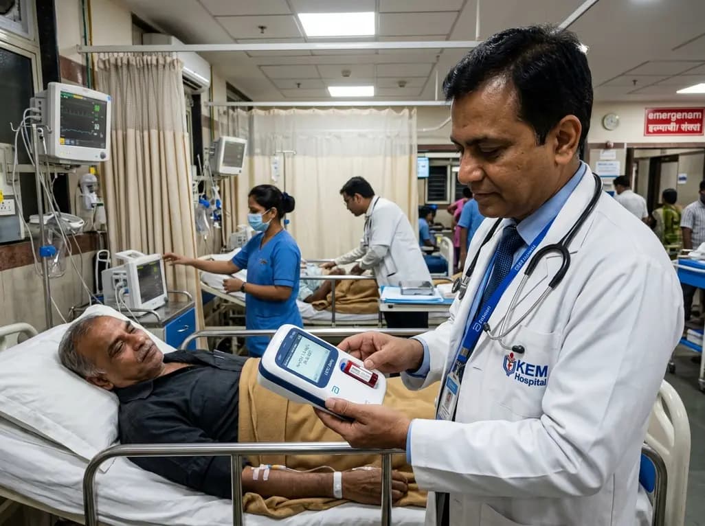
A real-world emergency department evaluation demonstrates that the Abbott i-STAT Alinity point-of-care high-sensitivity troponin I assay achieves excellent analytical precision and safely rules out myocardial infarction without false negatives, offering rapid bedside results to decongest acute care pathways.
Today

Integrating adult genetic services into reproductive and pregnancy care substantially alters clinical management. A study reveals that diagnostic genetic evaluations directly inform maternal, fetal, and neonatal care in nearly 57% of tested patients, highlighting the value of preconception and antenatal genomics.
Today

A cross-sectional study in Nepal evaluates cardiovascular risk factors among adults working above 4,000 meters. The findings reveal unexpected rates of diabetes, hypertension, and mountain sickness, underscoring the urgent need for targeted cardiometabolic screening in high-altitude occupational cohorts.
Today

A recent scoping review highlights the efficacy, usability, and patient satisfaction of virtual reality pain management for chronic musculoskeletal pain, emphasizing psychotherapy-based VR protocols.
Today

Preclinical findings demonstrate that dietary flavonoid hyperoside ameliorates high-fat diet-induced type 2 diabetes by stimulating brown adipose tissue thermogenesis, promoting white fat browning, dampening adipose macrophage inflammation, and restoring gut microbiota homeostasis.
Today
