.webp&w=384&q=75)

Loading, please wait...
Loading, please wait...
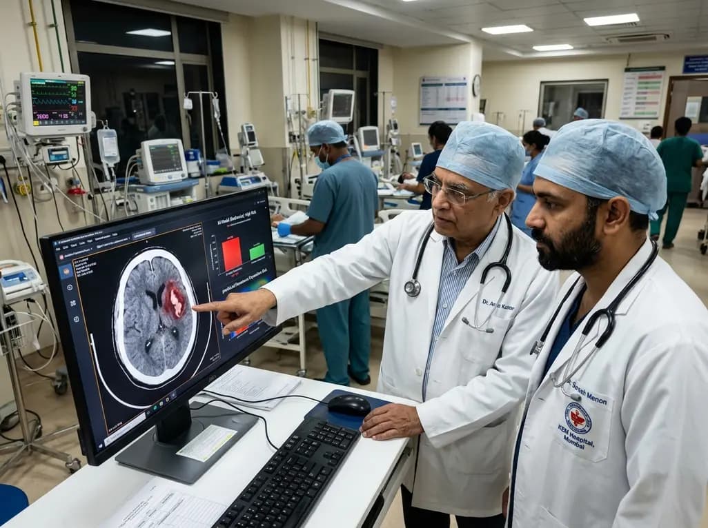
Nontraumatic intracerebral hemorrhage represents one of the most lethal forms of stroke, often leading to rapid neurological deterioration and permanent disability. A primary determinant of early clinical worsening is early hematoma enlargement, which typically occurs within the initial hours following symptom onset. Therefore, accurately predicting hematoma expansion remains essential for selecting patients who may benefit from intensive blood pressure lowering, targeted hemostatic therapies, or urgent surgical evacuation. However, human visual inspection of non-contrast head computed tomography scans often fails to reliably capture subtle radiological markers that precede significant bleeding progression.
Spontaneous intracerebral hemorrhage accounts for substantial global morbidity and mortality. Hematoma growth occurs in approximately one-third of these patients during the hyperacute phase, dramatically worsening functional outcomes and increasing early mortality. Clinicians routinely assess admission non-contrast computed tomography images to identify high-risk radiological features, such as the swirl sign, black hole sign, blend sign, and island sign. Although these qualitative imaging markers offer helpful visual clues, their overall sensitivity remains modest, and subjective interpretation varies widely among practitioners. Furthermore, clinical variables such as baseline blood pressure, admission Glasgow Coma Scale score, and onset-to-imaging time provide additional context, yet they rarely deliver precise individual risk stratification on their own. Consequently, acute care teams often face substantial diagnostic uncertainty when determining which patients require the most aggressive monitoring and early preemptive interventions.
To address this clinical dilemma, researchers evaluated whether artificial intelligence could enhance prognostic accuracy over traditional clinical assessment. Utilizing data from 900 participants enrolled in the Antihypertensive Treatment of Acute Cerebral Hemorrhage 2 trial, investigators developed and validated multiple machine learning models. The study analyzed baseline non-contrast head computed tomography scans and comprehensive clinical variables collected at admission. The investigators trained the algorithms on an initial cohort of 621 participants and subsequently tested their performance on an independent validation cohort of 279 patients. The primary endpoint was hematoma expansion, defined as an increase in hematoma volume greater than 33 percent or at least 6 milliliters at 24 hours. The researchers tested standalone deep learning imaging models, handcrafted radiomics algorithms, and hybrid models that integrated both quantitative imaging features and clinical variables.
In a direct comparison, the investigators surveyed expert clinicians and asked them to predict hematoma enlargement using the exact same baseline computed tomography scans and admission clinical data. The expert clinicians achieved an area under the receiver operating characteristic curve of only 0.591, demonstrating performance barely superior to chance. Moreover, interrater reliability among the clinicians was notably poor, yielding a kappa value of 0.156. In contrast, the deep learning imaging model analyzing raw computed tomography data achieved an area under the receiver operating characteristic curve of 0.680. Similarly, a random forest classifier integrating both radiomic features and clinical admission data achieved an area under the receiver operating characteristic curve of 0.677. Additionally, the intraclass correlation coefficient between clinician judgment and the top-performing machine learning model was 0.47, highlighting substantial divergence between human heuristics and algorithmic pattern recognition.
The superior performance of computational models stems from their unique capacity to extract high-dimensional quantitative data that lie beyond human visual perception. Handcrafted radiomics captures minute variations in voxel intensity, spatial heterogeneity, structural shape, and subtle boundary gradients within the acute hemorrhage. Meanwhile, deep convolutional neural networks autonomously learn complex representations of perihematomal tissue density and parenchymal disruption without requiring manual feature engineering. Because these algorithmic systems evaluate thousands of quantitative parameters simultaneously, they detect microscopic indicators of active microvascular extravasation that human observers inevitably miss. Consequently, computational algorithms provide consistent, objective, and reproducible risk assessments across diverse patient profiles, avoiding the subjective biases and perceptual limitations inherent in manual radiological review.
Although an area under the receiver operating characteristic curve of approximately 0.68 represents moderate absolute predictive power, it establishes a vital relative benchmark in a clinical task where expert judgment frequently falters. Integrating automated radiomics and deep learning pipelines into picture archiving and communication systems could transform hyperacute stroke workflows. When a patient with acute intracerebral hemorrhage arrives at the emergency department, automated algorithms could rapidly calculate expansion probability within seconds of scan completion. Clinicians could then leverage this objective risk score to prioritize intensive care unit bed allocation, personalize aggressive blood pressure targets, or enroll suitable candidates into clinical trials evaluating novel hemostatic agents. Thus, artificial intelligence serves not as a replacement for clinical acumen, but as an indispensable cognitive adjunct.
Moving forward, enhancing the predictive accuracy of artificial intelligence tools in neurocritical care will require the incorporation of comprehensive multimodal inputs. Future architectures should ideally combine serial neuroimaging, continuous physiological monitoring, point-of-care coagulation parameters, and genetic markers to build dynamic prognostic models. Furthermore, validating these tools across diverse global populations and community hospital settings will ensure broad generalizability. As healthcare systems increasingly adopt digital health solutions, standardized and interpretable machine learning models will play a pivotal role in refining emergency neurovascular care and improving functional recovery for stroke survivors worldwide.
Hematoma expansion is conventionally defined as an increase in hematoma volume by more than 33 percent or an absolute growth of at least 6 milliliters on follow-up imaging within 24 hours of symptom onset. This growth strongly correlates with early neurological worsening, functional dependence, and elevated mortality.
Clinicians rely on visual assessment of non-contrast scans, which reveals only gross morphological features like density heterogeneity or irregular borders. Because subtle microvascular leakage patterns remain imperceptible to the human eye, manual predictions suffer from substantial interobserver variability and yield poor diagnostic accuracy across clinical settings.
Deep learning radiomics models automatically extract thousands of subvisual voxel-level features from baseline computed tomography scans. By generating rapid, reproducible risk estimates, these algorithms help acute care teams stratify patients, tailor intensive blood pressure management, allocate intensive monitoring resources, and identify candidates for targeted anti-expansion therapies.
Disclaimer: This content is for informational and educational purposes only and should not be considered medical advice. Always consult a qualified healthcare provider for diagnosis and treatment decisions. Refer to the latest local and national guidelines for clinical practice.
References

Read summarized clinical updates, watch expert medical content, and earn CME certifications right from your smartphone.
Open your smartphone camera to scan and install.





A new study reveals that deep learning radiomics models significantly outperform expert clinicians in predicting hematoma expansion after acute intracerebral hemorrhage, establishing an objective benchmark to enhance acute neurocritical care decision-making.
last month

According to WHO data, average lifespan reached 73.3 years in 2023, recovering from pandemic lows. However, healthy life expectancy lags at 62.8 years due to surging noncommunicable conditions like cardiovascular disease and dementia, demanding proactive primary screening and sustained chronic care.
Today

A nationwide South Korean study shows underreporting of smoking among women dropped from 57.4% to 36.4% between 2008 and 2021. However, over one-third of female smokers still conceal their habit, highlighting the critical role of cotinine verification in clinical risk assessment.
Today

Medical robotics is transitioning beyond operative suites into diagnostics, cardiology, and endocrinology. Explore how innovations in tele-robotics, AI integration, and nanomedicine are overcoming geographical barriers and transforming precision healthcare delivery across India's evolving disease landscape.
Today

A new study evaluates the rheological stability of decellularized peripheral nerve matrix hydrogels under varied storage conditions. Findings show -80°C storage and freeze-drying preserve gelation kinetics and mechanical stiffness, offering key insights for shipping and translational clinical applications.
Today

A qualitative meta-synthesis highlights the complex interplay between gender dysphoria, minority stress, and eating disorders in transgender and gender-diverse individuals. Clinicians must recognize these distinct drivers to offer supportive, gender-affirming, and interdisciplinary eating disorder care.
Today
