.webp&w=384&q=75)

Loading, please wait...
Loading, please wait...

Radiation therapy remains a fundamental cornerstone for treating both primary and metastatic intracranial neoplasms. However, long-term survivors frequently experience debilitating delayed adverse events that degrade their daily quality of life. Specifically, late cranial irradiation toxicity manifests as progressive neurocognitive impairment, white matter damage, and neuroendocrine axis hypofunction. Although modern stereotactic and conformal techniques reduce collateral radiation exposure, clinicians still struggle to predict which patients will develop irreversible post-treatment injury. Consequently, identifying robust pretreatment biomarkers is critical for risk stratification and individualized clinical management.
Recent investigations demonstrate that patient-specific biological factors strongly dictate tissue vulnerability following therapeutic radiation. In a pivotal retrospective cohort study, researchers analyzed eighty adult patients receiving brain radiotherapy to evaluate late toxic sequelae. Notably, the study cohort comprised individuals with both malignant primary brain tumors and secondary brain metastases. The investigators evaluated putative vascular risk factors alongside genotypic profiles to determine their prognostic influence on post-radiation neurocognitive function. Furthermore, multivariate regression models identified two critical independent predictors of cognitive decline. Specifically, elevated pre-treatment homocysteine and the apolipoprotein E epsilon 4 (ApOE4) allele predicted significant Mini-Mental State Examination drops. Elevated homocysteine increased the odds of cognitive deterioration nearly fourfold. Simultaneously, the presence of an ApOE4 allele nearly tripled this clinical risk. Consequently, these findings highlight that microvascular susceptibility and impaired neural repair pathways drive progressive tissue injury. Therefore, baseline biological profiling provides essential prognostic clarity that radiation dosimetry alone cannot capture. In addition, clinicians can utilize these measurable pre-treatment biological metrics to identify highly susceptible patients prior to starting radiotherapy sessions. Hence, personalized risk assessment becomes an achievable clinical goal.
Hyperhomocysteinemia represents an established systemic risk factor for endothelial dysfunction, atherosclerosis, and microvascular thrombosis. Therefore, its pronounced correlation with post-radiation neurocognitive decline reflects accelerated cerebrovascular injury within irradiated neural tissue. Cranial radiation causes acute endothelial apoptosis, blood-brain barrier disruption, and persistent local neuroinflammation. When combined with elevated baseline homocysteine levels, radiation exacerbates small vessel vasculopathy and chronic microvascular ischemia. Consequently, this pathological cascade manifests radiographically as extensive confluent white matter hyperintensities on magnetic resonance imaging. In the clinical trial cohort, elevated pre-treatment homocysteine correlated significantly with progressive white matter changes on follow-up neuroimaging. Moreover, both treating oncologists and patients reported noticeable functional declines when homocysteine levels were abnormal before therapy. In India, nutritional deficiencies such as low dietary vitamin B12 and folate intake frequently cause elevated plasma homocysteine. Accordingly, oncologists must recognize that widespread micronutrient deficiencies in Indian patients could amplify neurovascular vulnerability to therapeutic radiation. Clinicians can readily screen for hyperhomocysteinemia in routine biochemical workups to guide early neuroprotective strategies. Furthermore, aggressive correction of elevated homocysteine through targeted vitamin supplementation warrants prospective clinical exploration. Thus, identifying hyperhomocysteinemia before cranial radiation offers an actionable opportunity to preserve long-term cognitive health.
The apolipoprotein E epsilon 4 allele remains the most thoroughly characterized genetic risk factor for sporadic neurodegenerative diseases. However, oncologists rarely evaluate this genetic marker before initiating cranial radiotherapy in neuro-oncology patients. The recent cohort findings demonstrate that ApOE4 carriers endure significantly greater radiation-induced neurocognitive deterioration than non-carriers. Biochemically, the ApOE4 isoform exhibits impaired antioxidant defense mechanisms and diminished lipid transport efficiency across damaged cellular membranes. Consequently, neural progenitor cells in the subgranular zone of the hippocampus display severely blunted regenerative capacity following ionizing radiation exposure. In addition, the ApOE4 genotype compromises microglial clearance of neurotoxic debris, sustaining a chronic inflammatory microenvironment. The clinical investigation demonstrated that ApOE4 presence correlated with objective Mini-Mental State Examination drops and subjective physician assessments of clinical decline. Furthermore, genetic testing revealed that ApOE4 carriers faced heightened vulnerability to radiation-induced white matter rarefaction. Therefore, identifying carrier status can refine clinical prognostication for brain tumor patients facing whole-brain or focal cranial irradiation. Clinicians can utilize this genomic insight to counsel families realistically regarding long-term functional expectations. Moreover, understanding genetic susceptibility encourages closer multidisciplinary monitoring between oncologists and neurologists throughout the survivorship period.
Radiation therapy directed at the cranial vault frequently damages the delicate hypothalamic-pituitary axis over time. Consequently, late endocrine deficiencies represent a common yet frequently underdiagnosed complication among long-term cancer survivors. Radiation triggers microvascular thrombosis and progressive ischemic atrophy within hypothalamic nuclei and pituitary anterior lobe tissue. In the recent Penn State cohort study, investigators tracked new endocrine failures across long-term follow-up intervals. Notably, the ApOE4 genotype demonstrated a statistically significant association with the emergence of secondary endocrine deficiencies. This intriguing finding suggests that genetic repair deficiencies worsen radiation-induced neuroendocrine organ injury alongside cognitive networks. In general, growth hormone deficiency and hypogonadism emerge first, followed sequentially by secondary hypothyroidism and adrenal insufficiency. Because post-radiation fatigue, emotional blunting, and cognitive slowing mimic hormonal deficits, delayed diagnosis often occurs in clinical practice. Therefore, multidisciplinary teams must implement structured neuroendocrine surveillance protocols following cranial radiation. Routine hormonal panels should evaluate thyroid, adrenal, and gonadal axes at regular six-month intervals. As a result, prompt hormone replacement therapy can dramatically alleviate fatigue and improve quality of life. Furthermore, early endocrinological intervention prevents debilitating metabolic consequences and cardiovascular morbidity in vulnerable cancer survivors.
Traditional statistical analyses frequently struggle to capture the complex non-linear interactions between diverse radiation toxicity predictors. To address this limitation, researchers developed machine-learning algorithms incorporating demographic factors, radiation dosimetry, and metabolic biomarkers. Specifically, the study team implemented gradient boosting and random forest models to rank predictive variables by relative importance. These artificial intelligence architectures successfully identified elevated homocysteine and ApOE4 status as top-tier predictors of post-treatment neurocognitive decline. In addition, the investigators translated these computational models into an accessible online risk nomogram for neuro-oncology clinicians. This digital tool integrates baseline patient variables to estimate personalized probabilities across five distinct late toxicity outcomes. Consequently, radiation oncologists can simulate different treatment scenarios and tailor radiation delivery techniques accordingly. For instance, high-risk patients may benefit substantially from hippocampal-sparing intensity-modulated radiotherapy or early radiosurgery rather than whole-brain radiation. Furthermore, computational risk stratification facilitates proactive enrollment into cognitive rehabilitation programs before severe functional decline manifests. Ultimately, integrating predictive machine learning into clinical oncology workflows bridges the gap between empirical evidence and precision medicine. Thus, modern algorithmic tools empower oncologists to balance tumor control against long-term neurological preservation with unprecedented quantitative accuracy.
Implementing routine biomarker screening before cranial irradiation holds immense clinical relevance for healthcare systems across India. In the Indian subcontinent, widespread vegetarian diets and nutritional deficits contribute to high population prevalence rates of hyperhomocysteinemia. Therefore, baseline homocysteine testing offers a cost-effective, readily available tool that standard clinical biochemistry laboratories can perform. In addition, identifying high-risk individuals allows Indian oncologists to consider hippocampal-avoidance radiation techniques and proactive memantine neuroprotection. Clinicians should also consider checking serum vitamin B12 and folate levels to correct reversible metabolic deficits promptly. Similarly, oncologists can partner closely with neurosurgeons, endocrinologists, and rehabilitation therapists to construct comprehensive survivorship care plans. Routine follow-up visits must feature standardized cognitive screening tests alongside serial pituitary hormone evaluations. Consequently, early therapeutic interventions, including cognitive rehabilitation exercises and hormone substitution, can preserve patient independence and vocational productivity. Moreover, family counseling regarding anticipated cognitive changes helps reduce psychological caregiver burden significantly. Ultimately, proactive risk stratifying ensures that brain tumor survivors in resource-diverse Indian settings achieve optimal functional outcomes. Hence, adopting baseline biomarker profiling represents a vital practical step toward modernizing neuro-oncology protocols across cancer centers.
Elevated homocysteine triggers endothelial dysfunction, enhances vascular inflammation, and promotes microvascular thrombosis. When brain tissue receives ionizing radiation, hyperhomocysteinemia exacerbates small-vessel radiation vasculopathy and blood-brain barrier disruption. Consequently, patients suffer accelerated cerebral ischemia and progressive white matter demyelination, leading to measurable cognitive and functional deterioration over time.
The ApOE4 isoform exhibits deficient antioxidant capacity and impaired membrane lipid transport compared to other apolipoprotein isoforms. Following cranial irradiation, ApOE4 carriers experience sustained neuroinflammation and reduced regenerative proliferation of hippocampal neural stem cells. Therefore, defective neurovascular repair mechanisms prevent damaged brain networks from recovering, causing significant late neurocognitive decline.
Clinicians can adopt hippocampal-avoidance intensity-modulated radiotherapy and stereotactic radiosurgery to minimize radiation dose to memory structures. Furthermore, prescribing neuroprotective agents like memantine during treatment preserves cognitive reserve. Routine pre-treatment screening and correction of hyperhomocysteinemia, along with structured cognitive rehabilitation and hormonal replacement, significantly safeguard survivorship quality.
Disclaimer: This content is for informational and educational purposes only... Refer to the latest local and national guidelines for clinical practice.
References

Read summarized clinical updates, watch expert medical content, and earn CME certifications right from your smartphone.
Open your smartphone camera to scan and install.





A study in the Journal of Neuro-Oncology reveals that pre-treatment elevated homocysteine and the ApOE4 allele independently predict late radiation-associated neurocognitive decline and endocrine deficiency in brain tumor patients, offering novel avenues for individualized risk stratification and intervention.
Today
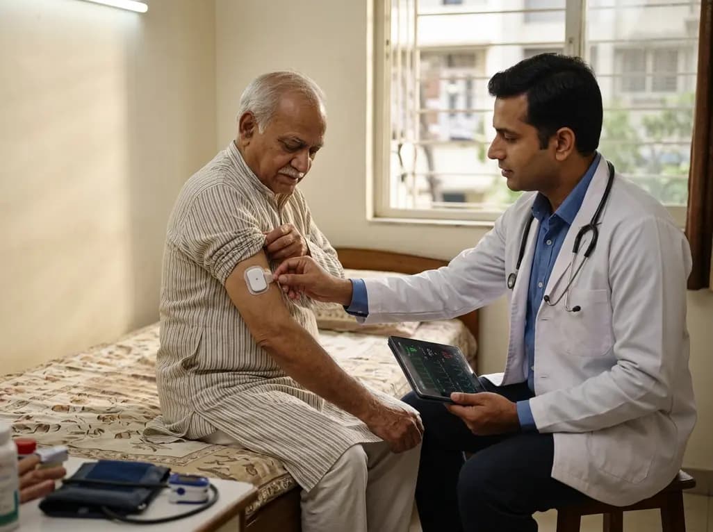
A feasibility study evaluates remote vital sign monitoring in acute hospital at home patients. Continuous wearable patches captured heart rate and respiratory rate with high adherence (89% and 75%), significantly exceeding manual spot checks for blood pressure, oxygen saturation, and temperature.
Today

A scoping review evaluates 116 studies on AI in women's reproductive health, finding that 83.6% carry high risk of bias. While ensemble models show promise, disparities and lack of validation hinder LMIC deployment.
Today

Differential oxygenation during peripheral TA-NRP can cause undetected coronary hypoxemia due to arch vessel clamping. A novel circuit with continuous aortic root monitoring detects desaturation early, facilitating left ventricular unloading and preventing allograft dysfunction and unnecessary heart discard.
Today

A longitudinal study reveals that patients undergoing staged bilateral hip arthroscopy experience progressive contralateral labral deterioration during surgical intervals. However, structural degradation does not compromise 2-year postoperative clinical outcomes or patient-reported satisfaction scores.
Today

A post hoc analysis of the PARM-T2D study shows pemafibrate exerts baseline-dependent effects on LDL-C and particle size in type 2 diabetes and hypertriglyceridemia. It lowers triglycerides and remnants while favorably remodeling atherogenic LDL particles based on initial lipid control status.
Today
