

Loading, please wait...
Loading, please wait...
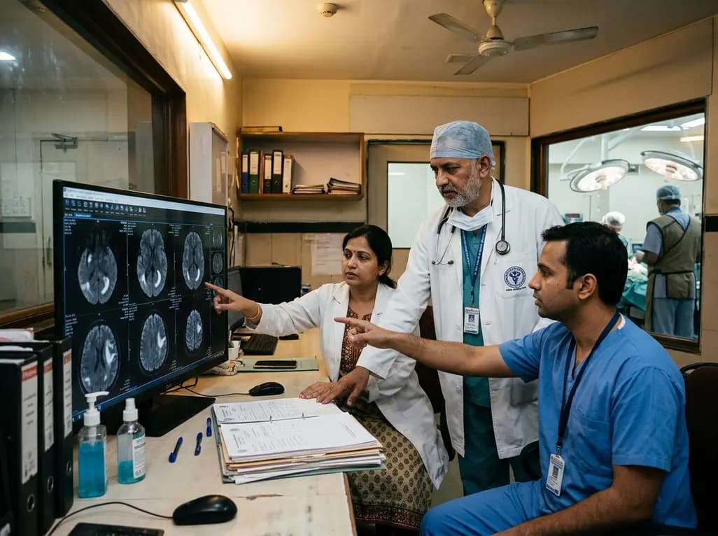
Resection of supratentorial gliomas within eloquent cortical and subcortical regions demands an intricate balance between maximizing oncological cytoreduction and preserving vital functional pathways. Neurosurgeons routinely obtain early diffusion-weighted magnetic resonance imaging within forty-eight hours after resection to evaluate acute ischemic complications. Clinicians, however, often struggle to correlate bright signals on diffusion scans with immediate clinical deterioration. Crucially, recent evidence demonstrates that analyzing postoperative DWI morphology offers superior diagnostic clarity compared to binary reporting. In a cohort of 139 patients, investigators evaluated how distinct lesion geometries correlate with neurological worsening. Consequently, evaluating the shape of signal abnormalities provides meaningful prognostic insights into patient functional trajectories. Conventional imaging identifies restricted water diffusion resulting from acute cytotoxic edema, but morphology reveals the true underlying mechanism of parenchymal disruption. Therefore, modern neuro-oncological management requires surgical teams to transition from binary scan interpretations toward nuanced spatial analyses. Understanding these morphological distinctions enables clinicians to distinguish benign, self-limiting tissue manipulation from severe, irreversible vascular compromise. Consequently, neurosurgeons can better anticipate rehabilitation needs, adjust medical therapies promptly, and deliver transparent prognostic guidance to patients and families.
The geometric classification of ischemic signals on postoperative neuroimaging directly informs functional prognosis. Specifically, investigators distinguish between sector-shaped wedge defects and thin marginal rim enhancements along the resection cavity. Sector-shaped lesions typically extend deeply into adjacent subcortical white matter, reflecting arterial branch territory ischemia or substantial venous infarction. In contrast, rim-shaped restricted diffusion represents localized mechanical compression, minor thermal injury from bipolar electrocautery, or benign hemostatic changes. The cohort data demonstrated that sector-shaped lesions carried an impressive positive predictive value of nearly sixty percent for acute neurological deterioration. Moreover, multivariable regression models confirmed that patients harboring sector-shaped infarcts faced nearly fourfold higher odds of developing functional decline. Conversely, thin rim lesions rarely predicted permanent functional loss, as these superficial changes spared critical projection pathways. Therefore, radiologists and neurosurgeons must meticulously document lesion architecture rather than treating all restricted diffusion uniformly. When surgical teams detect wedge-shaped ischemic signatures, they should initiate vigilant clinical monitoring and tailored neuroprotective protocols immediately. In addition, recognizing that peripheral rim lesions carry a benign trajectory prevents unnecessary clinical alarm and reduces overtreatment in neuro-intensive units.
Perhaps the most sobering insight from recent neurosurgical investigations involves the substantial discordance between neuroimaging findings and clinical deficits. Remarkably, more than sixty percent of postoperative neurological worsening occurred in patients who exhibited no restricted diffusion whatsoever. Consequently, surgical teams must recognize that acute ischemia represents only one component of perioperative neural compromise. Several alternative pathophysiological mechanisms explain functional deterioration when postoperative imaging appears completely normal. For example, mechanical traction on fragile subcortical pathways during deep tumor removal frequently induces transient axonal dysfunction. Furthermore, localized vasogenic edema, regional cerebral autoregulation changes, and neurochemical disruptions can acutely impair synaptic conduction without causing cytotoxic swelling. In addition, intraoperative cortical manipulation may trigger transient supplementary motor area syndromes or speech arrests that resolve spontaneously. Therefore, clinicians must never assume that a clean diffusion scan guarantees preserved functional intactness. Instead, neurosurgical teams should recognize that subcortical pathway integrity depends on delicate mechanical and microvascular preservation. Accordingly, multidisciplinary teams must implement comprehensive clinical bedside testing rather than relying exclusively on neuroimaging to evaluate functional status after complex eloquent operations.
Beyond surgical mechanics and ischemic configurations, tumor molecular genetics heavily dictate whether a patient recovers from postoperative functional worsening. Specifically, molecular classification reveals significant disparities in neuroplasticity and functional resilience between distinct glioma cohorts. Patients diagnosed with IDH-wildtype glioblastoma experience significantly higher rates of persistent neurological deficits compared to those with IDH-mutant lower-grade gliomas. Biologically, aggressive IDH-wildtype neoplasms exhibit diffuse infiltrative growth patterns, destructive microvascular invasion, and robust surrounding inflammatory responses. Consequently, these malignant tumors exhaust regional physiological reserves and compromise the neuroplastic capacity of neighboring eloquent circuits. In contrast, slower-growing IDH-mutant tumors often foster extensive cerebral reorganization over years, allowing adjacent cortex to assume redundant functional duties. Therefore, when surgical manipulation or minor ischemic insults affect peritumoral tissues, IDH-wildtype patients face greater difficulty re-establishing functional connectivity. Furthermore, elevated cellular turnover and pre-existing vascular dysregulation exacerbate parenchymal vulnerability to intraoperative hemodynamic fluctuations. Additionally, baseline neurocognitive assessments help clinicians map preoperative functional deficits and stratify operative risk profiles effectively. Accordingly, neuro-oncologists must integrate molecular diagnostic profiles alongside postoperative imaging when forecasting functional rehabilitation. This integrated approach ensures realistic rehabilitation goal-setting and informs timely multidisciplinary supportive care tailored to specific tumor subtypes.
These nuanced neuroimaging and molecular insights provide practical strategies for modern surgical planning and postoperative patient management. Neurosurgeons operating near eloquent structures should refine microdissection techniques to preserve small perforating arterioles and deep draining veins. Furthermore, applying advanced intraoperative neurophysiological monitoring, including dynamic subcortical stimulation, helps prevent excessive traction on eloquent tracts. When postoperative scans reveal concerning sector-shaped restricted diffusion, clinicians must optimize cerebral perfusion pressure and maintain adequate normovolemia. In addition, medical teams should initiate early, intensive physical, speech, and occupational therapy regimens before hospital discharge. Conversely, when patients exhibit neurological deficits despite normal imaging, teams can reassure families that non-ischemic axonal stunning often improves steadily. Consequently, structured communication regarding expected recovery timelines alleviates anxiety and enhances rehabilitation engagement. Surgical teams must also establish standardized imaging review protocols that incorporate detailed morphological assessments into multidisciplinary tumor board discussions. Moreover, integrating high-field intraoperative imaging and tractography validation offers real-time anatomical feedback to avert vascular compromise during late resection phases. Ultimately, synthesizing structural lesion geometry, intraoperative technical nuances, and tumor genomics optimizes patient-centered care. This holistic paradigm empowers clinicians to navigate the challenging landscape of eloquent-area brain tumor surgery safely and effectively.
Postoperative DWI morphology differentiates deep vascular disruption from superficial surgical trauma. Sector-shaped lesions reflect arterial branch or major venous occlusion, carrying a high positive predictive value for persistent neurological deficits. Conversely, thin rim-like restricted diffusion along the resection cavity represents minor local tissue reaction or electrocautery injury, typically correlating with favorable recovery and transient deficits.
Over sixty percent of early postoperative deficits occur without visible restricted diffusion on magnetic resonance imaging. These clinical declines stem from non-ischemic pathophysiological mechanisms, including mechanical traction on subcortical fiber tracts, regional brain edema, transient neurotransmitter exhaustion, and microvascular dysregulation. Consequently, preserved imaging integrity does not eliminate the risk of acute functional impairment.
When clinicians identify sector-shaped lesions on early postoperative imaging, they should immediately optimize cerebral perfusion by avoiding systemic hypotension and hypovolemia. Furthermore, surgical teams should initiate early multidisciplinary rehabilitation, including physical, occupational, and speech therapy. Multidisciplinary teams must also closely monitor patients for secondary edema and adjust clinical expectations regarding functional recovery timelines.
Disclaimer: This content is for informational and educational purposes only... Refer to the latest local and national guidelines for clinical practice.
References

Read summarized clinical updates, watch expert medical content, and earn CME certifications right from your smartphone.
Open your smartphone camera to scan and install.





A retrospective cohort study evaluates postoperative DWI morphology in eloquent-area glioma resection, finding sector-shaped ischemic lesions strongly predict neurological worsening, while 61% of deficits occur without acute DWI lesions, highlighting non-ischemic mechanisms.
Today

Kenya's confirmation of its eighth Ebola infection highlights the acute risk of secondary transmission in hospital environments. Clinicians must maintain high vigilance, enforce strict infection prevention protocols, and implement rapid triage mechanisms for viral hemorrhagic fevers across healthcare settings.
Today
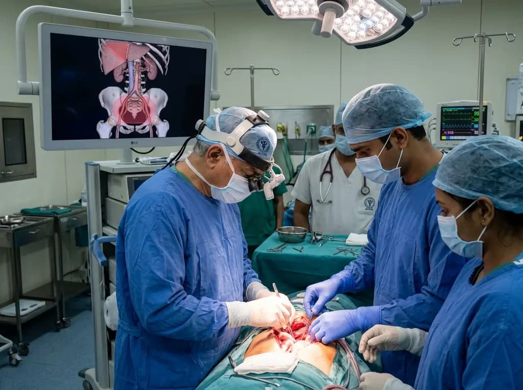
Surgical oncologists at AIIMS Delhi have mapped two critical anatomical landmarks: Ray's Zone in the deep pelvis and Ray's Hepatocaval Triangle near the liver. These predefined hazard boundaries guide dissection, dramatically reduce severe intraoperative bleeding, and preserve vital nerves and visceral organs.
Today
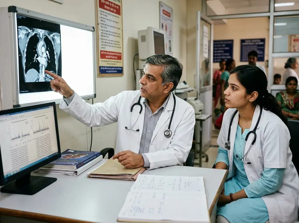
A study reveals that the MYLK c.4079G>A, p.(Gly1360Asp) variant is linked to high-penetrance heritable thoracic aortic disease and early-onset aortic dissection, showing a founder effect in Finnish cohorts. Findings emphasize early cascade screening and vigilant surveillance.
Today
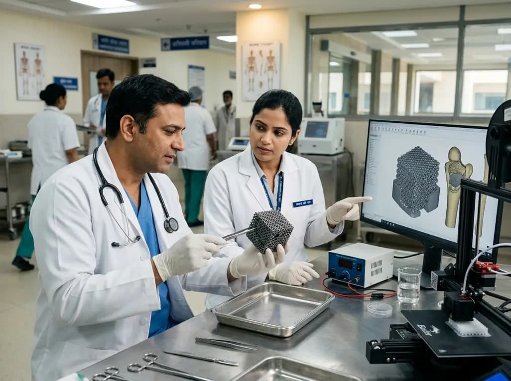
A novel sacrificial-template technique combines FDM polymer 3D printing and micro-arc oxidation to fabricate orientation-controlled porous titanium scaffolds. These bioactive constructs offer tailored stiffness, exceptional multidirectional load resistance, and robust osseointegration for bone defect repair.
Today
An animal model study reveals that gestational exposure to polystyrene microplastics causes persistent intrauterine growth restriction, testicular hypoplasia, Leydig cell depletion, and an atypical low-testosterone, low-LH endocrine profile in male offspring from birth into adulthood.
Today
