

Loading, please wait...
Loading, please wait...

Congenital lower urinary tract obstruction remains a significant cause of long-term pediatric morbidity. Among the various etiologies, posterior urethral valves represent the most frequent cause of anatomical urinary outflow obstruction in male infants and young boys. Historically, clinicians evaluated these patients primarily through the lens of renal preservation, detrusor pressure dynamics, and subsequent voiding patterns. However, contemporary pediatric urology increasingly recognizes that urinary dysfunction does not exist in isolation. The intimate anatomical, embryological, and neurological relationship between the lower urinary tract and the gastrointestinal system frequently produces concurrent symptomatology. While functional bladder and bowel dysfunction is well-documented in otherwise neurologically intact children, the impact of primary mechanical infravesical obstruction on bowel motility has remained poorly investigated. Consequently, evaluating how relieving mechanical outflow resistance influences colorectal transit provides valuable clinical insight for clinicians managing these complex pediatric patients.
The embryological origins and neurological innervation of the lower urinary tract and hindgut are intertwined. Both organ systems share common sacral nerve roots, specifically S2 through S4, which regulate autonomic and somatic control over micturition and defecation. Furthermore, the pelvic floor musculature acts as a shared physical diaphragm supporting both the urinary bladder and the rectum. In children born with posterior urethral valves, severe mechanical resistance in the posterior urethra forces the detrusor muscle to generate abnormally high voiding pressures. Over time, this chronic outflow resistance produces severe bladder wall hypertrophy, hyperreflexia, and elevated pelvic floor tone. Consequently, the child experiences heightened muscle spasms across the entire pelvic floor complex. This hypertonicity directly impedes the normal relaxation of the external anal sphincter during defecation. Therefore, the physiological mechanical barrier created by the urethral valve radiates across adjacent pelvic networks, secondary rectal stasis, and subsequent functional fecal retention.
A recent prospective observational study conducted at the Santobono-Pausilipon Children's Hospital in Naples, Italy, systematically evaluated this gastrointestinal-urinary link. Investigators enrolled 101 male children aged 0 to 6 years diagnosed with posterior urethral valves between January 2021 and March 2024. The research team rigorously categorized bowel habits using standardized Rome IV criteria at baseline and following definitive surgical intervention. Remarkably, the baseline analysis revealed that 26.7 percent of the enrolled cohort met the diagnostic criteria for chronic functional constipation. These children exhibited typical signs of severe withholding, infrequent bowel movements, and painful defecation. Moreover, many patients demonstrated secondary encopresis and abdominal discomfort alongside classic lower urinary tract symptoms. Historically, clinicians frequently attributed such gastrointestinal distress to dietary patterns, behavioral withholding, or fluid restrictions. Instead, these findings establish that functional colorectal delays occur with high prevalence among boys suffering from anatomical infravesical obstruction.
The therapeutic cornerstone for congenital urinary obstruction is prompt transurethral endoscopic valve ablation. Following surgical resection of the obstructing mucosal leaflets, investigators reassessed bowel function during standardized follow-up evaluations. The post-procedural outcomes were striking and statistically robust. Among the 27 patients diagnosed with baseline chronic constipation, bowel symptoms resolved completely in 19 children following successful endoscopic ablation. This represents a remarkable resolution rate exceeding 70 percent. Furthermore, this dramatic improvement occurred without introducing escalating doses of osmotic laxatives or radical dietary modifications. By relieving the severe mechanical obstruction at the bladder outlet, endoscopic surgery alleviated compensatory detrusor strain and normalized elevated pelvic floor pressures. Consequently, the surrounding pelvic striated musculature regained normal compliance, allowing physiological rectal emptying. Thus, removing the primary urological obstruction directly restored functional gastrointestinal dynamics in the majority of affected boys.
These findings carry profound implications for general pediatricians, pediatric urologists, pediatric gastroenterologists, and pediatric nephrologists. Bladder and bowel dysfunction represents an interconnected axis where failure to address one component frequently exacerbates the other. Chronic constipation distends the rectum, which subsequently compresses the bladder base, impairs ureterovesical junction competency, and worsens post-void residual urine volumes. In children with anatomical urethral valves, untreated constipation can perpetuate recurrent urinary tract infections, worsen hydronephrosis, and accelerate renal damage. Therefore, clinicians must thoroughly assess bowel habits in all children presenting with suspected or confirmed posterior urethral valves. Incorporating validated tools, such as the Rome IV criteria and stool consistency scales, should become routine during initial evaluations. Similarly, pediatricians encountering refractory constipation in young boys with atypical voiding symptoms should maintain vigilance for underlying anatomical lower urinary tract obstruction.
Comprehensive care for children with posterior urethral valves necessitates a collaborative multidisciplinary strategy. While endoscopic ablation effectively relieves the anatomical urethral barrier, long-term bladder health requires vigilant surveillance. Many boys continue to manifest myogenic bladder failure or detrusor instability despite successful valve resection. In these persistent cases, unresolved gastrointestinal dysfunction can severely undermine bladder recovery. Therefore, post-ablation management must incorporate combined urological and gastroenterological protocols. Clinicians should evaluate stool frequency, toilet training progress, and urinary continence at every follow-up visit. If constipation persists after endoscopic ablation, physicians must initiate prompt behavioral therapy, adequate hydration, and osmotic laxatives like polyethylene glycol. Additionally, pelvic floor physiotherapy can help retrain dysfunctional voiding and defecation habits. By adopting an integrated treatment philosophy, healthcare providers can safeguard renal function, prevent recurrent infections, and significantly enhance the quality of life for these pediatric patients.
Posterior urethral valves generate chronic urinary outflow resistance, forcing the bladder detrusor and pelvic floor muscles to contract against high pressures. This sustained pelvic floor hypertonicity impairs the normal relaxation of the external anal sphincter during defecation. Additionally, shared sacral nerve roots between the bladder and rectum create shared neurofunctional cross-talk, leading to functional stool withholding and secondary chronic constipation.
Evidence shows that endoscopic ablation resolves chronic constipation in over seventy percent of affected patients. Relieving the mechanical outflow obstruction reduces detrusor strain, decreases baseline pelvic floor muscle hypertonicity, and restores normal defecation reflexes. However, patients with long-standing behavioral withholding, rectal dilatation, or permanent detrusor remodeling may still require supplemental laxative therapy, dietary guidance, and structured pelvic floor retraining.
Severe chronic constipation causes rectal distension that directly compresses the bladder wall and trigone. This mechanical compression increases intravesical pressures, prevents complete bladder emptying, and aggravates secondary vesicoureteral reflux. Elevated urinary storage pressures and chronic urinary stasis dramatically increase the risk of recurrent febrile urinary tract infections, which can accelerate irreversible renal parenchymal scarring and chronic kidney disease.
Disclaimer: This content is for informational and educational purposes only and should not be taken as professional medical advice. Always seek the advice of your physician or other qualified health provider with any questions you may have regarding a medical condition. Refer to the latest local and national guidelines for clinical practice.
References
Manganaro V et al. Posterior urethral valve and chronic constipation: Association and clinical outcome following endoscopic ablation in children. J Pediatr Gastroenterol Nutr. 2026 Oct 18. doi: 10.1002/jpn3.70622. PMID: 42893903.
Dos Santos J, et al. Bladder and bowel dysfunction in children with posterior urethral valves: Systematic screening and modern management strategies. J Pediatr Urol. 2021;17(4):485-492.
Koff SA. Bladder-bowel dysfunction: Understanding the interconnected pelvic organs in pediatric urinary tract disorders. Nat Clin Pract Urol. 2008;5(6):321-329.

Read summarized clinical updates, watch expert medical content, and earn CME certifications right from your smartphone.
Open your smartphone camera to scan and install.





A prospective study reveals that over a quarter of boys with posterior urethral valves suffer from chronic constipation. Crucially, endoscopic valve ablation resolves gastrointestinal symptoms in over 70% of these patients, highlighting the deep neurofunctional cross-talk within bladder-bowel dysfunction.
Today

Kenya's confirmation of its eighth Ebola infection highlights the acute risk of secondary transmission in hospital environments. Clinicians must maintain high vigilance, enforce strict infection prevention protocols, and implement rapid triage mechanisms for viral hemorrhagic fevers across healthcare settings.
Today
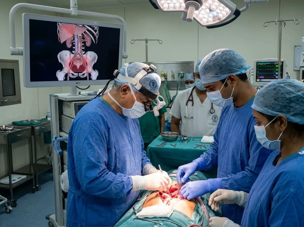
Surgical oncologists at AIIMS Delhi have mapped two critical anatomical landmarks: Ray's Zone in the deep pelvis and Ray's Hepatocaval Triangle near the liver. These predefined hazard boundaries guide dissection, dramatically reduce severe intraoperative bleeding, and preserve vital nerves and visceral organs.
Today
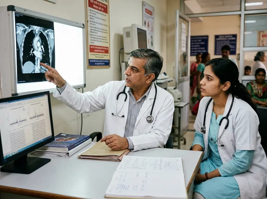
A study reveals that the MYLK c.4079G>A, p.(Gly1360Asp) variant is linked to high-penetrance heritable thoracic aortic disease and early-onset aortic dissection, showing a founder effect in Finnish cohorts. Findings emphasize early cascade screening and vigilant surveillance.
Today
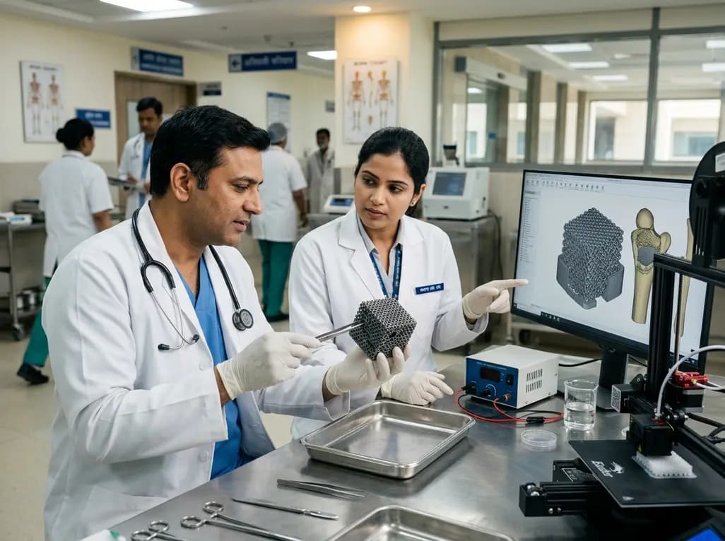
A novel sacrificial-template technique combines FDM polymer 3D printing and micro-arc oxidation to fabricate orientation-controlled porous titanium scaffolds. These bioactive constructs offer tailored stiffness, exceptional multidirectional load resistance, and robust osseointegration for bone defect repair.
Today

A novel label-free electrochemical biosensor utilizing magnetic nanocomposites enables ultrasensitive TERT C228T mutation detection (LOD: 0.67 pM) for rapid risk stratification in papillary thyroid cancer, offering promising point-of-care utility for endocrine oncology.
Today
