.webp&w=384&q=75)

Loading, please wait...
Loading, please wait...

Modern spine surgery frequently utilizes cervical disc arthroplasty to treat radiculopathy while preserving normal segmental motion. Consequently, surgical teams perform this motion-sparing procedure with increasing frequency across diverse patient cohorts. However, a distinct subset of patients inevitably develops persistent or recurrent radicular symptoms at the treated index level. In these complex scenarios, revision anterior surgery presents considerable technical challenges. Specifically, anterior scar formation, device extraction risks, and the frequent need to convert mobility to spinal fusion elevate patient morbidity. Therefore, clinicians urgently require reliable motion-preserving salvage options. Performing a posterior cervical foraminotomy serves as a targeted decompression strategy in these demanding situations. This focused dorsal approach directly accesses the neural foramen without disturbing the anterior prosthetic implant. Moreover, surgeons decompress the affected nerve root while completely preserving the existing arthroplasty construct. Recent clinical investigations demonstrate that secondary posterior foraminotomy effectively addresses recurrent neural compression following primary disc replacement. Accordingly, spine surgeons increasingly employ this targeted posterior decompression to rescue symptomatic patients. This strategic technique circumvents difficult anterior revision procedures while consistently maintaining motion preservation goals.
Surgeons implement posterior cervical foraminotomy through either minimally invasive tubular retractors or modern endoscopic visualization systems. During this targeted procedure, the surgeon identifies the classic V-shaped interlaminar junction overlying the compressed nerve root. Subsequently, precision high-speed diamond burrs and micro-punches unroof the lateral recess and neural canal. Clinicians must preserve at least fifty percent of the native facet joint complex throughout the decompression. Biomechanical studies confirm that preserving the facet architecture prevents iatrogenic segmental instability after anterior arthroplasty. Furthermore, this posterior route completely avoids the previous anterior surgical corridor, eliminating risks to the esophagus and recurrent laryngeal nerve. Operating from a dorsal corridor allows direct visualization of the exit zone and axilla of the cervical nerve root. Consequently, the surgical team can precisely resect recurrent uncinate osteophytes or foraminal soft disc fragments. Laboratory kinematic analyses demonstrate that performing unilateral or bilateral posterior foraminotomies induces negligible alteration in spinal motion profiles. Therefore, surgeons safely expand the neural aperture without destabilizing the biomechanical equilibrium established by the artificial disc prosthesis.
Multicenter clinical research underscores substantial therapeutic benefits when utilizing posterior cervical foraminotomy after cervical disc arthroplasty. In recent multicenter cohort analyses, patients underwent same-level secondary decompression at an average interval of eighty-seven weeks following index arthroplasty. Researchers tracked these individuals over an average follow-up extending past one hundred and four weeks postoperatively. Significantly, arm pain visual analog scores decreased markedly from a preoperative mean of 5.1 down to 2.4. Moreover, seventy-five percent of these individuals successfully achieved the minimal clinically important difference for arm pain relief. Similarly, mean axial neck pain scores improved significantly from 4.9 preoperatively to 3.4 at final evaluation. Approximately sixty-nine percent of patients surpassed the clinical threshold for meaningful neck pain reduction. Although general Neck Disability Index scores demonstrated modest numerical improvements without achieving statistical significance, patients reported high satisfaction with symptom resolution. Thus, targeted nerve decompression reliably resolves recalcitrant radicular agony caused by progressive foraminal narrowing. In addition, these quantitative outcome measures illustrate that posterior decompression provides consistent, meaningful relief for properly selected candidates.
Maintaining physiological spinal motion represents the foundational objective of initial cervical disc arthroplasty. Fortunately, performing a secondary posterior cervical foraminotomy does not destabilize the functional motion segment. In recent clinical series, researchers observed zero cases of secondary mechanical instability or progressive kyphosis following dorsal decompression. Biomechanical stability remains robust because the anterior prosthesis maintains intervertebral disc height while unroofing the neural canal leaves crucial posterior structures intact. However, surgeons must exercise meticulous technique during lateral bone removal to avoid excessive facet joint resection. Preserving the integrity of the lateral facet capsule ensures continued resistance against pathological translational and rotational forces. Furthermore, avoiding segmental fusion protects adjacent cervical motion segments from accelerated degenerative wear and compensatory hypermobility. Longitudinal investigations verify that patients maintain satisfactory flexion, extension, and lateral bending arcs following this dual anterior-posterior treatment strategy. Therefore, combining an anterior artificial disc with a posterior laminoforaminotomy preserves kinetic spinal behavior effectively. In contrast to fusion revision, this motion-sparing approach respects native biomechanics, delivering durable decompression without placing excessive stress on neighboring levels.
Although posterior cervical foraminotomy demonstrates substantial clinical success, spine specialists must remain vigilant regarding revision pathways. In published multicenter data, only two out of seventeen patients, representing approximately twelve percent, required subsequent revision surgery. Specifically, both non-responders underwent anterior explantation of the artificial disc and conversion to anterior cervical discectomy and fusion. Importantly, initial posterior decompression does not compromise surgical landmarks or increase the technical difficulty of subsequent anterior revisions. Because dorsal decompression avoids anterior scarring, surgeons encounter a pristine surgical plane if anterior revision ultimately becomes necessary. Furthermore, clinicians can reserve major revision arthroplasty or arthrodesis for individuals failing less invasive posterior decompression. Careful preoperative radiographic assessment remains vital to exclude hardware loosening, significant subsidence, or prosthetic displacement before selecting posterior decompression. When implant position remains pristine and radiculopathy stems purely from foraminal osteophytes, posterior decompression serves as an ideal initial step. Consequently, this stepped-care algorithmic approach optimizes clinical outcomes while avoiding unnecessary anterior hardware removal. Thus, surgeons systematically resolve recurrent radicular compression while protecting future revision alternatives.
Surgeons consider posterior cervical foraminotomy when patients present with persistent or recurrent unilateral or bilateral radiculopathy after index arthroplasty. Specifically, cross-sectional imaging must verify that the anterior prosthetic implant remains well-positioned without loosening, subsidence, or migration. In addition, magnetic resonance imaging or computed tomography should demonstrate foraminal stenosis due to recurrent uncovering, uncovertebral osteophytes, or residual disc fragments. Consequently, dorsal decompression addresses nerve entrapment directly without compromising anterior device stability.
Clinical and biomechanical studies demonstrate that posterior cervical foraminotomy does not cause segmental cervical instability when surgeons preserve at least fifty percent of the facet joint. Furthermore, preserving facet integrity maintains translational stiffness and rotational resistance across physiological movement arcs. Clinical investigations reveal zero cases of delayed subluxation or kyphosis after same-level posterior decompression. Consequently, patients safely retain motion preservation benefits from their initial disc arthroplasty while achieving effective neural decompression.
If radicular pain persists after posterior foraminotomy, spine specialists can safely convert the index level to an anterior cervical discectomy and fusion. Importantly, initial dorsal decompression preserves anterior tissue planes, preventing scar-related complications during anterior exposure. Surgeons simply remove the artificial disc implant, thoroughly decompress the anterior space, and place a fusion cage with plate stabilization. Consequently, posterior foraminotomy functions as an effective intermediate intervention that preserves future reconstructive options.
Disclaimer: This content is for informational and educational purposes only... Refer to the latest local and national guidelines for clinical practice.
References

Read summarized clinical updates, watch expert medical content, and earn CME certifications right from your smartphone.
Open your smartphone camera to scan and install.





Posterior cervical foraminotomy provides significant pain relief for persistent or recurrent radiculopathy after cervical disc arthroplasty. This motion-preserving salvage decompression avoids complex revision surgeries and segmental instability while achieving high rates of clinical improvement.
Today
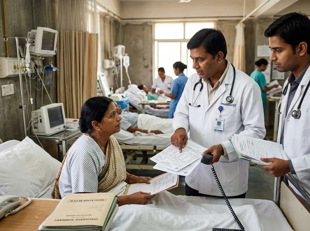
A retrospective cohort study demonstrates that comprehensive pharmacy transitions of care—including admission medication history, discharge review, and 72-hour telephone follow-up—significantly reduced 30-day readmission rates in acute care patients, highlighting the value of pharmacist-led clinical interventions.
Today

Meghalaya recorded a historic 55% reduction in maternal deaths, dropping from 244 to 107 over five years. Through the MOTHER app, dedicated transit homes, and expanded infrastructure like the 150-bed Tura Maternity Hospital with its first public NICU, the state demonstrates an effective public health model.
Today

Explore modern perioperative management protocols for clinical xenotransplantation. Learn how costimulation blockade, anti-inflammatory perfusion, and coagulopathy rescue overcome interspecies hurdles.
Today

At Homa CME 2026 at Hyderabad's T-Hub, healthcare leaders emphasized moving beyond routine metrics through digital calculators and precision stratification. Discover why characterizing individual insulin secretion, resistance patterns, and vascular risks transforms chronic metabolic care in India.
Today
