

Loading, please wait...
Loading, please wait...
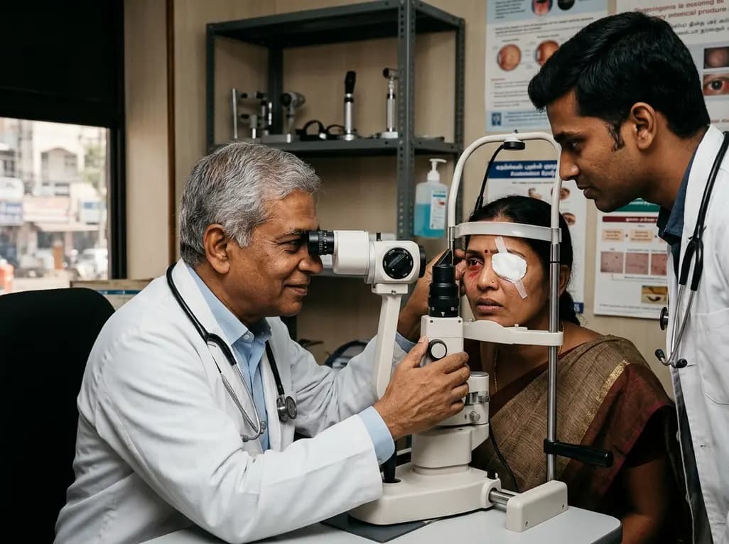
Solid organ transplantation offers life-saving benefits for end-stage organ failure, yet long-term graft survival mandates continuous immunomodulatory therapy. Although maintenance immunosuppression effectively suppresses allograft rejection, clinicians increasingly observe new autoinflammatory conditions in these patients. Among these unexpected presentations, post-transplant ocular inflammation represents an emerging clinical dilemma. When transplant recipients present with sudden red eye, ocular discomfort, or visual impairment, doctors often suspect opportunistic intraocular infections. However, compelling clinical evidence indicates that sterile inflammatory disorders such as uveitis and scleritis can also arise de novo despite ongoing systemic immunosuppressive regimens.
Patients undergoing solid organ transplantation require potent immunomodulatory agents, such as calcineurin inhibitors, antimetabolites, and mammalian target of rapamycin inhibitors. Curiously, ophthalmologists frequently employ these exact drug classes to manage chronic non-infectious uveitis in the general population. Consequently, the appearance of de novo uveitis or scleritis in transplant recipients presents a striking paradox.
In a landmark multi-year cohort study, researchers analyzed solid organ recipients who developed presumed non-infectious ocular inflammation while adhering to systemic maintenance therapy. The study cohort included recipients of kidney transplants, combined kidney-pancreas transplants, and liver transplants. The mean age of affected patients was approximately forty-seven years. Interestingly, the ocular manifestations emerged at a median interval of four years following organ transplantation.
The clinical spectrum in these patients included both anterior uveitis and scleritis. Furthermore, six out of nine patients experienced an isolated, single inflammatory episode, while three patients suffered recurrent disease. Every documented episode responded favorably to local or systemic corticosteroid therapy. Therefore, prompt ophthalmic assessment remains vital for identifying sterile inflammation early and avoiding unnecessary toxicity from empirical antimicrobial treatments.
Why do transplant recipients develop ocular inflammatory disease despite systemic immunomodulatory treatment? While immunosuppressive regimens broadly silence specific effector pathways, they can inadvertently disrupt delicate immunoregulatory networks. Solid organ recipients experience altered immune homeostasis, characterized by shifting balances between effector T helper cells and regulatory T lymphocytes.
Specifically, calcineurin inhibitors like tacrolimus and cyclosporine target interleukin-2 transcription, which suppresses conventional T cells. However, regulatory T cells also rely heavily on interleukin-2 signaling for their survival and suppressive competence. As a result, chronic immunosuppressive exposure may impair regulatory T cell function, permitting escape pathways for autoreactive clones. Furthermore, peripheral lymphopenia often stimulates homeostatic proliferation, which can inadvertently activate dormant, self-reactive immune responses against ocular antigens.
In addition, subclinical viral reactivations or environmental exposures can stimulate innate immune pathways through pattern recognition receptors. This innate signaling triggers robust local cytokine cascades within the immune-privileged eye. Even when systemic T-cell responses remain suppressed, local ocular tissues may experience inflammatory damage driven by alternative cytokine circuits. Understanding these molecular mechanisms helps physicians appreciate that systemic immunosuppression does not guarantee total protection against focal, organ-specific autoimmunity.
When evaluating an immunosuppressed transplant recipient presenting with ocular complaints, clinicians face an urgent diagnostic challenge. Because systemic immunosuppression elevates the risk of opportunistic pathogens, physicians must meticulously exclude infectious etiologies before diagnosing non-infectious uveitis. Infectious uveitis in transplant recipients can progress rapidly and cause irreversible blindness if clinicians misclassify it as autoimmune disease.
Opportunistic ocular pathogens include cytomegalovirus, herpes simplex virus, varicella-zoster virus, and fungal entities such as Candida and Aspergillus. In addition, protozoal infections like toxoplasmosis can mimic sterile anterior or posterior uveitis. Therefore, comprehensive diagnostic workups require dilated funduscopic examination, high-resolution optical coherence tomography, and, when indicated, anterior chamber paracentesis for molecular polymerase chain reaction testing.
Furthermore, systemic laboratory evaluations should include serological assays, blood cultures, and targeted screening for latent tuberculosis and syphilis. If diagnostic testing definitively excludes active infectious agents, clinicians can confidently diagnose presumed non-infectious ocular inflammation. Establishing this distinction prevents catastrophic visual outcomes, because administering high-dose corticosteroids to an unrecognized intraocular infection can induce rapid retinal necrosis or fulminant endophthalmitis.
Managing non-infectious uveitis and scleritis in solid organ transplant recipients requires careful calibration of anti-inflammatory therapy. Because these patients already receive complex maintenance immunosuppression, adding further systemic agents demands close collaboration between ophthalmologists and transplant specialists. Fortunately, clinical observations demonstrate that post-transplant ocular inflammation responds remarkably well to standard corticosteroid protocols.
For isolated anterior uveitis, intense topical corticosteroid drops, such as prednisolone acetate one percent, typically induce rapid remission without perturbing systemic transplant dynamics. Clinicians should concurrently prescribe cycloplegic drops to relieve ciliary spasm and prevent posterior synechiae formation. In cases involving scleritis, intermediate uveitis, or posterior segment involvement, systemic oral corticosteroids or periocular triamcinolone injections may become necessary.
However, systemic corticosteroid escalation carries significant risks in transplant patients, including worsened glycemic control, hypertension, avascular necrosis, and elevated systemic infection risks. Consequently, physicians must design rapid tapering schedules once clinical control occurs. Furthermore, transplant physicians must monitor renal function, hepatic enzymes, and therapeutic drug levels of baseline calcineurin inhibitors, ensuring that adjunctive treatments do not precipitate allograft dysfunction.
The emergence of de novo ocular inflammation underscores the critical need for coordinated multidisciplinary care among ophthalmologists, nephrologists, and transplant surgeons. Because transplant patients often prioritize graft survival above subtle visual symptoms, mild ocular discomfort can easily escape early detection. Therefore, healthcare teams must educate transplant recipients to recognize early warning signs, including ocular redness, photophobia, floaters, and blurred vision.
In addition, routine ophthalmic evaluations should become an integral component of comprehensive post-transplant health maintenance. Although routine screening protocols typically focus on metabolic complications like diabetic retinopathy or steroid-induced cataracts, clinicians must maintain vigilance for inflammatory phenomena. Documenting baseline visual acuity and intraocular pressure helps detect late-onset complications, including secondary glaucoma and macular edema.
Moreover, multidisciplinary communication proves indispensable during disease recurrences. When transplant recipients experience recurrent uveitis, clinicians may need to optimize their systemic maintenance immunosuppressive regimen. For example, switching from one antimetabolite to another or adjusting mammalian target of rapamycin inhibitor dosing might simultaneously protect the allograft and stabilize intraocular inflammation. Through synchronized co-management, healthcare teams can safeguard both patient survival and lifelong visual function.
Yes, solid organ transplant recipients can certainly develop non-infectious uveitis or scleritis while receiving maintenance immunosuppressive therapy. Although systemic medications effectively prevent organ graft rejection, they can selectively disrupt immune regulatory networks and permit localized inflammatory cascades to activate. Recent clinical evidence demonstrates that patients may experience sterile ocular inflammation several years after transplantation, which requires prompt ophthalmic evaluation, precise differential diagnosis, and appropriate anti-inflammatory therapy.
Clinicians differentiate non-infectious ocular inflammation from opportunistic infections by conducting detailed slit-lamp examinations, dilated fundus evaluations, and targeted imaging. Because immunosuppressed transplant recipients face significant risks from herpesviruses, cytomegalovirus, and opportunistic fungi, doctors often perform molecular polymerase chain reaction analysis on anterior chamber fluid or vitreous samples. Once thorough microbiological testing excludes active ocular infection, physicians can safely diagnose sterile inflammatory conditions and initiate corticosteroid treatment.
Steroid therapy remains the primary intervention for managing sterile post-transplant ocular inflammation. Anterior uveitis usually resolves rapidly with intensive topical corticosteroid drops, whereas deeper intraocular inflammation or scleritis often necessitates oral corticosteroids or localized periocular injections. Physicians must carefully taper these steroids once inflammation subsides, while diligently monitoring intraocular pressure, systemic blood pressure, serum glucose levels, and maintenance immunosuppressive drug concentrations to avoid systemic toxicity.
Disclaimer: This content is for informational and educational purposes only and is intended for healthcare professionals. It does not constitute medical advice or establish a doctor-patient relationship. Clinicians should exercise independent clinical judgment and verify treatment regimens with current pharmacological resources. Refer to the latest local and national guidelines for clinical practice.
References
Katragadda K et al. Ocular Inflammation in Solid Organ Transplant Patients Receiving Maintenance Immunosuppression. Ocul Immunol Inflamm. 2026 Nov 12. doi: 10.1080/09273948.2026.2751588. PMID: 43019528.
Giorgakis E et al. Herpetic ocular infections in solid organ transplantation: A narrative review. Transplant Rev (Orlando). 2026 Apr;40(2):101005. doi: 10.1016/j.trre.2026.101005.
Nelson J et al. Consensus Recommendations for Use of Maintenance Immunosuppression in Solid Organ Transplantation. Pharmacotherapy. 2022 Aug;42(8):599-633. doi: 10.1002/phar.2718.
Jabs DA. Immunosuppression for the Uveitides. Ophthalmology. 2018 Jun;125(2):193-202. doi: 10.1016/j.ophtha.2017.08.007.

Read summarized clinical updates, watch expert medical content, and earn CME certifications right from your smartphone.
Open your smartphone camera to scan and install.





A new study reveals that solid organ transplant recipients can paradoxically develop presumed non-infectious uveitis and scleritis while on maintenance immunosuppression. Clinicians must recognize these steroid-responsive inflammatory episodes and differentiate them from opportunistic ocular infections.
Today

Ascertainment bias in cancer case-control studies inflates odds ratios and penetrance estimates when cases are drawn from high-risk clinics. A landmark modeling study demonstrates substantial bias ranging up to 23.72, highlighting the critical need for ascertainment adjustment in genetic risk counseling.
Today
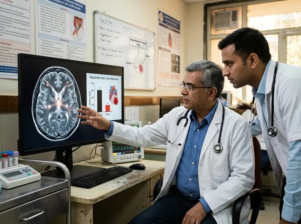
Researchers demonstrate that central induction of synthetic torpor in non-hibernating rats provides striking cardioprotection, reducing myocardial infarct size by ~40% during ischaemia-reperfusion injury through endogenous survival signaling, independent of hypothermia.
Today
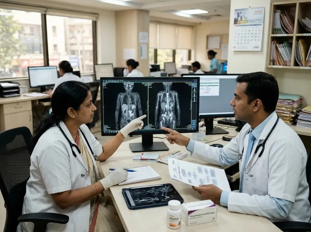
Chronic nonbacterial osteomyelitis (CNO) is a pediatric autoinflammatory bone disorder. This review covers the French protocol, emphasizing whole-body MRI as the diagnostic gold standard, prudent biopsy avoidance, first-line NSAID treatment, and second-line biologic or bisphosphonate therapies for spinal lesions.
Today
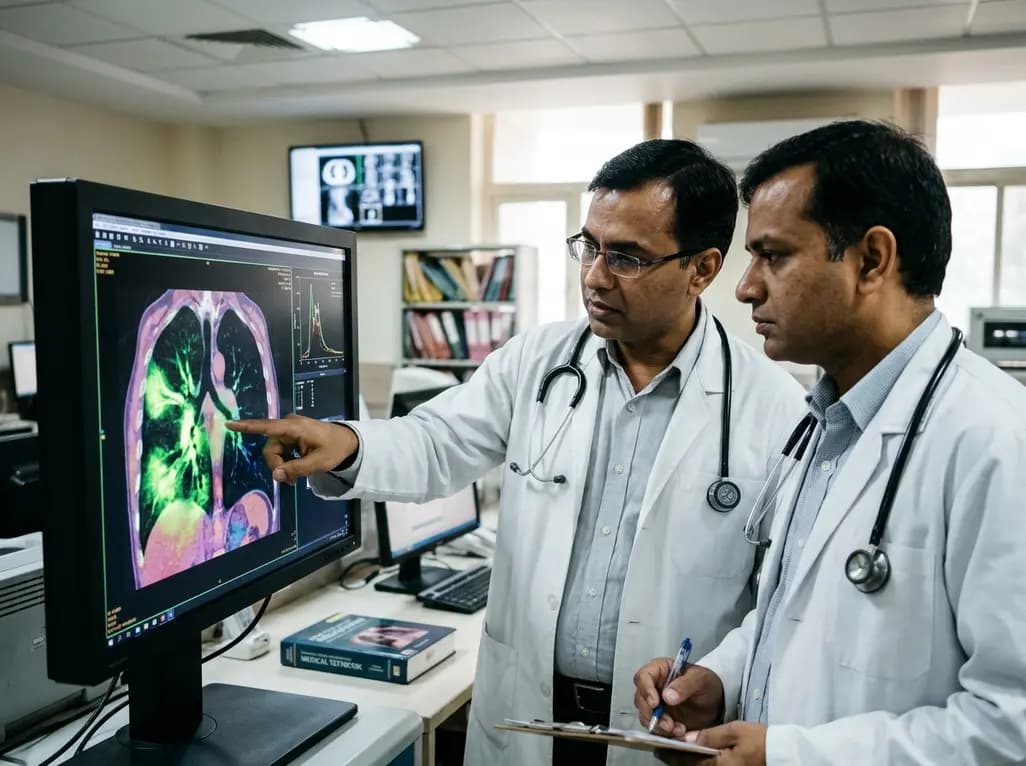
A novel fluorine-functionalized near-infrared copper fluorescent probe selectively illuminates lung tissue, revealing critical links between hyperglycemia, ER stress, and pulmonary malignancy for improved early diagnosis.
Today
