.webp&w=384&q=75)

Loading, please wait...
Loading, please wait...
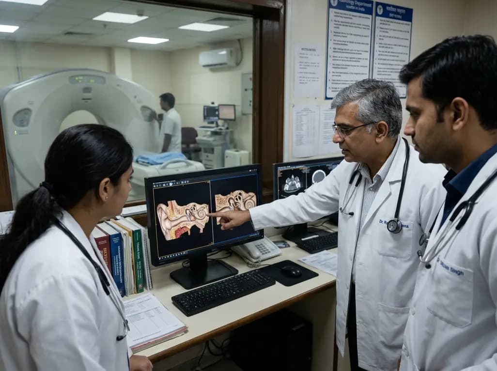
The field of medical imaging is currently witnessing a transformative shift with the introduction of photon-counting detector CT (PCD-CT). This technology represents the most significant leap in computed tomography since the advent of multi-detector systems. Unlike conventional energy-integrating detectors (EID) that convert X-rays into light before turning them into electrical signals, PCD-CT uses semiconductor materials to directly convert X-ray photons into electrical pulses. This direct conversion process eliminates the need for reflective septa within the detector, allowing for much smaller detector elements. Consequently, photon-counting detector CT can achieve ultra-high spatial resolution that was previously unattainable in clinical practice. For specialists dealing with the temporal bone, where critical anatomical structures are often less than a millimeter in size, this advancement offers unprecedented clarity. The ability to visualize the fine details of the middle and inner ear is not just a technical triumph but a potential game-changer for diagnostic precision in complex otologic cases.
The primary advantage of photon-counting detector CT lies in its ability to resolve minute details. Conventional CT scanners often struggle with the submillimeter components of the temporal bone, such as the stapes footplate, the incudostapedial joint, and the delicate partitions of the cochlea. PCD-CT provides a limiting spatial resolution of approximately 150 micrometers or less, which is significantly superior to standard EID-CT systems. This enhanced resolution allows radiologists to identify subtle pathologies that might have been missed previously. For instance, the visualization of the modiolus and the individual turns of the cochlea becomes much sharper, aiding in the preoperative assessment for cochlear implantation. Furthermore, the increased resolution facilitates the detection of superior semicircular canal dehiscence, where even a tiny gap in the bone can lead to debilitating vestibular symptoms. By providing clearer images of these small gaps, PCD-CT helps clinicians differentiate between true dehiscence and "near-dehiscence," thereby refining surgical planning and improving patient counseling.
In addition to superior resolution, photon-counting detector CT offers a better signal-to-noise ratio and increased dose efficiency. Traditional EID systems suffer from electronic noise, especially at low radiation doses, which can obscure fine bony details. PCD-CT effectively eliminates this electronic noise by using an energy threshold that ignores low-energy pulses typically associated with noise. This technical feature is particularly beneficial in temporal bone imaging, where high-resolution kernels naturally increase image graininess. With PCD-CT, clinicians can maintain excellent image quality while reducing the radiation dose by 30% to 50% compared to conventional protocols. This is a crucial benefit for pediatric patients or individuals requiring multiple follow-up scans. For children with congenital ear anomalies or chronic inflammatory conditions, the ability to obtain diagnostic-grade images at a fraction of the traditional radiation dose aligns with the principle of ALARA (As Low As Reasonably Achievable) while providing superior diagnostic information.
Metal artifacts have long been a challenge in temporal bone imaging, particularly in patients with ossicular prostheses or cochlear implants. The high density of metallic components causes streaking and blooming artifacts that often obscure the interface between the device and the surrounding bone. Photon-counting detector CT addresses this issue through its inherent spectral capabilities and high-resolution sampling. Recent studies have demonstrated that PCD-CT significantly reduces blooming artifacts, allowing for a more accurate assessment of prosthesis positioning and stability. For example, after a stapedotomy, it is vital to confirm that the piston is correctly seated within the oval window. Conventional CT might show a blurred metallic mass, whereas PCD-CT can clearly delineate the piston's relationship to the surrounding structures. This clarity is equally valuable for evaluating cochlear implant electrode arrays, ensuring they are properly positioned within the scala tympani without causing traumatic injury to the delicate cochlear partitions.
While the technical superiority of photon-counting detector CT is well-established, its impact on routine clinical outcomes is currently being defined. The transition from "better images" to "better outcomes" requires robust clinical evidence. Currently, PCD-CT is proving highly valuable in select clinical scenarios where high-resolution is non-negotiable. These include the evaluation of conductive hearing loss of unknown etiology, complex revision middle ear surgeries, and precise mapping of the internal auditory canal. As the technology becomes more widely available in large academic centers and specialized ENT clinics, we expect to see a shift in the standard of care. The ability to characterize tissue at a spectral level may also open new doors for identifying inflammatory versus neoplastic processes within the petrous bone. Although most current data come from phantom and cadaveric studies, early patient-centric research suggests that the improved diagnostic confidence provided by PCD-CT can lead to more targeted surgical interventions and better post-operative monitoring.
The integration of photon-counting detector CT into clinical workflows requires an understanding of its unique reconstruction requirements. To leverage the full potential of the ultra-high-resolution mode, radiologists must use specific sharp kernels and thinner slice thicknesses, often down to 0.2 mm. This creates a massive amount of data, necessitating advanced storage solutions and powerful diagnostic workstations. However, the trade-off is a dataset that can be reconstructed in any plane with near-isotropic resolution, which is essential for the complex anatomy of the ear. Surgeons can now view the temporal bone in Pöschl or Stenvers planes with a level of detail that mirrors the view through a surgical microscope. As we move forward, the combination of PCD-CT with artificial intelligence and deep-learning denoising algorithms will further enhance image quality, making this technology the cornerstone of future otolaryngological diagnostics and personalized treatment planning in patients with complex hearing and balance disorders.
Conventional CT uses energy-integrating detectors that first convert X-rays to light, leading to light scatter and loss of resolution. Photon-counting detector CT uses semiconductor materials to directly convert X-ray photons into electrical signals. This eliminates the need for septa between detector elements, allowing for much smaller pixels and significantly higher spatial resolution. This is particularly useful for visualizing the tiny, submillimeter structures found within the temporal bone.
Yes, photon-counting detector CT is highly dose-efficient because it effectively eliminates electronic noise. Research indicates that PCD-CT can provide superior image quality of the temporal bone at radiation doses 30% to 43% lower than conventional EID-CT. In some pediatric subgroups, dose reductions of over 70% have been achieved while still maintaining excellent diagnostic clarity, making it a safer option for children who may need multiple imaging studies over time.
Absolutely. One of the major challenges with conventional CT is metal artifact or blooming from ear prostheses and cochlear implants. Photon-counting detector CT significantly reduces these artifacts due to its smaller detector size and better sampling. This allows clinicians to see exactly where a prosthesis is located and how it interacts with the surrounding bone, which is critical for checking if an implant has shifted or if it was correctly placed during surgery.
Disclaimer: This content is for informational and educational purposes only and does not constitute medical advice. Always seek the advice of a qualified healthcare provider regarding any medical condition or treatment. The findings discussed are based on emerging research and may not yet represent the standard of care in all clinical settings. Refer to the latest local and national guidelines for clinical practice.
References
Epperson MV et al. Photon-counting detector computed tomography for temporal bone: does higher resolution matter? Curr Opin Otolaryngol Head Neck Surg. 2026 Jul 07. doi: 10.1097/MOO.0000000000001145. PMID: 42406515.
Pellby D et al. Comparison of photon-counting CT with energy-integrating CT in temporal bone imaging: an anthropomorphic phantom study. Acta Radiol. 2026 Apr 17. doi: 10.1177/02841851261431600. PMID: 41995623.
Rajendran K et al. Improved visualization of temporal bone structures with photon-counting detector CT: An intra-patient comparison. Eur J Radiol. 2025 Aug 26. doi: 10.1016/j.ejrad.2025.111111.
"
Read summarized clinical updates, watch expert medical content, and earn CME certifications right from your smartphone.
Open your smartphone camera to scan and install.





Photon-counting detector CT (PCD-CT) is transforming temporal bone imaging by offering ultra-high spatial resolution and reduced radiation doses. This technology enhances the visualization of submillimeter structures like the ossicles and inner ear, potentially improving diagnostic accuracy in otology.
3 months ago

Novel diimide and hydrazone scaffolds, particularly compound #C4, synergistically reactivate latent HIV-1 when combined with JQ1 or PKC activators. Demonstrating donor-matched proviral transcription ex vivo and non-canonical signaling, these findings advance shock and kill strategies toward reservoir eradication.
Today

A cross-sectional study reveals that maternal RSV vaccination coverage reached only 43.7% in Argentina, with maternal vaccine knowledge being the sole independent predictor of uptake. Clinicians play a crucial role in eliminating informational and structural barriers to optimize infant health.
Today

Review how tailored exercise programs benefit frail older patients with heart failure. Explore pathophysiological mechanisms, individualized training prescriptions, and strategies to overcome adherence barriers in clinical practice.
Yesterday

This comparative study evaluates posterior labral tears with and without glenohumeral instability, highlighting distinct demographic patterns, MRI underestimation in stable shoulders, and successful arthroscopic repair outcomes.
Yesterday

A clinical study demonstrates that early intervention with tocilizumab, either as monotherapy or bridged with corticosteroids, achieves significant proptosis reduction and resolves active inflammation in treatment-naive thyroid eye disease.
Yesterday
