.webp&w=384&q=75)

Loading, please wait...
Loading, please wait...
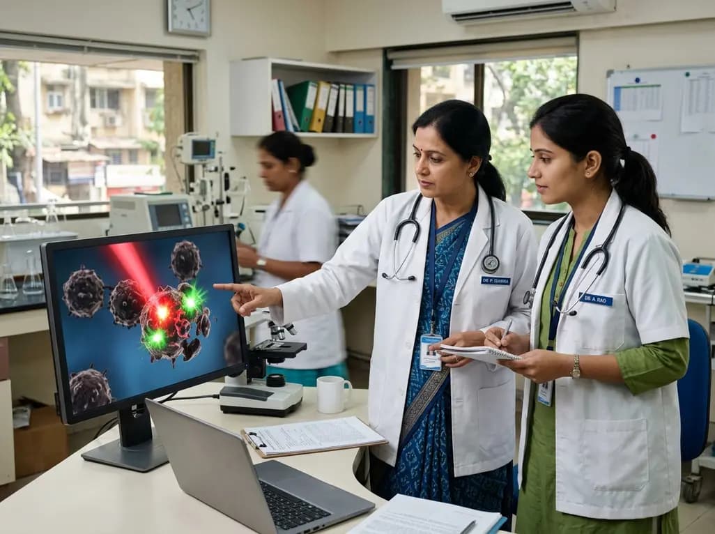
Photodynamic therapy represents an innovative oncological modality that destroys malignant tissue without compromising structural connective tissue integrity. Clinicians utilize this strategy across various neoplastic conditions, including cutaneous, oral, and visceral malignancies. Crucially, understanding photodynamic therapy cell death requires deciphering the interactions among three nontoxic elements: a photosensitizer, specific light wavelengths, and molecular oxygen. Upon illumination, these components generate cytotoxic species that trigger regulated cell death. Consequently, this photocytotoxic action dismantles tumors while priming systemic host immunity.
Photodynamic action requires a photosensitizer, light of an activating wavelength, and tissue oxygen. When light irradiates the targeted tissue, the photosensitizer absorbs photons and transitions from its ground singlet state to an excited singlet state. Subsequently, rapid intersystem crossing converts the molecule into a longer-lived triplet state. From this reactive state, two competing photochemical reactions generate lethal cytotoxic molecules.
Type I pathways transfer electrons directly to cellular biomolecules or molecular oxygen. Consequently, this electron exchange generates superoxide anions, hydrogen peroxide, and highly toxic hydroxyl radicals. Conversely, Type II pathways transfer energy directly to ground-state triplet oxygen, generating singlet oxygen. Because singlet oxygen has an ultra-short half-life under forty nanoseconds, its diffusion distance is strictly confined to twenty nanometers. Therefore, oxidative damage occurs exclusively within the immediate vicinity of the intracellular photosensitizer. Furthermore, singlet oxygen oxidizes unsaturated lipids, structural proteins, and nucleic acids. Ultimately, this intense oxidative stress overwhelms endogenous antioxidant defenses, accelerates organellar breakdown, and triggers cellular collapse.
Once reactive oxygen species accumulate, they trigger distinct regulated cell death cascades. Photodynamic therapy cell death encompasses apoptosis, necroptosis, and regulated autophagy. When photosensitizers localize within mitochondria, photodynamic activation induces immediate opening of mitochondrial permeability transition pores. Consequently, cytochrome c escapes into the cytosol, binds apoptotic protease activating factor 1, and forms the apoptosome. This complex activates caspase-9 and downstream executioner caspases, systematically dismantling structural cytoskeletal proteins.
Additionally, oxidative stress at the endoplasmic reticulum disrupts calcium homeostasis and induces severe protein misfolding. Consequently, sustained reticular stress activates caspase-12, driving apoptotic pathways. However, when cancer cells exhibit defective apoptotic machinery or severe adenosine triphosphate depletion, photodynamic therapy triggers regulated necroptosis. Necroptosis proceeds through receptor-interacting protein kinases RIPK1 and RIPK3 alongside mixed lineage kinase domain-like pseudokinase. Furthermore, photodynamic damage stimulates macroautophagy. While early autophagy can act as a cytoprotective survival mechanism, prolonged autophagic flux precipitates lethal autophagic destruction. Similarly, extensive lipid peroxidation can activate ferroptosis, expanding the repertoire of malignant cell eradication.
High selectivity toward neoplastic tissue represents a major therapeutic advantage of phototherapy. Importantly, tumor selectivity does not depend on a single biological property. Instead, it arises from dynamic interactions among vascular architecture, cellular transport, and spatial illumination. Neoplastic lesions feature defective endothelium, wide fenestrations, and poor lymphatic drainage. Consequently, this enhanced permeability and retention effect facilitates selective macromolecular photosensitizer extravasation and parenchymal entrapment.
Furthermore, malignant cells show elevated metabolic demands and overexpress low-density lipoprotein and transferrin receptors. Because lipophilic photosensitizers bind circulating endogenous lipoproteins, neoplastic cells internalize these compounds via receptor-mediated endocytosis. Additionally, altered transporter expression, including decreased ATP-binding cassette transporter clearance, promotes intracellular retention within malignant cells. Moreover, the acidic extracellular microenvironment characteristic of malignant tissue facilitates the uptake of amphiphilic photosensitizer molecules. Clinicians complement these physiological mechanisms through targeted physical light delivery via flexible optical fibers. Thus, dual biological and physical selectivity spares surrounding normal structures from irreversible damage.
The precise subcellular localization of a photosensitizer governs the resulting death pathway. Because singlet oxygen cannot diffuse far from its origin, initial photodamage remains compartmentalized. Positively charged lipophilic photosensitizers preferentially accumulate along inner mitochondrial membranes due to high negative mitochondrial transmembrane potential. Consequently, illumination selectively degrades anti-apoptotic Bcl-2 proteins, triggering rapid caspase activation.
In contrast, amphiphilic photosensitizers often localize within lysosomes via endocytic pathways. Photoactivation causes lysosomal membrane permeabilization, releasing hydrolytic cathepsins into the cytoplasm. Subsequently, these proteases cleave Bid into truncated Bid, indirectly provoking secondary mitochondrial destruction. Moreover, photosensitizers targeted to the endoplasmic reticulum release sequestered calcium ions into the cytoplasm, provoking severe reticular collapse. Photosensitizers that adhere to the plasma membrane directly induce rapid loss of ionic balance and swelling, promoting necrosis. Crucially, most therapeutic photosensitizers avoid nuclear localization, which prevents DNA mutations and secondary carcinogenesis. Therefore, chemical modifications directing photosensitizers toward specific organelles allow oncologists to tailor cell death outcomes effectively.
Despite notable clinical benefits, the tumor microenvironment presents significant biological barriers. Because the generation of cytotoxic singlet oxygen requires molecular oxygen, severe intratumoral hypoxia diminishes therapeutic efficacy. Furthermore, rapid photochemical reactions consume ambient oxygen during illumination, temporarily intensifying hypoxia. To overcome this limitation, clinicians utilize fractionated light illumination protocols. Consequently, dark intervals allow microvascular blood flow to replenish oxygen within the illuminated field. Additionally, researchers develop oxygen-carrying nanocarriers and near-infrared photosensitizers that penetrate deeper into dense tumors.
Importantly, photodynamic therapy also triggers potent systemic antitumor immunity by inducing immunogenic cell death. Stressed cancer cells express calreticulin on their outer surface and release high-mobility group box 1 alongside adenosine triphosphate. Consequently, these damage-associated molecular patterns recruit and activate dendritic cells, which present tumor neoantigens to CD8+ cytotoxic T lymphocytes. Furthermore, photodynamic damage destroys tumor microvasculature, cutting off nutrient supplies to residual malignant clones. Ultimately, combining photodynamic regimens with modern immune checkpoint inhibitors fosters durable systemic antitumor immunity against distant metastases.
Photodynamic therapy combines non-ionizing light with non-toxic photosensitizers to generate localized oxidative cytotoxicity. Unlike systemic chemotherapy, it causes minimal off-target toxicities and avoids systemic bone marrow suppression. Furthermore, unlike ionizing radiation, photodynamic treatment does not induce primary DNA double-strand breaks or permanent connective tissue scarring. Therefore, clinicians can safely repeat photodynamic treatments multiple times at the same anatomical site without reaching cumulative dose toxicity thresholds.
Intratumoral hypoxia poses a major clinical barrier because Type II photochemical reactions require molecular oxygen to generate singlet oxygen. When oxygen levels drop within dense tumor cores, photodynamic efficacy declines. Consequently, clinicians use fractionated illumination protocols with dark intervals that permit microvascular reoxygenation between light pulses. Additionally, advanced delivery formulations incorporate oxygen-carrying nanoparticles or catalyze endogenous peroxide decomposition, sustaining cytotoxic efficacy under hypoxic conditions.
Yes, photodynamic therapy can induce systemic abscopal responses through the activation of immunogenic cell death. When illuminated malignant cells die, they release damage-associated molecular patterns and tumor-specific antigens. Consequently, mature host dendritic cells prime cytotoxic T lymphocytes against these antigens. These activated lymphocytes then travel through the systemic circulation to eradicate distant unilluminated tumor deposits. Combining photodynamic protocols with immune checkpoint blockade further magnifies this abscopal effect.
Disclaimer: This content is for informational and educational purposes only and does not constitute medical advice. Refer to the latest local and national guidelines for clinical practice.
References

Read summarized clinical updates, watch expert medical content, and earn CME certifications right from your smartphone.
Open your smartphone camera to scan and install.





Photodynamic therapy combines a photosensitizer, light, and oxygen to produce reactive oxygen species that induce regulated cancer cell death. Tumor selectivity, subcellular localization, and immunogenic signaling collectively drive tumor destruction and systemic immunity.
Today

A fatal case involving a research technician at a Siberian anti-plague facility has triggered global surveillance inquiries. While tests rule out high-threat pathogens like plague and show no onward spread, the missing definitive diagnosis highlights crucial challenges in laboratory biosafety and clinical workups.
Today

A systematic review and transcriptomic cross-study consensus analysis illuminates the role of necroptosis in cervical cancer. By promoting anti-tumor immunogenicity and resolving apoptosis resistance, necroptotic pathways offer actionable prognostic signatures and promising avenues for combination immunotherapy.
Today

A systematic review and meta-analysis of 33 studies evaluates the effectiveness of LDL apheresis techniques in familial hypercholesterolemia. Double-filtration plasmapheresis demonstrated the greatest reductions in total cholesterol and LDL-C, while safety and clinical feasibility guide individual therapy.
Today

A scoping review of 964 studies on warm-up in athletes reveals that research heavily emphasizes acute performance proxies while significant gaps remain in injury prevention, dosage parameters, and real-world implementation.
Today

In severe acute pancreatitis, initial normal calcium levels can mask primary hyperparathyroidism due to saponification. This case report highlights how serial calcium monitoring unmasked severe hypercalcemia, guiding life-saving emergency medical therapy and curative parathyroidectomy.
Today
