.webp&w=384&q=75)

Loading, please wait...
Loading, please wait...

Evaluating phase-contrast MRI shunt function represents a significant advancement and a major improvement in neurosurgical diagnostics. Furthermore, this rapid imaging technique allows clinicians to directly quantify cerebrospinal fluid (CSF) flow because it uses velocity-sensitive sequences. Traditionally, doctors rely on ventricular size and invasive shunt taps to assess patency, but these methods often yield inconclusive results in complex postoperative scenarios. Therefore, by incorporating this brief 60-second sequence, surgeons can make informed decisions and also avoid unnecessary revisions since the resulting data is objective and reliable.
Radiologists use bipolar gradients to distinguish flowing protons from stationary tissue. Subsequently, this process generates quantitative data since it measures phase shifts across the shunt tube. In addition, clinicians can compare these values to external ventricular drain (EVD) outputs to confirm patency. For example, in a recent case involving a 19-year-old with Dandy-Walker syndrome, researchers measured a shunt flow of 21.2 mL/hr. Consequently, this value significantly exceeded the patient’s prior mean drain output, so the medical team confirmed shunt patency. As a result, they safely removed the invasive drain while maintaining clinical stability and also avoiding further surgery.
Moreover, phase-contrast MRI offers several benefits because it is entirely non-invasive. First, this reduces the risk of infection compared to needle-based shunt tapping. Second, it provides objective numerical data instead of subjective visual interpretations of ventricular size. Third, the brief acquisition time makes it suitable for emergency settings and routine surveillance alike. Finally, implementing this tool can enhance patient safety and also refine postoperative management while reducing overall healthcare costs. In conclusion, PC-MRI is a vital adjunct since it provides definitive proof of flow, although clinical correlation remains necessary for final decisions.
Phase-contrast MRI utilizes velocity encoding (VENC) to measure the phase shift of moving protons. This shift is directly proportional to the velocity of the cerebrospinal fluid, allowing for the precise calculation of flow volume in mL/hr.
Yes, research indicates that PC-MRI can detect flow in the parenchymal portion of the shunt, even in the presence of magnetic adjustable valves. However, radiologists must plan the sequences carefully to minimize artifacts from the valve itself.
Disclaimer: This content is for informational and educational purposes only and does not constitute medical advice or a professional relationship. Always seek the advice of a physician or other qualified health provider with any questions regarding a medical condition. Refer to the latest local and national guidelines for clinical practice.
References
Ha JH et al. Using phase-contrast MRI to assist in assessment of shunt function following postoperative intraventricular hemorrhage: illustrative case. J Neurosurg Case Lessons. 2026 Feb 16. doi: undefined. PMID: 41698194.
PerK R et al. Phase-Contrast MRI Detection of Ventricular Shunt CSF Flow: Proof of Principle. J Neuroimaging. 2020;30(6):761-767.
Sharma A et al. Phase-Contrast Magnetic Resonance Imaging of Intracranial Shunt Tube: A Valuable Adjunct in the Diagnosis of Ventriculoperitoneal Shunt Malfunction. Oper Neurosurg (Hagerstown). 2017;13(5):565-571.

Read summarized clinical updates, watch expert medical content, and earn CME certifications right from your smartphone.
Open your smartphone camera to scan and install.





The use of phase-contrast MRI shunt function assessment provides a non-invasive, quantitative way to evaluate CSF flow and guide shunt management decisions....
6 months ago

A comparative analysis shows that older age, elevated blood glucose, and abdominal adiposity are primary drivers of hypertension. Comprehensive evaluation of cardiometabolic risk profiles enables clinicians to detect overlapping metabolic dysfunctions early and optimize targeted prevention strategies.
Today

A premature neonate developed upper limb compartment syndrome after uterine rupture extruded the arm through a scar defect. Conservative management with continuous monitoring yielded complete functional recovery and normal limb growth at 10-year follow-up, highlighting non-operative safety in selected cases.
Today

Left ventricular thrombus is a severe complication of acute decompensated heart failure. This clinical review explores the pathophysiology, imaging challenges, and pharmacotherapeutic considerations of laminated thrombi, evaluating the evolving role of direct oral anticoagulants versus traditional warfarin.
Today
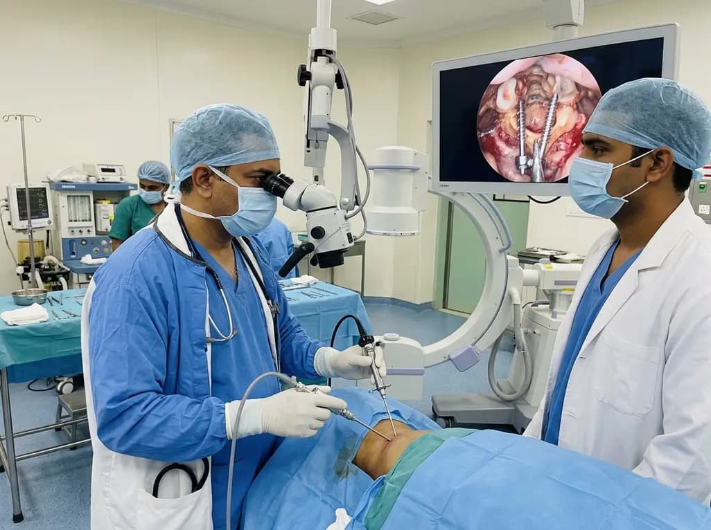
Endoscopic posterior cervical fusion combines minimally invasive decompression, joint preparation, and rigid screw-rod fixation for atlantoaxial pathologies. Early clinical findings demonstrate solid bony union, excellent symptom relief, and minimal soft-tissue morbidity without significant vascular compromise.
Yesterday

Atherosclerosis involves extensive glycometabolic reprogramming across immune and vascular cells. This review examines how glycolysis, the pentose phosphate pathway, and lactate-driven epigenetic shifts fuel plaque vulnerability, while highlighting novel therapeutic targets like PFKFB3 and LDHA.
Today
