.webp&w=384&q=75)

Loading, please wait...
Loading, please wait...

Fluorine-18 fluorodeoxyglucose positron emission tomography integrated with computed tomography, commonly termed PET/CT in paediatric lymphoma, represents a cornerstone in contemporary pediatric hematology-oncology. Malignant lymphoma constitutes one of the most frequent neoplasms affecting children and adolescents, encompassing both Hodgkin lymphoma and diverse non-Hodgkin lymphoma subtypes. Historically, conventional anatomical imaging modalities such as standalone computed tomography and magnetic resonance imaging struggled to distinguish active tumor from post-treatment fibrous scar tissue. Consequently, clinicians encountered high rates of diagnostic uncertainty and unnecessary overtreatment. Today, the integration of functional metabolic imaging has revolutionized clinical pathways across initial staging, interim evaluation, and definitive end-of-therapy assessments.
Moreover, PET/CT provides unparalleled sensitivity for identifying occult extranodal disease, including subtle splenic, skeletal, and bone marrow involvement. Therefore, modern clinical protocols increasingly rely on baseline metabolic scans to establish accurate staging without the mandatory invasive marrow biopsies of previous decades. In addition, the functional assessment offered by fluorodeoxyglucose uptake establishes an objective metabolic baseline. This baseline enables clinicians to measure early tumor kinetics during induction chemotherapy. As pediatric oncology continually strives to optimize cure rates while limiting lifelong toxicity, functional imaging serves as an essential guide for tailoring therapy intensity to real-time biological responses.
The primary clinical success of PET/CT in paediatric lymphoma stems from its central role in response-adapted therapeutic protocols. International cooperative groups, including EuroNet-PHL and the Children's Oncology Group, have successfully integrated interim PET/CT into standard treatment algorithms. Specifically, clinicians evaluate metabolic response after one or two cycles of initial chemotherapy using standardized interpretive frameworks like the five-point Deauville scale. Patients who demonstrate complete metabolic response early in their treatment trajectory often qualify for de-escalation strategies. Most notably, early responders can safely omit consolidation radiotherapy without compromising overall survival rates.
Consequently, omitting radiotherapy spares growing pediatric patients from debilitating long-term sequelae, such as premature cardiovascular disease, thyroid dysfunction, musculoskeletal hypoplasia, and secondary radiation-induced malignancies. Conversely, patients exhibiting inadequate metabolic clearance or disease progression can undergo timely treatment escalation. Such treatment intensification may incorporate intensified chemotherapy regimens or targeted novel therapies before refractory disease develops. Furthermore, this dynamic risk stratification guarantees that high-intensity regimens are reserved strictly for patients with aggressive, therapy-resistant disease. Thus, response-adapted strategies deliver individualized care, ensuring that pediatric survivors enjoy both excellent cure rates and superior long-term health and vitality.
Beyond traditional visual scoring and maximum standardized uptake value (SUVmax), quantitative volumetric biomarkers are transforming risk stratification in pediatric lymphomas. Although SUVmax remains widely accessible in routine practice, it merely reflects the highest metabolic intensity within a single image voxel. Therefore, SUVmax frequently fails to capture total systemic disease burden or intratumoral heterogeneity. In contrast, advanced quantitative parameters such as metabolic tumour volume (MTV) and total lesion glycolysis (TLG) offer a comprehensive assessment of total metabolic burden throughout the entire body.
Recent studies demonstrate that high baseline metabolic tumour volume strongly correlates with inferior event-free survival across both pediatric Hodgkin lymphoma and aggressive non-Hodgkin subtypes. By quantifying both the volumetric expanse and the metabolic intensity of all active lesions, TLG and MTV provide superior prognostic granularity compared to conventional Ann Arbor staging alone. Moreover, integrating these quantitative parameters into initial staging allows clinicians to identify vulnerable pediatric patients who may fail standard first-line therapies. While manual segmentation historically limited the clinical application of these biomarkers, semi-automated and automated segmentation algorithms are rapidly streamlining quantitative workflows. Ultimately, incorporating volumetric biomarkers into routine diagnostic reports promises to refine frontline risk stratification and enhance precision oncology.
Because children exhibit elevated biological vulnerability to ionizing radiation and longer expected lifespans, minimizing cumulative diagnostic radiation exposure remains a major priority in pediatric oncology. Traditional sequential CT scans and repeated PET evaluations expose pediatric patients to substantial lifetime radiation doses, slightly increasing secondary cancer risks. Consequently, nuclear medicine departments are implementing advanced dose-reduction protocols. These protocols combine low-dose computed tomography protocols with highly sensitive digital PET detectors and time-of-flight technology, thereby significantly lowering tracer activity requirements.
Additionally, the emergence of integrated hybrid positron emission tomography and magnetic resonance imaging (PET/MRI) represents a major breakthrough in pediatric care. PET/MRI completely eliminates the ionizing radiation associated with computed tomography while providing superior soft-tissue contrast resolution. Specifically, PET/MRI excels in evaluating head, neck, abdominal, and musculoskeletal lymphomatous lesions where anatomical delineation is critical. Furthermore, advanced diffusion-weighted imaging (DWI) paired with metabolic data enhances diagnostic specificity. Although the widespread adoption of PET/MRI faces logistical obstacles, such as extended scanning times and limited scanner availability, it represents the gold standard for reducing lifetime radiation burdens in pediatric patients requiring longitudinal oncologic surveillance.
The modern therapeutic landscape for pediatric lymphoma is expanding beyond conventional cytotoxic regimens to include innovative immunotherapies, such as immune checkpoint inhibitors and chimeric antigen receptor (CAR) T-cell therapies. However, evaluating immune-based treatments using conventional fluorodeoxyglucose PET/CT presents unique diagnostic dilemmas. Immune activation frequently induces transient inflammatory tumor flare, known as pseudoprogression, which can mimic true disease progression on standard FDG imaging. Therefore, oncologists and nuclear medicine specialists must implement specialized response criteria, such as LYRIC and RECIL, to prevent premature discontinuation of effective immunotherapy.
Furthermore, researchers are investigating novel radiotracers to overcome the inherent limitations of non-specific glucose metabolism. Proliferation-targeted agents, such as fluorothymidine (FLT), provide direct insight into active cellular division, distinguishing immune-mediated inflammation from viable neoplastic tissue. Similarly, immuno-PET radiotracers targeting specific cell-surface receptors (such as CD19, CD20, or CD30) enable non-invasive molecular characterization and real-time biodistribution tracking. Although pediatric clinical trials exploring these novel tracers remain in early stages, molecular targeted imaging holds immense potential for predicting immunotherapy responsiveness, monitoring receptor expression, and personalizing targeted cellular therapies in refractory pediatric lymphoma.
The convergence of artificial intelligence, radiomics, and deep learning is rapidly accelerating the analytical power of PET/CT in paediatric lymphoma. Advanced radiomic pipelines extract high-throughput quantitative features from metabolic images, capturing complex spatial relationships, textural heterogeneity, and morphological patterns invisible to the naked human eye. When combined with machine learning algorithms, baseline radiomic signatures can accurately predict early treatment failure, biological aggressiveness, and molecular subtypes. In addition, deep learning algorithms now facilitate fully automated tumor segmentation, substantially reducing inter-observer variability and saving valuable clinical time.
Nevertheless, significant hurdles remain before these cutting-edge digital innovations can achieve widespread translation into routine pediatric practice. Foremost among these challenges is the urgent need for international standardization across imaging protocols, reconstruction parameters, and segmentation algorithms. Pediatric malignancies are relatively rare, which naturally limits single-institution sample sizes and hinders robust machine learning validation. Therefore, international collaborative networks, data harmonisation initiatives, and multicenter trials are critical for standardizing quantitative imaging biomarkers. By establishing rigorous multicenter validation, pediatric oncology can ensure that artificial intelligence tools and quantitative PET innovations translate into safe, reproducible, and equitable improvements for children battling lymphoma worldwide.
Interim PET/CT evaluates real-time metabolic response after initial chemotherapy cycles. Pediatric patients who achieve a rapid complete metabolic response can safely omit consolidation radiotherapy. Because radiation therapy increases the risk of secondary malignancies, cardiac disease, and growth abnormalities, omitting it significantly reduces long-term toxicity. Consequently, response-adapted protocols preserve curative efficacy while protecting the child's future quality of life and physiological development.
Standard SUVmax only reflects the highest metabolic uptake within a single voxel, failing to measure total systemic disease burden. In contrast, metabolic tumour volume (MTV) and total lesion glycolysis (TLG) quantify the entire volumetric expanse and total biological activity of all active tumors. Therefore, these quantitative parameters offer superior prognostic stratification, helping clinicians identify high-risk pediatric patients who require treatment intensification.
PET/MRI combines metabolic evaluation with superior soft-tissue contrast, significantly enhancing visualization in complex anatomical regions like the head, neck, and pelvis. Crucially, PET/MRI completely eliminates the ionizing radiation associated with computed tomography. This substantial dose reduction is vital for pediatric oncology patients, who face lifelong risks of radiation-induced secondary cancers from repeated longitudinal imaging and ongoing surveillance scans.
Disclaimer: This content is for informational and educational purposes only and does not constitute medical advice, diagnosis, or treatment. It is intended for healthcare professionals only. Healthcare professionals should make clinical decisions based on their independent clinical judgment and individual patient circumstances. Always consult official guidelines and primary sources before initiating or modifying any medical treatment. Refer to the latest local and national guidelines for clinical practice.
References

Read summarized clinical updates, watch expert medical content, and earn CME certifications right from your smartphone.
Open your smartphone camera to scan and install.





FDG PET/CT has become central to managing paediatric lymphoma, enabling response-adapted therapy and selective radiotherapy omission. Emerging advances in volumetric biomarkers, PET/MRI, radiomics, and immunotherapy are expanding its precision while reducing long-term radiation exposure in children.
last month

Ceftriaxone therapy can lead to biliary sludge and gallstone formation due to calcium-ceftriaxone precipitation. A recent cohort study shows that the noninvasive Fibrosis-4 (FIB-4) index, alongside treatment duration, independently predicts lithogenesis, enabling proactive risk stratification.
Today

A multiparametric MRI approach combining quantitative T2 shading, apparent diffusion coefficient (ADC) measurements, and morphological signs provides superior diagnostic accuracy over subjective evaluation to differentiate ovarian endometriomas from benign hemorrhagic cysts.
Today

A landmark case report reveals a donor heart beating for a century in a 63-year-old recipient, challenging conventional age limits in cardiac transplantation and offering critical lessons on organ longevity, donor selection, and long-term graft survival.
Today
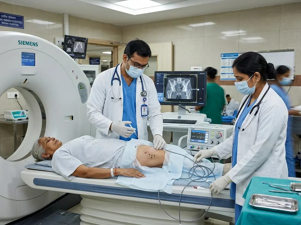
CT-guided pulsed radiofrequency ablation of the obturator nerve delivers durable pain relief up to 12 months in mild-to-moderate hip osteoarthritis, while offering a safe short-term palliative bridge for severe grade 4 disease without procedural complications.
Today

New research reveals that M1 macrophage-derived exosomes aggravate diabetic nephropathy by transferring WTAP to stabilize S1PR2 mRNA. Silencing WTAP in these vesicles attenuates endothelial injury and renal fibrosis, pointing toward innovative nanomedicine therapies.
Yesterday
