.webp&w=384&q=75)

Loading, please wait...
Loading, please wait...
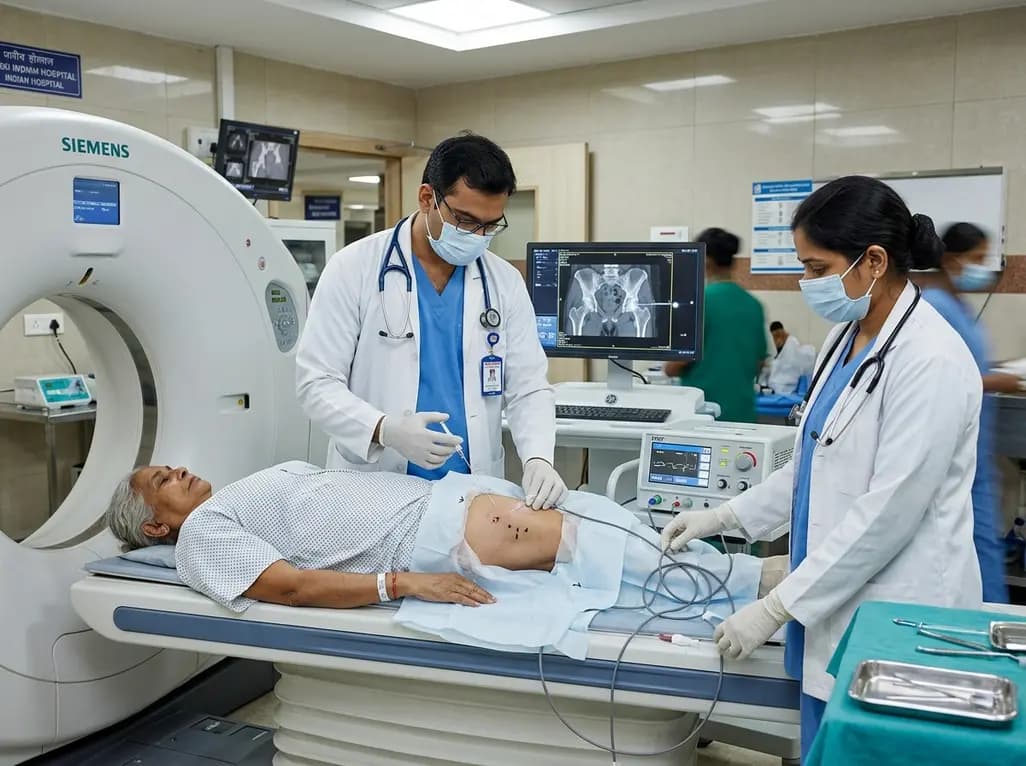
Chronic hip osteoarthritis causes substantial disability and functional impairment among aging populations worldwide. While total hip arthroplasty provides definitive treatment for end-stage joint degeneration, many patients face contraindications or prolonged surgical waiting periods. Consequently, interventional pain physicians actively seek minimally invasive neuromodulatory techniques to alleviate refractory discomfort. Recent clinical investigations demonstrate that percutaneous pulsed radiofrequency ablation targeting the articular branches of the obturator nerve offers meaningful relief for symptomatic patients.
Hip joint innervation derives primarily from sensory branches of the femoral, obturator, and sciatic nerves. Specifically, the anterior and medial joint capsule receives rich nociceptive supply from the articular branches of the obturator nerve. Traditional continuous thermal radiofrequency neurotomy destroys neural tissue through high-temperature coagulation necrosis. However, this thermal destruction creates risks of deafferentation pain, motor weakness, and post-procedural neuritis. In contrast, pulsed radiofrequency ablation delivers short bursts of high-frequency electrical current separated by silent intervals. This pulsing mechanism maintains the tissue temperature below 42 degrees Celsius, thereby preventing coagulative tissue necrosis. Furthermore, the generated electrical field alters synaptic transmission, reduces microglial activation, and modulates c-Fos expression in the dorsal horn. As a result, the modality disrupts nociceptive signaling without compromising structural integrity. Clinicians frequently encounter older individuals in India and globally who exhibit multiple comorbidities, making general anesthesia hazardous. Therefore, non-destructive neuromodulation provides an appealing intermediate strategy. Patients avoid prolonged opioid therapy, reduce reliance on non-steroidal anti-inflammatory agents, and experience sustained functional improvements during daily weight-bearing activities.
Accurate needle placement determines the therapeutic success of sensory denervation around the hip joint. Historically, interventional practitioners relied on landmark-guided approaches or fluoroscopic visualization. However, fluoroscopy fails to display surrounding soft tissue margins, neurovascular bundles, and variable muscular planes. In contrast, computed tomography provides cross-sectional anatomical visualization with superior spatial resolution. Physicians can track the radiofrequency cannula in real time, steering it precisely toward the obturator notch. Consequently, operators position the active tip adjacent to the articular sensory fibers while avoiding major motor branches and regional vascular structures. In a recently reported retrospective study, interventionalists treated thirty-three hips in thirty patients presenting with radiographically confirmed osteoarthritis. The patient cohort had a mean age of sixty-nine years, spanning twenty-five to ninety years. Practitioners delivered pulsed radiofrequency electrical fields continuously for twelve minutes under direct CT guidance. Furthermore, rigorous post-procedure monitoring according to Cardiovascular and Interventional Radiological Society of Europe criteria confirmed an exceptional safety profile. Thus, CT navigation significantly eliminates trajectory errors and guarantees consistent, reproducible electrode placement.
The therapeutic trajectory of joint neuromodulation correlates closely with baseline radiographic degeneration. In the documented study, researchers stratified participants according to Kellgren-Lawrence grades two through four. Immediately following the twelve-minute ablation, patients experienced profound analgesia, showing an average visual analog scale drop of nine points. However, long-term durability diverged sharply between structural severity subgroups. Patients exhibiting mild-to-moderate osteoarthritis (grades two and three) demonstrated enduring analgesic benefits. Specifically, this mild-to-moderate cohort maintained an average pain score reduction of six points at six months. Moreover, these patients retained a clinically substantial four-point decrease at twelve months post-procedure. In contrast, individuals suffering from grade four end-stage joint destruction encountered rapid pain recurrence at approximately three months. Severe structural collapse, subchondral cyst formation, and mechanical bone-on-bone friction ultimately overwhelm purely sensory neuromodulatory suppression. Therefore, clinicians must educate patients with terminal osteoarthritis that pulsed radiofrequency ablation functions primarily as a temporary analgesic bridge. This bridge provides critical symptom relief while patients optimize medical fitness before definitive joint arthroplasty.
Safety remains paramount when selecting interventional therapies for elderly and fragile populations. Fortunately, the study recorded zero intraprocedural or post-procedural complications across all thirty-three treated joints. Continuous thermal neurotomy produces tissue lesions heating beyond eighty degrees Celsius, which frequently risks sensory dysesthesia or adjacent muscular denervation. Because pulsed radiofrequency energy keeps local tissue temperatures strictly under forty-two degrees, the cellular architecture experiences negligible thermal destruction. In addition, preserving motor branch conduction prevents quadriceps or adductor weakness, which protects against balance disturbance and fall injuries. This safety margin proves especially relevant for geriatric patients living with osteoporosis. Furthermore, the percutaneous technique requires only local anesthesia and modest sedation, completely avoiding hemodynamic instability associated with neuraxial blocks. Patients ambulate promptly following brief recovery monitoring. As a result, the outpatient intervention minimizes hospital stay durations and reduces overall procedural costs. Healthcare teams in high-volume orthopedic centers can integrate this safe protocol into routine day-care interventional services without requiring intensive care backup.
Managing degenerative hip disease requires a progressive, multidisciplinary treatment continuum. Initial conservative therapy combines structured physiotherapy, weight management, and oral analgesics. However, chronic reliance on pharmacotherapy frequently leads to gastrointestinal, renal, and cardiovascular toxicity. When intra-articular corticosteroid or visco-supplementation injections lose therapeutic efficacy, clinicians often confront a difficult management void. Pulsed radiofrequency ablation effectively bridges this gap, establishing durable sensory suppression without systemic adverse effects. Moreover, effective analgesia breaks the debilitating cycle of kinesiophobia and muscle atrophy, empowering patients to engage actively in rehabilitation exercises. Orthopedic surgeons can also strategically deploy this neuromodulation technique for patients on prolonged surgical waiting lists. In addition, the procedure serves individuals who carry prohibitive cardiopulmonary risks that preclude elective joint replacement surgery. By reducing baseline nociceptive input, the intervention stabilizes joint function and significantly elevates health-related quality of life. Thus, incorporating CT-guided neuromodulation into modern musculoskeletal protocols delivers a versatile, patient-centered option across varied clinical settings.
Pulsed radiofrequency delivers brief bursts of high-voltage electrical current that maintain tissue temperatures below forty-two degrees Celsius, avoiding thermal coagulation necrosis. In contrast, continuous radiofrequency applies sustained thermal energy exceeding eighty degrees Celsius to ablate nerves. Therefore, pulsed radiofrequency modulates synaptic transmission and dampens pain signaling while completely preserving motor function. This biological mechanism significantly reduces the risk of post-procedural neuritis, deafferentation dysesthesia, and muscle weakness.
Patients with mild-to-moderate hip osteoarthritis, classified as Kellgren-Lawrence grades two or three, experience the most durable pain relief. Clinical investigations demonstrate sustained analgesic efficacy extending up to twelve months in this cohort. In contrast, patients exhibiting end-stage grade four disease typically experience pain recurrence within three months. Consequently, clinicians utilize this modality in advanced cases primarily as a short-term palliative bridge before surgical joint replacement.
Computed tomography guidance provides cross-sectional anatomical visualization with superior tissue contrast compared to conventional two-dimensional fluoroscopy. Consequently, interventionalists can navigate the radiofrequency cannula with sub-millimeter precision directly to the articular sensory branches of the obturator nerve. This cross-sectional view prevents unintended injury to adjacent vascular channels and motor nerves. As a result, CT guidance ensures optimal electrode placement, improves therapeutic outcomes, and completely eliminates severe intraoperative procedural complications.
Disclaimer: This content is for informational and educational purposes only and does not constitute formal medical advice, diagnosis, or treatment. Healthcare professionals should evaluate individual patient factors, clinical circumstances, and institutional protocols before initiating any interventional pain procedures. Refer to the latest local and national guidelines for clinical practice.
References

Read summarized clinical updates, watch expert medical content, and earn CME certifications right from your smartphone.
Open your smartphone camera to scan and install.





CT-guided pulsed radiofrequency ablation of the obturator nerve delivers durable pain relief up to 12 months in mild-to-moderate hip osteoarthritis, while offering a safe short-term palliative bridge for severe grade 4 disease without procedural complications.
Today

Ceftriaxone therapy can lead to biliary sludge and gallstone formation due to calcium-ceftriaxone precipitation. A recent cohort study shows that the noninvasive Fibrosis-4 (FIB-4) index, alongside treatment duration, independently predicts lithogenesis, enabling proactive risk stratification.
Today

A multiparametric MRI approach combining quantitative T2 shading, apparent diffusion coefficient (ADC) measurements, and morphological signs provides superior diagnostic accuracy over subjective evaluation to differentiate ovarian endometriomas from benign hemorrhagic cysts.
Today

A landmark case report reveals a donor heart beating for a century in a 63-year-old recipient, challenging conventional age limits in cardiac transplantation and offering critical lessons on organ longevity, donor selection, and long-term graft survival.
Today

New research reveals that M1 macrophage-derived exosomes aggravate diabetic nephropathy by transferring WTAP to stabilize S1PR2 mRNA. Silencing WTAP in these vesicles attenuates endothelial injury and renal fibrosis, pointing toward innovative nanomedicine therapies.
Yesterday
