.webp&w=384&q=75)

Loading, please wait...
Loading, please wait...

Pediatric otolaryngologists increasingly prefer intracapsular tonsillotomy over complete extracapsular tonsillectomy for managing pediatric sleep-disordered breathing. This tissue-sparing technique reduces immediate postoperative pain, accelerates dietary resumption, and dramatically decreases secondary hemorrhage. However, partial tonsil resection deliberately preserves lymphoid parenchyma and the underlying capsule. Consequently, clinicians are now observing rare suppurative complications, specifically the emergence of peritonsillar abscess after tonsillotomy in older children. Although overall complication rates remain modest, physicians must recognize this potential late surgical sequel. Early recognition enables prompt drainage and prevents severe pharyngeal compromise.
Historically, complete extracapsular tonsillectomy served as the definitive surgical standard for pediatric tonsillar hypertrophy and recurrent infection. During extracapsular dissection, surgeons excise both palatine tonsils entirely along the muscular fascial plane. Nevertheless, exposing the pharyngeal constrictor bed triggers intense postoperative pain and elevates the danger of post-tonsillectomy bleeding. Because of these recognized morbidities, intracapsular tonsillotomy emerged as a preferred alternative for pediatric airway obstruction. Surgeons employ microdebriders or coblation wands to vaporize obstructing tonsillar mass while leaving the tonsil capsule intact. As a result, children experience faster functional recovery and consume fewer opioid analgesics. Furthermore, large institutional registries show that tonsillotomy reduces secondary postoperative hemorrhage rates by more than fivefold compared to total excision. Therefore, parents and surgical teams favor tonsillotomy for uncomplicated tonsillar enlargement. Nonetheless, partial ablation carries an inherent anatomical consequence because viable lymphoid remnants persist in the pharyngeal fossa. While extracapsular surgery permanently clears the tonsillar bed, subcapsular tissue retained during tonsillotomy can slowly proliferate over subsequent years. Thus, surgical teams must balance rapid initial recovery against the remote potential for late parenchymal complications.
To evaluate long-term infectious outcomes, investigators conducted a comprehensive fifteen-year retrospective case series at a tertiary pediatric hospital. Researchers examined medical records of 25,410 pediatric patients who underwent tonsil surgery between January 2010 and January 2025. Among 5,576 children treated with intracapsular tonsillotomy, nine patients subsequently developed a confirmed peritonsillar abscess, yielding an incidence of 0.16%. In contrast, among 19,834 patients who underwent extracapsular tonsillectomy, only a single individual developed an abscess, representing an incidence of 0.005%. Consequently, patients receiving tonsillotomy demonstrated a significantly higher relative likelihood of experiencing quinsy during long-term follow-up. This clinical disparity highlights how persistent lymphoid architecture alters infectious susceptibility. Even though an overall risk of 0.16% remains acceptably low, the thirty-fold relative increase carries substantial clinical importance. Moreover, seven of the ten affected surgical patients displayed documented residual tonsillar tissue upon clinical examination. These institutional data demonstrate that tonsillotomy does not completely eliminate deep pharyngeal infections. Otolaryngologists must understand this statistical distinction when counseling families preoperatively regarding surgical selection.
The pathological mechanism leading to late abscess development involves persistent tonsillar tissue within the pharyngeal fossa. When surgeons perform intracapsular tonsillotomy, they spare the capsule and deep follicular layers to protect the muscular wall. Over time, hormonal shifts, recurrent viral infections, and environmental antigens can stimulate hyperplasia within these retained tonsillar islands. Additionally, postoperative healing creates scarring and fibrous bands across the mucosal surface. This cicatricial tissue can obstruct residual crypt openings and adjacent minor salivary glands, known as Weber glands. Once drainage pathways become occluded, retained secretions foster bacterial proliferation and localized microabscess formation. Common pathogens include Streptococcus pyogenes, Streptococcus anginosus, and various oral anaerobic species. When the infection breaches the remaining parenchymal limits, purulent exudate collects between the tonsillar remnant and the pharyngeal wall. In contrast, total extracapsular tonsillectomy creates a fibrotic scar bed that lacks glandular parenchyma, preventing typical abscess formation. Therefore, residual lymphoid stroma acts as a crucial bacterial reservoir. Recognizing this underlying pathophysiology explains why retaining tonsillar tissue leaves patients vulnerable to delayed infectious complications.
One crucial clinical insight from recent data is the prolonged latency between surgery and abscess onset. In the reviewed pediatric cohort, the mean interval from surgical resection to abscess diagnosis was 8.2 years. Furthermore, patients presented at a mean age of 13.7 years, with presentations spanning from 10 to 18 years. Because young children typically undergo tonsillotomy for obstructive symptoms, they reach teenage years before developing severe infections. Consequently, adolescent patients and their parents may not recall their specific surgical history accurately. Many families report that the patient had tonsils removed, leading emergency providers to overlook quinsy. Emergency physicians might assume that past tonsil surgery renders a peritonsillar abscess impossible. As a result, patients may experience delayed antimicrobial therapy or late surgical intervention. Clinicians evaluating adolescents with severe unilateral throat pain, trismus, and uvular deviation must maintain high suspicion for peritonsillar collections. In addition, physical examination should carefully inspect the tonsillar pillars for residual soft tissue bulging. Cross-sectional imaging or point-of-care ultrasound can definitively confirm fluid collections when clinical signs remain ambiguous.
Managing peritonsillar abscesses after previous tonsillotomy requires acute drainage combined with strategic long-term planning. During acute presentation, clinicians must administer intravenous fluids, analgesics, and broad-spectrum antimicrobial agents immediately. Furthermore, therapeutic needle aspiration or formal incision and drainage under local or general anesthesia relieves airway distress quickly. Once the acute infection resolves, surgeons should assess the patient for persistent tonsillar hypertrophy. In the tertiary hospital series, five of the nine tonsillotomy patients underwent revision extracapsular tonsillectomy to remove troublesome residual lymphoid tissue. Revision tonsillectomy permanently eradicates the infectious focus, preventing recurrent quinsy or chronic sore throat. Nevertheless, revision dissection in scarred pharyngeal tissue carries higher technical difficulty and requires meticulous surgical control. Beyond surgical management, these findings provide essential guidance for preoperative discussions. Otolaryngologists must explain the risk profiles of intracapsular versus extracapsular approaches transparently to parents. While tonsillotomy delivers exceptional short-term recovery benefits, residual tissue retains a small, measurable risk of future infection. Maintaining accurate surgical documentation ensures that future providers interpret recurring pharyngeal symptoms correctly.
Intracapsular tonsillotomy preserves the fibrous capsule and a peripheral rim of lymphoid tissue to reduce surgical bleeding and postoperative pain. Over time, these retained tissue remnants can undergo lymphoid hyperplasia. Subsequent viral or bacterial infections, combined with scar-induced obstruction of tonsillar crypts or minor salivary ducts, promote bacterial stasis. Pathogens like Streptococcus pyogenes proliferate within the trapped tissue, eventually forming a purulent abscess collection adjacent to the pharyngeal wall.
Recent long-term clinical data reveal that peritonsillar abscess occurs significantly more often following tonsillotomy than after total tonsillectomy. In a 15-year institutional study of pediatric patients, the abscess incidence was 0.16% after intracapsular tonsillotomy compared to only 0.005% after complete extracapsular tonsillectomy. Although the absolute risk after tonsillotomy remains remarkably low, the relative risk increases roughly thirty-fold because residual lymphoid tissue provides a persistent anatomical nidus for deep pharyngeal infection.
Surgeons generally recommend revision extracapsular tonsillectomy when clinical examination confirms substantial residual lymphoid tissue following an acute peritonsillar abscess. Revision surgery completely excises the remaining tonsillar capsule and parenchymal remnants, eliminating the anatomical substrate for recurring quinsy. While acute drainage resolves the immediate infection, revision tonsillectomy provides definitive protection against subsequent episodes, especially in adolescents who experience chronic unilateral pharyngeal inflammation, obstructive symptoms, or recurrent tonsillar infections.
Disclaimer: This content is for informational and educational purposes only and should not be construed as medical advice, diagnosis, or treatment. Always consult a qualified healthcare provider for specific health concerns. The views expressed are those of the authors and do not necessarily reflect official policy. Refer to the latest local and national guidelines for clinical practice.
References

Read summarized clinical updates, watch expert medical content, and earn CME certifications right from your smartphone.
Open your smartphone camera to scan and install.





A 15-year comparative study reveals a rare but notable long-term risk of peritonsillar abscess after intracapsular tonsillotomy compared to total tonsillectomy in pediatric patients, with a mean latency of 8.2 years due to residual lymphoid tissue, emphasizing the need for comprehensive preoperative counseling.
Today
A comprehensive meta-analysis of 19 trials shows altitude and hypoxic exposure significantly improve running time-trial performance by enhancing oxygen transport, running economy, and hematological markers in endurance athletes.
Today
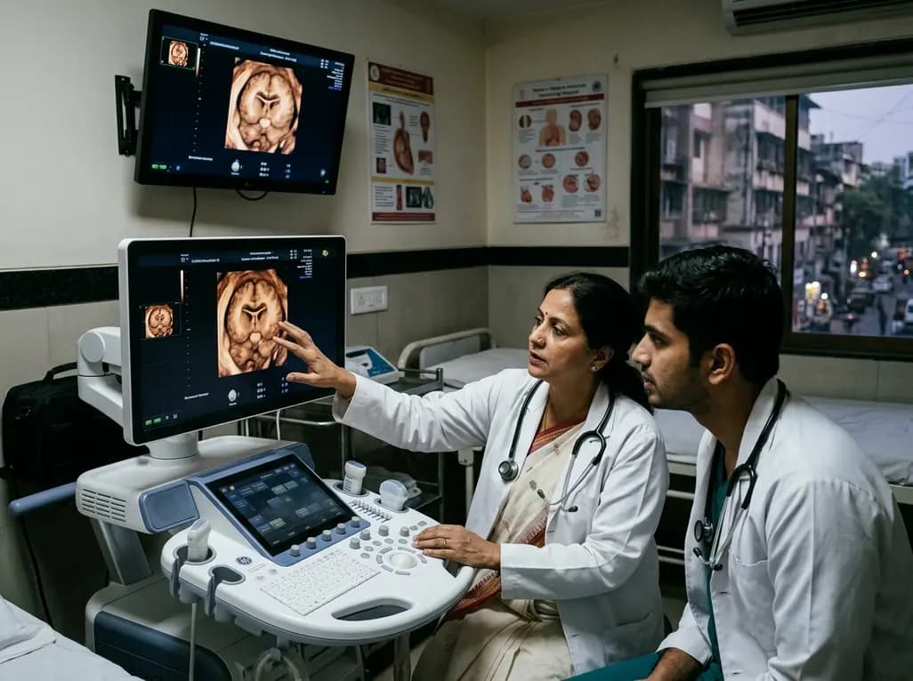
A 3D transvaginal neurosonography study shows the fetal cavum septi pellucidi develops fluid cavitation around 19 weeks and a corpus callosal length of 11.25 mm. These insights help clinicians prevent premature false-positive diagnoses of absent septum pellucidum during second-trimester anomaly scans.
Today

The STADIA-HFpEF trial evaluated whether dapagliflozin improves left ventricular distensibility in phenotypically stratified HFpEF patients without significant myocardial fibrosis. While primary diastolic endpoints were neutral, exploratory improvements in left atrial conduit strain and biomarkers emerged.
Yesterday

An 8-week integrative neuromuscular training protocol significantly improves unilateral jump symmetry, balance, and squat strength in adolescent female sprinters. By correcting lower-extremity asymmetry, this evidence-based intervention reduces injury risk while optimizing athletic sprint performance.
Today

The randomized GLARIF trial evaluated early evening versus bedtime insulin glargine administration in type 1 diabetes during Ramadan. The study showed similar hypoglycemia rates and glycemic metrics between both regimens, highlighting the critical need for proactive monitoring and individualized dose management.
Today
