.webp&w=384&q=75)

Loading, please wait...
Loading, please wait...

Perioperative Visual Loss spine surgery remains one of the most devastating complications that a patient and a clinical team can face. Although the incidence is relatively rare, the impact on a patient's quality of life is profound, often resulting in permanent and irreversible blindness. Clinical research indicates that while surgical techniques have advanced, the risk of visual compromise persists, particularly in complex spinal reconstructions. This condition typically manifests shortly after the patient awakens from general anesthesia. Because the etiology is multifactorial, understanding the underlying mechanisms is essential for any clinician involved in perioperative care. It is not merely a surgical complication but a systemic challenge that requires a deep understanding of hemodynamic stability and patient positioning. Consequently, the medical community has focused increasingly on identifying high-risk scenarios and improving the communication of these risks to patients during the preoperative phase.
Historically, the occurrence of visual loss after non-ocular surgery was poorly understood. However, modern registries and multicenter studies have shed light on the specific vulnerabilities of the optic nerve during prolonged procedures. For patients undergoing major spinal fusions, the risk is often linked to the physiological stress of the prone position. Therefore, it is imperative for both surgeons and anaesthetists to recognize that POVL is a material risk. This means it is a complication that a reasonable patient would want to know about before consenting to surgery. Addressing this risk requires a unified approach, where all members of the perioperative team are on the same page regarding prevention and disclosure. By prioritizing patient education and safety, clinicians can better navigate the complex ethical and clinical landscape surrounding this rare but catastrophic event.
A significant challenge in managing Perioperative Visual Loss spine surgery is the disparity in how different specialists perceive the risk. Recent survey data reveals that anaesthetists are generally more likely to categorize POVL as a material risk compared to their surgical counterparts. For instance, approximately 79.7% of anaesthetists view it as a critical discussion point, while only 57.7% of surgeons share this view. This discrepancy is further highlighted by the estimated incidence rates cited by each group. Anaesthetists often estimate the frequency of POVL to be higher, ranging from 0.03% to 0.2%, whereas many surgeons believe the risk is as low as 0.0001%. Such wide variations in perception can lead to inconsistent patient counseling and may leave patients feeling underinformed about potential outcomes.
Furthermore, the experience of having a patient suffer from POVL significantly alters a clinician's future practice. Those who have witnessed this complication firsthand are much more likely to incorporate detailed visual risk discussions into their routine consent process. This suggests that personal experience often drives clinical vigilance more than statistical data alone. To bridge this gap, there is a clear need for standardized education and national guidelines. When surgeons and anaesthetists align their understanding of incidence and materiality, they provide a more cohesive and reassuring message to the patient. Ultimately, the goal is to ensure that every patient undergoing high-risk spine surgery receives a balanced and evidence-based explanation of all potential neurological complications, including those affecting vision.
Understanding the pathophysiology of Perioperative Visual Loss spine surgery is vital for developing effective preventive strategies. The most common cause of POVL in this setting is Ischemic Optic Neuropathy (ION), which can be further classified into anterior and posterior types. Posterior Ischemic Optic Neuropathy (PION) is particularly prevalent in spine surgery and is thought to result from decreased oxygen delivery to the retrobulbar portion of the optic nerve. Several factors contribute to this ischemia, including prolonged operative times, significant blood loss, and intraoperative hypotension. Additionally, the prone position itself plays a major role by increasing venous pressure in the head and neck. This elevated pressure can lead to interstitial edema within the optic nerve's confined spaces, further compromising microvascular perfusion.
Beyond ION, other causes include Central Retinal Artery Occlusion (CRAO) and cortical blindness. CRAO is often the result of direct external pressure on the globe, which can occur if the head is not properly positioned or if the support frames shift during the procedure. This highlight the absolute necessity of meticulous positioning and frequent eye checks throughout the surgery. Cortical blindness, though rarer, involves ischemia in the visual cortex of the brain and is often associated with global hemodynamic instability or embolic events. Each of these mechanisms underscores the complexity of the perioperative environment. Clinicians must manage a delicate balance of blood pressure, fluid volume, and physical positioning to protect the patient's sight. Therefore, a comprehensive understanding of these vascular and mechanical risks is a prerequisite for any high-volume spine program.
Not every patient undergoing a back procedure has the same risk for Perioperative Visual Loss spine surgery. Identifying high-risk individuals is a cornerstone of modern perioperative medicine. Data from clinical registries suggest that certain patient factors significantly increase susceptibility. These include male sex, obesity, and pre-existing vascular conditions such as hypertension or diabetes. Patients with a history of smoking or those with a small cup-to-disc ratio in their optic nerve anatomy may also be at higher risk. When these patient-specific factors are combined with long-duration surgeries, such as multi-level spinal fusions, the cumulative risk rises. Specifically, procedures lasting longer than six hours or involving an estimated blood loss exceeding one liter are considered high-risk scenarios.
In addition to patient factors, the use of specific surgical equipment can influence risk levels. For example, the use of a Wilson frame has been associated with higher abdominal pressure and subsequently higher venous pressures, which may contribute to optic nerve edema. In contrast, using a three-pin head holder can eliminate the risk of direct ocular compression entirely. Therefore, the preoperative assessment must involve a detailed review of both the patient's medical history and the planned surgical approach. By stratifying patients based on these known variables, the clinical team can tailor their anesthetic and surgical plans. This proactive approach not only allows for better risk mitigation but also ensures that the consent process is focused on the patients who need the most detailed information.
The timing and setting of the discussion regarding Perioperative Visual Loss spine surgery are just as important as the content itself. Most experts agree that the optimal time to discuss POVL is in the outpatient or pre-assessment clinic. Discussing such a devastating risk on the day of surgery can significantly increase patient anxiety and may not allow for adequate reflection. In the calm environment of a clinic, clinicians can use visual aids and simplified language to explain why vision loss is a concern. It is also a best practice for both the surgeon and the anaesthetist to participate in this discussion. Since the causes of POVL are both surgical and anesthetic, having both specialists confirm the risk reinforces the importance of the topic to the patient.
Effective consent involves more than just listing a complication; it involves explaining what the team will do to prevent it. Clinicians should outline the steps they take, such as specialized positioning, careful blood pressure management, and intraoperative monitoring. This transparency helps build trust and demonstrates a commitment to patient safety. Moreover, documentation of this specific discussion is crucial from a medicolegal perspective. In many jurisdictions, the failure to mention a rare but catastrophic risk like blindness can be grounds for legal action if the complication occurs. Therefore, integrating POVL into standardized consent forms for high-risk spine surgery is a prudent step for any institution. By making this discussion a routine part of preoperative care, we ensure that patients are truly empowered to make informed decisions about their health.
Once the patient is in the operating room, the focus shifts to the active prevention of Perioperative Visual Loss spine surgery. Maintaining hemodynamic stability is paramount; clinicians should aim to keep the mean arterial pressure within a range that ensures adequate perfusion to the optic nerve. This often requires the use of invasive blood pressure monitoring and careful titration of fluids and vasopressors. While excessive crystalloid administration has been linked to increased tissue edema, a balanced approach using colloids may be beneficial in certain cases. Furthermore, periodic checks of the patient's head position are mandatory to ensure that there is no pressure on the eyes. The head should be kept at or above the level of the heart whenever possible to facilitate venous drainage from the head.
In addition to these physiological measures, the surgical team should work to minimize operative time without compromising the quality of the procedure. For very long cases, some centers consider staged procedures to reduce the duration of prone positioning. Postoperatively, the patient's vision should be assessed as soon as they are awake and alert. If any deficit is noted, an urgent ophthalmology consultation and possibly an MRI are required to differentiate between various causes of visual loss. While treatment options for established ION are unfortunately limited, early recognition allows for the optimization of blood pressure and oxygenation, which might prevent further progression. Ultimately, preventing POVL is a continuous process that begins with patient selection and ends with vigilant postoperative care. By adhering to these practical strategies, the perioperative team can significantly reduce the likelihood of this tragic outcome.
The most common cause is Ischemic Optic Neuropathy (ION), particularly the posterior variant. It occurs when the blood supply to the optic nerve is compromised due to factors like prolonged prone positioning, low blood pressure, and significant blood loss. This condition is often irreversible and results in severe vision loss.
Ideally, both the spine surgeon and the anaesthetist should discuss the risk of POVL. Since the complication results from a combination of surgical positioning and anesthetic management, a collaborative discussion ensures the patient understands the full scope of the risk and the preventive measures being taken by the entire team.
Risk reduction involves maintaining stable blood pressure, minimizing surgical time, and ensuring meticulous head positioning to avoid pressure on the eyes. Using head-holding devices that keep the eyes free from contact and keeping the head slightly elevated to improve venous drainage are also critical steps in preventing this complication.
Disclaimer: This content is for informational and educational purposes only and does not constitute medical advice. It is not intended to be a substitute for professional medical advice, diagnosis, or treatment. Always seek the advice of your physician or other qualified healthcare provider with any questions you may have regarding a medical condition. Refer to the latest local and national guidelines for clinical practice.
References
Pitsika M et al. Perioperative visual loss (POVL) in spine surgery: a survey of the practice and views of spinal surgeons and anaesthetists. Br J Neurosurg. 2025 Aug. doi: 10.1080/02688697.2023.2275621. PMID: 37943103.
American Society of Anesthesiologists Task Force on Perioperative Visual Loss. Practice advisory for perioperative visual loss associated with spine surgery: an updated report. Anesthesiology. 2019;130(1):12-30.
Postoperative Visual Loss (POVL). Anesthesia Patient Safety Foundation (APSF). [Online]. Available at: https://www.apsf.org/article/postoperative-visual-loss-povl/.

Read summarized clinical updates, watch expert medical content, and earn CME certifications right from your smartphone.
Open your smartphone camera to scan and install.





Perioperative Visual Loss (POVL) is a rare but devastating complication of spine surgery. This article examines the differences in risk perception between surgeons and anaesthetists, identifies key risk factors like the prone position and blood loss, and outlines best practices for the consent process.
Last week
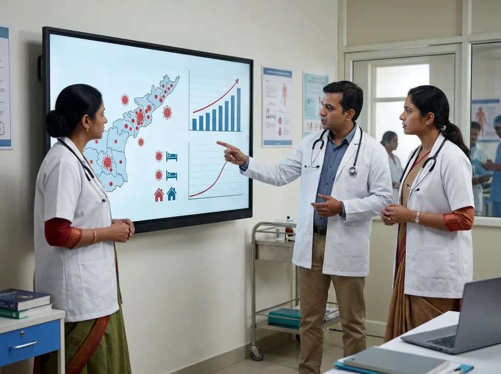
Andhra Pradesh reported 10 new Covid-19 cases, taking the state tally to 49 while deaths remain at four. With 24 patients hospitalized and 16 under home isolation, the Health Department has intensified monitoring. Medical professionals should review regional distribution, diagnostic protocols, and management plans.
Today

An 11-year Swedish registry study of 618 uterine sarcoma patients found that minimally invasive surgery yielded survival comparable to open surgery in early stages. However, adjuvant chemotherapy conferred no survival benefit in localized or advanced disease, highlighting stage and histology as key outcomes.
3 days back

A cross-sectional study evaluates post-intensive care syndrome in cardiac patients 2-4 weeks post-ICU discharge, highlighting cognitive, psychological, and functional impairments and the need for structured multidisciplinary rehabilitation.
3 days back

Anterior cruciate ligament reconstruction failure lacks uniform definition. A narrative review proposes an integrative framework incorporating objective and subjective instability, persistent pain, restricted motion, graft rupture, and secondary meniscal injury to standardize clinical reporting.
3 days back
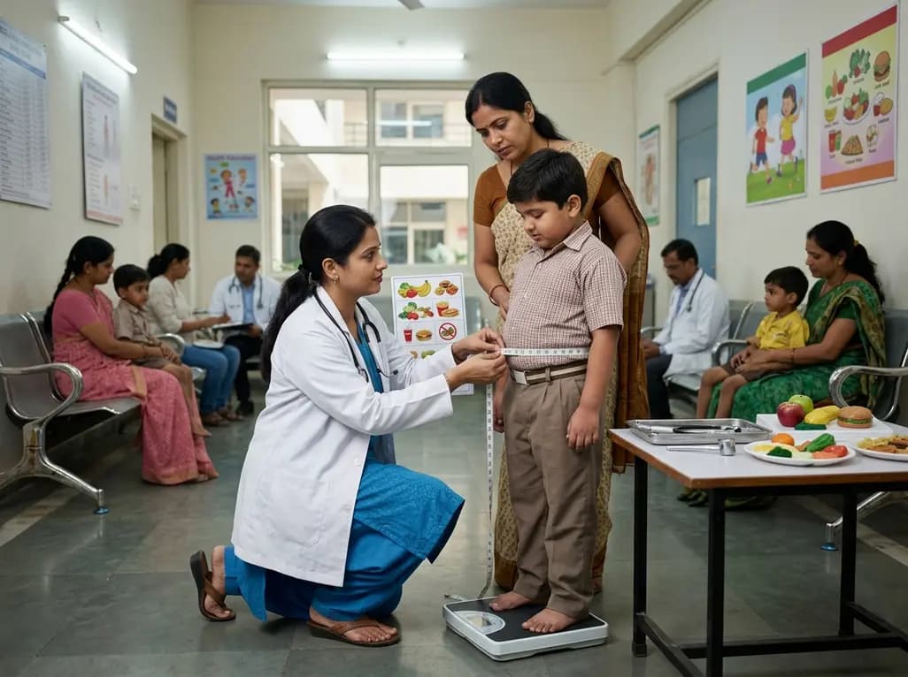
With World Obesity Atlas data warning that over 41 million Indian children are overweight or obese, ICMR and NIN have unveiled a 10-point policy roadmap. The initiative calls for mandatory front-of-pack labeling, HFSS taxes, strict marketing bans, and healthier school environments to curb non-communicable diseases.
Today
