.webp&w=384&q=75)

Loading, please wait...
Loading, please wait...
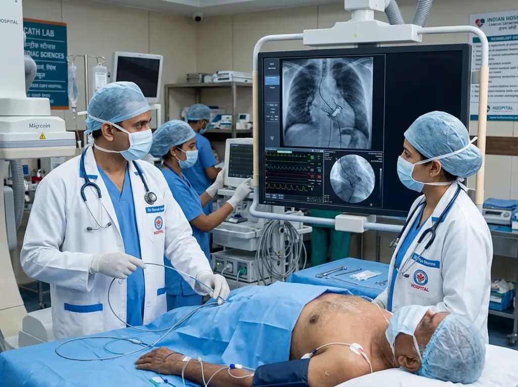
Bioprosthetic heart valves are widely utilized during cardiac surgical interventions; however, structural valve degeneration remains an inevitable complication over extended follow-up periods. When bioprosthetic degradation occurs on the right side of the heart, patients frequently present with progressive right ventricular decompensation and congestive organ damage. Historically, open reoperation served as the sole standard of care for addressing bioprosthetic failure. Nevertheless, repeat surgical intervention carries substantial perioperative morbidity and mortality, especially in individuals who have undergone multiple previous sternotomies. In recent years, percutaneous tricuspid valve-in-valve implantation has emerged as a groundbreaking transcatheter therapy. This minimally invasive strategy enables interventional cardiologists to restore native-like hemodynamics without the prohibitive hazards of open re-entry. Consequently, transcatheter solutions represent an increasingly vital therapeutic pathway for multimorbid patients suffering from failed tricuspid prostheses.
Bioprosthetic tricuspid valves encounter significant hemodynamic stressors that accelerate structural deterioration over time. In particular, calcification, leaflet thickening, tearing, and pannus formation eventually precipitate severe stenosis, severe regurgitation, or combined valvular dysfunction. Furthermore, patients with underlying rheumatic heart disease frequently require multiple sequential valve surgeries across their lifetimes. As a consequence of prior open procedures, dense mediastinal adhesions develop, which dramatically elevates the risk of severe hemorrhage and graft injury during repeat sternotomy. Additionally, long-standing right-sided valvular failure leads directly to intractable systemic venous congestion, congestive hepatopathy, and secondary liver cirrhosis. Therefore, conventional surgical re-replacement in these frail individuals carries an exceptionally high surgical mortality rate. Interventional structural cardiology teams must recognize these clinical hurdles early, because timely transcatheter intervention can halt irreversible multi-organ deterioration and restore systemic stability.
Transcatheter tricuspid valve-in-valve implantation provides an elegant and effective alternative to traditional redo open-heart surgery. Clinicians typically perform the procedure via transfemoral venous access under general anesthesia or conscious sedation, guided meticulously by transesophageal echocardiography and fluoroscopy. First, operators advance a steerable delivery catheter through the inferior vena cava into the right atrium. Subsequently, they cross the stenotic or regurgitant bioprosthetic valve with an atraumatic guidewire and position a robust, stiff rail into a distal pulmonary artery branch or the right ventricular apex. After confirming stable position, the team delivers a balloon-expandable or self-expanding transcatheter heart valve within the rigid sewing ring of the degraded surgical prosthesis. Because the existing surgical framework provides a stable anchoring zone, operators can achieve precise anatomical alignment. This targeted approach immediately eliminates structural valvular failure while avoiding the extensive systemic trauma associated with cardiopulmonary bypass.
Although transcatheter valve-in-valve procedures demonstrate excellent clinical success, specific anatomical and technical challenges demand rigorous operator awareness. Specifically, the risk of valve migration or displacement during balloon inflation, commonly termed "melon seeding," represents a notable technical pitfall. This phenomenon occurs when non-uniform radial forces push the expanding transcatheter valve axially out of the surgical bioprosthetic frame into either the right ventricle or the right atrium. Consequently, meticulous pre-procedural multislice computed tomography and three-dimensional echocardiography are essential to evaluate the true internal diameter and fluoroscopic markers of the surgical valve. Moreover, interventionalists often utilize slow, controlled balloon inflation or specialized landing techniques to prevent unseating. In addition, right ventricular pacing and stable wire support help mitigate abrupt translational shifts during deployment, thereby guaranteeing secure mechanical anchoring and preventing disastrous device malposition.
The successful placement of a transcatheter valve within a degenerated tricuspid bioprosthesis produces immediate and marked hemodynamic improvements. Echocardiographic evaluations consistently show significant reductions in mean transvalvular pressure gradients alongside the complete elimination of severe valvular regurgitation. As a direct result of restored forward flow, central venous pressures drop promptly, relieving systemic venous congestion. Over the subsequent weeks, patients frequently experience rapid resolution of refractory peripheral edema, ascites, and hepatic congestion. Furthermore, clinical evidence demonstrates sustained improvements in functional class, exercise tolerance, and overall quality of life. Even in challenging patients presenting with advanced congestive hepatopathy, such as Child-Pugh B cirrhosis, stabilizing right-sided hemodynamics helps mitigate further hepatic necrosis and promotes clinical recovery without subjecting the patient to the perils of cardiopulmonary bypass.
The successful management of complex tricuspid valve disease relies upon comprehensive evaluation by a multidisciplinary heart team. This team must include structural interventional cardiologists, cardiac surgeons, advanced heart failure specialists, cardiovascular imaging experts, and cardiac anesthesiologists. Together, clinicians systematically evaluate surgical risk calculators, frailty indices, anatomical suitability, and the functional status of left-sided prosthetic valves. Moreover, expanding clinical registries and emerging clinical trials continue to refine patient selection criteria and long-term anticoagulation strategies for right-sided transcatheter bioprostheses. As device technology and delivery systems advance, percutaneous tricuspid therapies will become even safer, more versatile, and widely accessible. Consequently, transcatheter valve-in-valve techniques are set to become the standard front-line therapeutic approach for failed tricuspid prostheses in high-risk surgical candidates worldwide.
Bioprosthetic valves deteriorate primarily due to progressive tissue calcification, mechanical fatigue, leaflet degeneration, and chronic inflammatory responses. Over time, these structural alterations lead to leaflet stiffening, tearing, or pannus ingrowth. Consequently, the affected bioprosthetic valve develops significant stenosis, severe insufficiency, or combined hemodynamic dysfunction, which eventually precipitates clinical heart failure and requires therapeutic reintervention.
Redo surgical valve replacement carries high perioperative mortality due to dense mediastinal adhesions from prior sternotomies, which elevate bleeding risks. Furthermore, prolonged right ventricular dysfunction and chronic venous congestion frequently induce liver cirrhosis and renal impairment. These systemic comorbidities significantly increase perioperative complications, making minimally invasive transcatheter options a much safer therapeutic alternative.
Operators prevent melon seeding through comprehensive pre-procedural computed tomography imaging, accurate valve sizing, and rigorous anatomical assessment. During deployment, clinicians utilize slow balloon inflation, rapid ventricular pacing, and dedicated high-support guidewires positioned stably in the pulmonary artery. These technical maneuvers ensure controlled radial expansion, stable coaxial alignment, and secure anchoring inside the preexisting surgical ring.
Disclaimer: This content is for informational and educational purposes only and does not constitute formal medical advice, diagnosis, or treatment recommendations. Clinical decisions should always be made by qualified healthcare professionals based on individual patient assessments and established clinical guidelines. Refer to the latest local and national guidelines for clinical practice.
References
Sellers AM et al. Percutaneous Tricuspid Valve-in-Valve Implantation With Melon Seeding During Deployment. JACC Case Rep. 2026 Aug 15. doi: undefined. PMID: 42603160.
O'Gara PT et al. 2026 ACC/AHA/ASE/HRS/STS Expert Consensus Decision Pathway for the Evaluation and Management of Patients With Tricuspid Regurgitation. J Am Coll Cardiol. 2026;83(14):1455-1489.
Webb JG et al. Transcatheter Valve-in-Valve Implantation for Degenerated Bioprosthetic Heart Valves: Practical Strategies and Clinical Outcomes. JACC Cardiovasc Interv. 2023;16(8):912-925.

Read summarized clinical updates, watch expert medical content, and earn CME certifications right from your smartphone.
Open your smartphone camera to scan and install.





Percutaneous tricuspid valve-in-valve implantation provides a life-saving, minimally invasive approach for high-risk patients experiencing severe bioprosthetic tricuspid valve failure and refractory right heart failure after multiple cardiac surgeries.
last month

A secondary analysis of an HIV cure-related trial in South Africa highlights the acceptability and safety of serial excisional lymph node and gut biopsies among young women. Clear communication, empathetic care, and altruistic motives helped mitigate initial apprehension and sustain longitudinal trial engagement.
Today

A nationwide study covering 3.7 million Swedish births reveals that prenatal exposure to TORCH pathogens significantly elevates long-term risks of intellectual disability and autism. Healthcare providers must recognize clinical indicators, promote prenatal prevention, and track early pediatric neurodevelopment.
Today
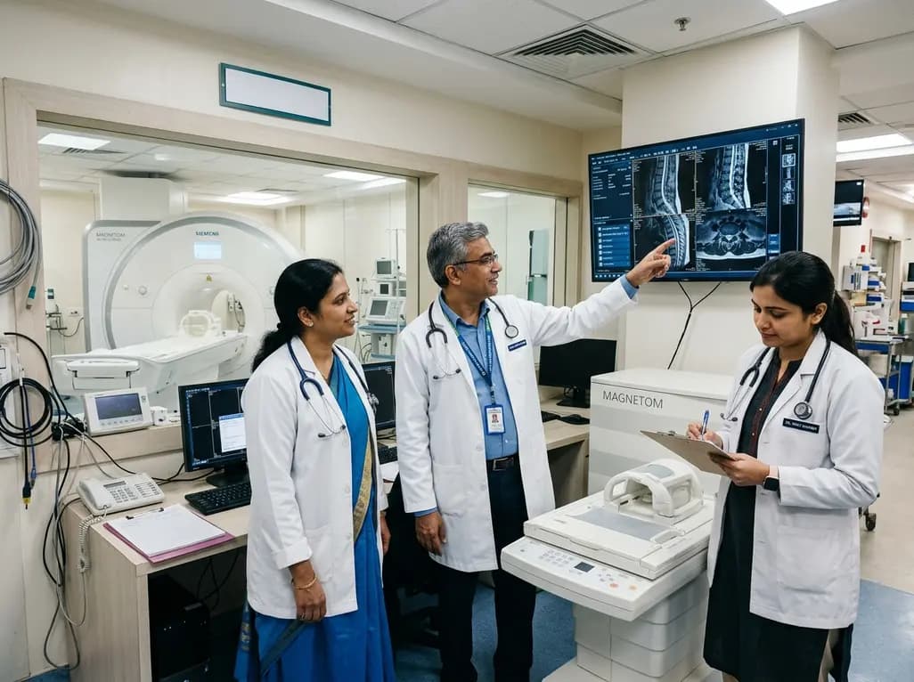
Under India's PLI initiative, domestic production of 57 critical medical devices has commenced, including MRI and CT scanners, cath labs, and heart valves. Global partnerships and local investments are expanding high-end technology, curbing import reliance, and strengthening clinical accessibility nationwide.
Today

A longitudinal study reveals that patients undergoing staged bilateral hip arthroscopy experience progressive contralateral labral deterioration during surgical intervals. However, structural degradation does not compromise 2-year postoperative clinical outcomes or patient-reported satisfaction scores.
Today

New immunological research reveals how PTPN22 forms a delayed inhibitory signaling complex to restrain T cell activation, explaining how the autoimmune-linked R620W variant disrupts feedback control and promotes persistent inflammation in conditions like rheumatoid arthritis and type 1 diabetes.
Today
