.webp&w=384&q=75)

Loading, please wait...
Loading, please wait...
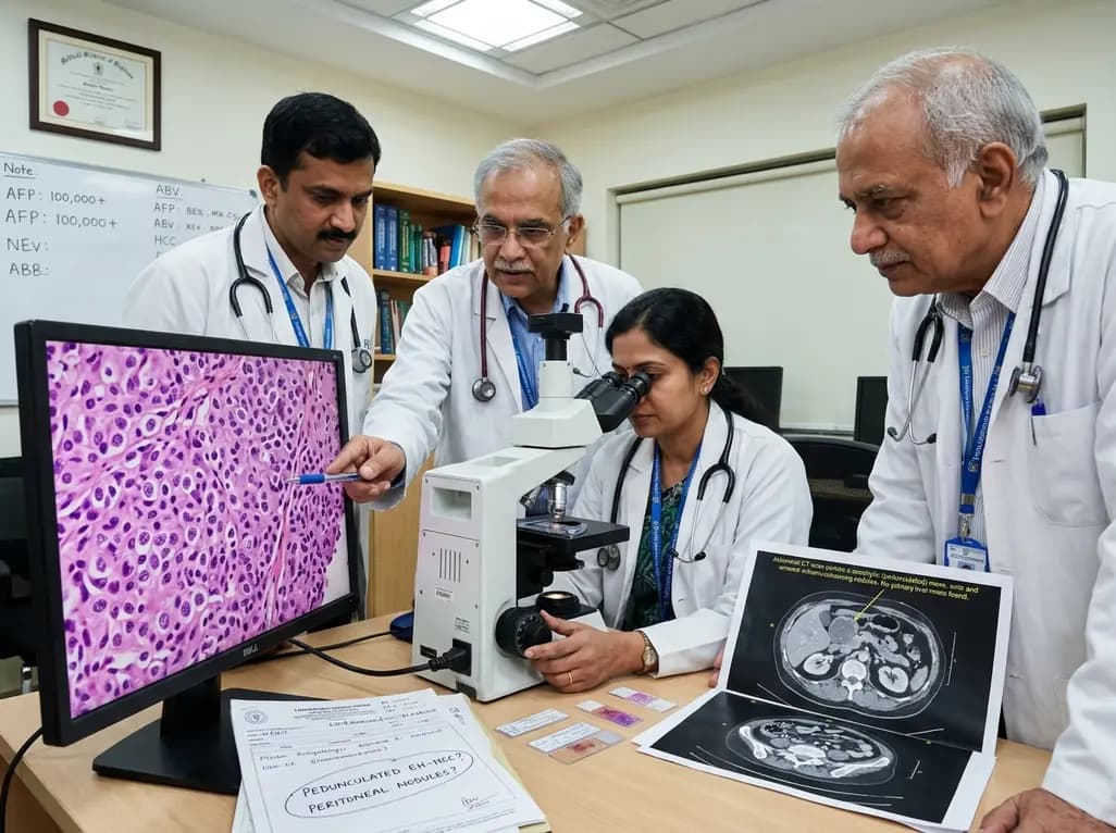
Hepatocellular carcinoma typically develops within cirrhotic hepatic parenchyma and displays characteristic vascular enhancement patterns on dynamic cross-sectional imaging. However, clinicians occasionally encounter atypical variants such as pedunculated extrahepatic HCC, which can mimic non-hepatic intra-abdominal neoplasms. In rare scenarios, an exophytic or pedunculated tumor can seed the peritoneal cavity before clinicians detect any obvious primary liver mass. A recently reported clinical case illustrates this diagnostic dilemma in a patient in his 40s who initially presented with isolated elevation of alpha-fetoprotein. Although baseline imaging failed to identify a distinct intrahepatic parenchymal lesion, aggressive peritoneal dissemination eventually manifested alongside soaring tumor markers.
The patient initially sought medical evaluation following routine laboratory screening that revealed mild elevations in serum alpha-fetoprotein. Early sonographic evaluations of the upper abdomen did not demonstrate any focal parenchymal defects, prompting standard observational follow-up. Over a 20-month surveillance window, the clinical picture changed dramatically when serum alpha-fetoprotein surged to 200,000 ng/mL and protein induced by vitamin K absence-II reached 1,524 mAU/mL. Subsequent contrast-enhanced computed tomography showed widespread peritoneal enhancement and ascites without an evident intrahepatic mass. Consequently, the clinical team pursued diagnostic laparoscopy, revealing extensive nodular seeding across the omentum and mesentery.
Histopathological evaluation coupled with immunohistochemical analysis confirmed metastatic hepatocellular carcinoma. A detailed retrospective review of cross-sectional imaging eventually identified a minute, hypervascular pedunculated nodule extending from the left lateral liver segment toward the left triangular ligament. This slender pedicle lesion exhibited classic arterial hyperenhancement and venous washout. Therefore, this subtle structural finding accounted for the disseminated intra-abdominal disease. Despite comprehensive therapeutic intervention, the patient succumbed to advanced disease 14 months after surgical diagnosis, highlighting the aggressive biology of extrahepatically growing tumors.
Serum biomarkers serve as indispensable surveillance adjuncts, particularly when imaging fails to capture subtle or occult disease. Alpha-fetoprotein remains the standard diagnostic biomarker, but clinicians increasingly pair it with protein induced by vitamin K absence-II to improve sensitivity. When these biomarkers demonstrate progressive or exponential increases, physicians must maintain a high index of suspicion for extrahepatic tumor spread or ectopic hepatic tissue malignancy. A normal intrahepatic ultrasound examination should not completely reassure the clinician when serological markers climb continuously.
Furthermore, discordant biomarker elevations demand comprehensive multiphase imaging of the entire abdominal cavity rather than focused liver parenchymal sweeps alone. In this case, the astronomical rise in tumor markers signaled extensive tumor burden that standard hepatic parenchyma protocols initially overlooked. Clinicians should establish clear escalation thresholds for advanced imaging whenever serological markers trend upward without an explanatory intrahepatic nidus. By integrating serial dual-biomarker monitoring with targeted full-field abdominopelvic imaging, clinical teams can identify occult extrahepatic lesions before widespread intraperitoneal implantation occurs.
Peritoneal carcinomatosis in hepatocellular carcinoma occurs uncommonly, historically documented in fewer than five percent of presentations. Generally, intra-abdominal tumor spillage arises from spontaneous capsular rupture of a subcapsular liver lesion or following iatrogenic instrumentation. However, pedunculated extrahepatic tumors possess unique anatomical vulnerabilities that predispose them to peritoneal dissemination without frank macro-rupture. Because these tumors grow along thin stalks extending beyond the hepatic capsule, they lack the protective barrier of normal liver parenchyma and Glisson capsule.
Additionally, repetitive mechanical friction between the mobile tumor nodule and surrounding visceral peritoneum can cause microscopic cell exfoliation directly into the peritoneal fluid. Once exfoliated malignant hepatocytes enter the peritoneal circulation, gravity and natural fluid dynamics distribute them across the omentum, mesentery, and dependent pelvic spaces. The high vascularity of these exophytic lesions also facilitates local micro-hemorrhages and tumor friability. Consequently, even tiny exophytic lesions originating near ligamentous attachments can seed the entire peritoneal surface, creating diffuse carcinomatosis while the hepatic parenchyma remains largely unremarkable.
Conventional abdominal ultrasonography often exhibits significant blind spots, particularly along the subdiaphragmatic surface, gastrohepatic ligament, and left triangular ligament. Therefore, clinicians must utilize multiphasic dynamic contrast-enhanced computed tomography or magnetic resonance imaging with hepatobiliary-specific contrast agents whenever tumor markers rise anomalously. Radiologists must scrutinize the peripheral hepatic attachments and contiguous ligaments during image review, looking specifically for small hypervascular projections that exhibit early washout kinetics.
When non-invasive imaging remains ambiguous despite strong clinical suspicion of peritoneal malignancy, diagnostic laparoscopy serves as an indispensable tool. Direct laparoscopic visualization enables thorough inspection of the peritoneal cavity, mesenteric leaves, and hepatic ligamentous attachments that cross-sectional imaging can easily miss. Furthermore, laparoscopy permits targeted tissue biopsies with minimal morbidity, providing adequate tissue architecture for decisive immunohistochemical profiling. Pathologists can confirm hepatocellular lineage using markers like HepPar-1, Arginase-1, and glypican-3, effectively differentiating extrahepatic hepatocellular lesions from primary peritoneal mesothelioma or gastrointestinal adenocarcinomas.
Managing hepatocellular carcinoma with diffuse peritoneal metastasis presents profound clinical difficulties, as systemic spread generally precludes curative surgical resection. Historically, multi-kinase inhibitors like sorafenib formed the backbone of therapy for advanced disease, but modern standards now prioritize combination immunotherapy regimens. Regimens incorporating programmed death-ligand 1 inhibitors alongside anti-angiogenic agents demonstrate superior objective response rates and overall survival in advanced hepatocellular carcinoma. Nevertheless, extensive peritoneal seeding and associated malignant ascites often complicate therapeutic tolerance and drug delivery.
In highly selected patients with isolated, oligometastatic peritoneal implants, multidisciplinary tumor boards may consider cytoreductive surgery combined with systemic therapy. However, when peritoneal dissemination is diffuse and associated with high tumor volumes, systemic therapy remains the primary intervention. Early multidisciplinary coordination between surgical oncologists, hepatologists, and medical oncologists ensures rapid transition to targeted systemic agents before clinical performance status declines. Clinicians must maintain realistic expectations regarding survival while actively managing symptomatic ascites and maintaining nutritional support throughout the treatment continuum.
A pedunculated extrahepatic tumor originates from the liver parenchyma but projects outward into the peritoneal cavity via a fibrovascular stalk. Unlike conventional intrahepatic tumors encased by hepatic parenchyma, this variant grows exophytically along peritoneal ligaments, making early ultrasound detection challenging.
Standard ultrasound primarily evaluates intrahepatic parenchyma and frequently misses small exophytic nodules located near peritoneal folds, such as the left triangular ligament. Additionally, small pedunculated lesions can easily blend into adjacent gastrointestinal structures or subdiaphragmatic spaces on sonography.
First-line systemic management for advanced hepatocellular carcinoma with extrahepatic metastasis typically involves immune checkpoint inhibitor combinations, such as atezolizumab plus bevacizumab or durvalumab plus tremelimumab. Multi-kinase inhibitors like sorafenib or lenvatinib serve as alternative first-line or second-line choices.
Disclaimer: This content is for informational and educational purposes only and should not be used as a substitute for professional medical advice, diagnosis, or treatment. Refer to the latest local and national guidelines for clinical practice.
References
Isomura K et al. Extrahepatically growing hepatocellular carcinoma diagnosed after presentation with peritoneal dissemination: a case report. Oxf Med Case Reports. 2026 Aug undefined. doi: 10.1093/omcr/omag157. PMID: 42633106.
Tanaka A, Takeda R, Yamamoto H, et al. Extrahepatic large hepatocellular carcinoma with peritoneal dissemination: multimodal treatment, including four surgical operations. J Hepatobiliary Pancreat Surg. 2000;7(3):339-344. doi:10.1007/s005340070060.
Nakayama H, Takayama T, Hemming AW, et al. Favorable control of hepatocellular carcinoma with peritoneal dissemination by surgical resection: a case report and review of the literature. Surg Today. 2023;53(4):450-458.

Read summarized clinical updates, watch expert medical content, and earn CME certifications right from your smartphone.
Open your smartphone camera to scan and install.





A rare case of pedunculated extrahepatic hepatocellular carcinoma presenting with peritoneal dissemination highlights crucial surveillance pitfalls, atypical radiological patterns, and diagnostic strategies in patients with elevated tumor markers and no obvious intrahepatic mass.
last month

A large UK cohort study of 1.4 million women shows that pregnancy complications, including gestational diabetes, hypertension, and preterm birth, substantially raise long-term risks for hypertension, chronic kidney disease, type 2 diabetes, and cardiovascular events, highlighting early postpartum intervention needs.
Today

A landmark single-cell study reveals that MEHP, a major phthalate metabolite, damages human ovarian tissue beyond follicles. By disrupting glial-stromal communication, cellular adhesion, and mitochondrial respiration, environmental plasticizers impair ovarian integrity at realistic exposure levels.
Today

Giant cardiac hemangiomas are rare benign vascular tumors that can cause severe hemodynamic compromise and arrhythmias through mass effect. This comprehensive review covers clinical presentation, advanced multimodality imaging, histopathological diagnosis, surgical resection, and long-term surveillance protocols.
Today
This clinical overview examines recent advancements in lumbar disc herniation surgery, comparing microscopic and full-endoscopic discectomy while highlighting biportal techniques and emerging motion-preserving innovations for spine specialists.
Today

The World Health Organization has issued its first global guidelines for managing child and adolescent obesity. The recommendations strongly endorse family-based dietary interventions, regular physical activity, and behavioral support as frontline therapy, reserving GLP-1 therapies and surgery for select cases.
Today
