.webp&w=384&q=75)

Loading, please wait...
Loading, please wait...

Primary cardiac tumours represent an exceptionally rare clinical entity in cardiovascular medicine. Benign neoplasms account for the overwhelming majority of primary cardiac lesions, with atrial myxomas observed most frequently. In contrast, primary vascular neoplasms of the heart remain extraordinarily unusual. Vascular malformations such as hemangiomas constitute only about two percent of all benign cardiac tumours. These vascular neoplasms stem from dysregulated endothelial proliferation and can develop within the endocardium, myocardium, or epicardium. Occasionally, clinicians identify a giant cardiac hemangioma that expands progressively within the pericardial cavity. Although these neoplasms remain histologically benign, their profound physical expansion poses serious physiological hazards. The expanding vascular lesion steadily compresses adjacent coronary vessels, cardiac chambers, or great mediastinal veins. Consequently, ventricular filling and systemic hemodynamic stability become significantly compromised. Clinicians must maintain high vigilance because these tumors frequently mimic other mediastinal or pericardial masses. Historically, pathologists discovered many cardiac hemangiomas unexpectedly during autopsy examinations. However, modern cardiovascular imaging now uncovers these vascular masses during earlier clinical stages. Timely diagnostic identification enables prompt surgical planning before devastating compressive cardiovascular events develop.
The clinical manifestations of cardiac hemangiomas vary widely depending on anatomical location, tumor size, and growth velocity. Small intramyocardial hemangiomas often remain clinically silent and escape detection for many decades. Conversely, extensive pericardial lesions cause overt symptoms by exerting direct mechanical mass effect upon neighboring thoracic organs. Patients frequently report progressive exertional dyspnea, non-anginal chest pressure, and reduced exercise tolerance. Furthermore, compression of adjacent diaphragmatic structures or abdominal viscera can trigger persistent epigastric discomfort. Moreover, sudden positional symptoms can occasionally signal mechanical dynamic obstruction of major vascular conduits. In complicated cases, myocardial involvement incites life-threatening ventricular arrhythmias, heart block, or progressive congestive heart failure. Massive pericardial extension may also produce recurrent pericardial effusions and acute cardiac tamponade. Unfortunately, non-specific constitutional and gastrointestinal symptoms frequently cause substantial diagnostic delays. Patients often undergo extensive abdominal or pulmonary evaluations before physicians order dedicated echocardiographic assessments. Therefore, clinicians must maintain heightened awareness when evaluating unexplained cardiomegaly or refractory exertional breathlessness. Prompt recognition of subtle hemodynamic abnormalities expedites targeted cardiovascular imaging and prevents catastrophic physiological decompensation.
Accurate preoperative diagnosis requires comprehensive multimodality cardiovascular imaging. Transthoracic echocardiography provides the primary initial evaluation for any suspected intracardiac or pericardial lesion. It quickly assesses tumor mobility, chamber compression, and valvular function. However, echocardiography often offers inadequate acoustic visualization for massive extracardiac pericardial masses. Consequently, advanced cross-sectional imaging techniques are crucial for definitive structural assessment. Contrast-enhanced computed tomography clearly delineates spatial relationships between the tumor and adjacent coronary anatomy. Furthermore, cardiac magnetic resonance imaging provides unrivaled soft-tissue characterization. Cardiac hemangiomas typically exhibit intermediate signal intensity on T1-weighted sequences and marked hyperintensity on T2-weighted images. In addition, contrast-enhanced sequences demonstrate heterogeneous, prolonged enhancement consistent with slow vascular pooling. Invasive coronary angiography occasionally assists surgical teams by identifying specific coronary feeding branches and characteristic tumor blushing. Similarly, computed tomography angiography helps surgeons visualize critical vascular margins around the resectable lesion. Together, these complementary imaging modalities establish an accurate anatomical map that facilitates precise operative planning and minimizes unexpected intraoperative complications.
Complete surgical resection remains the definitive treatment of choice for giant cardiac vascular tumors. Even in asymptomatic individuals, surgical intervention prevents catastrophic rupture, intractable arrhythmias, and cardiac tamponade. Performing surgery on highly vascular cardiac lesions requires meticulous operative planning and exceptional technical precision. Cardiac surgeons usually perform median sternotomy and institute cardiopulmonary bypass to maintain hemodynamic control. However, isolated pericardial masses without extensive myocardial invasion can occasionally undergo off-pump resection. The surgical team must carefully dissect the neoplasm while safeguarding critical coronary vessels and phrenic nerves. Additionally, prominent feeding arteries present substantial intraoperative bleeding risks. Surgeons systematically ligate major feeder vessels before mobilizing the primary tumor mass. Whenever surgical excision creates structural myocardial or pericardial defects, surgeons reconstruct the tissue using bovine pericardial patches. In difficult anatomic locations, selective cardioplegic arrest guarantees an immobile, bloodless field for delicate vascular dissection. Total tumor excision completely relieves mechanical compression and restores cardiovascular dynamics. Successful operative intervention prevents long-term morbidity and ensures excellent functional recovery.
Definitive tumor verification requires thorough histopathological evaluation of the resected specimen. Pathologists classify cardiac hemangiomas into cavernous, capillary, or arteriovenous subtypes. Cavernous hemangiomas, the most frequent subtype, feature dilated, thin-walled vascular spaces lined by mature endothelial cells. In contrast, capillary hemangiomas display tight aggregates of small-caliber vessels. Histopathologists must distinguish benign hemangiomas from lethal vascular malignancies such as cardiac angiosarcomas. Angiosarcomas exhibit significant cellular atypia, frequent mitotic figures, and destructive tissue invasion. Furthermore, standard immunohistochemical staining confirms vascular origin through positive CD31, CD34, and ERG expression. Following complete macroscopic resection, patient prognosis is universally outstanding. Most individuals achieve rapid functional recovery without residual cardiac impairment. Nevertheless, partial resections carry recognized risks of late local recurrence. Therefore, regular postoperative surveillance using transthoracic echocardiography remains essential. Moreover, cardiac magnetic resonance imaging provides reliable follow-up whenever echocardiographic acoustic windows prove suboptimal. Structured clinical follow-up ensures early detection of potential recurrences and maintains optimal long-term cardiovascular health.
Cardiac hemangiomas arise from abnormal proliferation and development of vascular endothelial cells within cardiac tissue. Although exact genetic drivers remain uncertain, scientists believe developmental malformations during embryonic vascular morphogenesis trigger these lesions. Growth factors, including vascular endothelial growth factor and basic fibroblast growth factor, likely stimulate continuous vascular channel expansion. Consequently, the lesion slowly enlarges over several decades, occasionally achieving giant dimensions before clinical discovery.
Spontaneous regression occurs occasionally in infantile hemangiomas, but adult cardiac hemangiomas almost never regress spontaneously. Instead, untreated adult vascular lesions typically persist or steadily expand over time. As these vascular lesions grow, they elevate risks of severe arrhythmias, mechanical outflow tract obstruction, and cardiac tamponade. Therefore, clinicians do not recommend watchful waiting for large lesions. Complete surgical resection remains the definitive treatment for preventing life-threatening cardiovascular complications.
The risk of tumor recurrence following complete surgical excision is extremely low. When surgeons achieve negative margins, patients typically achieve complete cure without recurrence. However, incomplete resection or subtotal tumor debulking increases recurrence risks because residual abnormal vascular channels continue growing. Consequently, cardiac teams prioritize complete macroscopic resection whenever anatomically feasible. Regular postoperative surveillance using echocardiography or magnetic resonance imaging ensures early detection if recurrence develops.
Disclaimer: This content is for informational and educational purposes only and does not constitute medical advice, diagnosis, or treatment. It is intended to support, not replace, the relationship between patients and their healthcare providers. Refer to the latest local and national guidelines for clinical practice.
References

Read summarized clinical updates, watch expert medical content, and earn CME certifications right from your smartphone.
Open your smartphone camera to scan and install.





Giant cardiac hemangiomas are rare benign vascular tumors that can cause severe hemodynamic compromise and arrhythmias through mass effect. This comprehensive review covers clinical presentation, advanced multimodality imaging, histopathological diagnosis, surgical resection, and long-term surveillance protocols.
Today

A large UK cohort study of 1.4 million women shows that pregnancy complications, including gestational diabetes, hypertension, and preterm birth, substantially raise long-term risks for hypertension, chronic kidney disease, type 2 diabetes, and cardiovascular events, highlighting early postpartum intervention needs.
Today
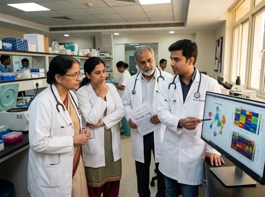
A landmark single-cell study reveals that MEHP, a major phthalate metabolite, damages human ovarian tissue beyond follicles. By disrupting glial-stromal communication, cellular adhesion, and mitochondrial respiration, environmental plasticizers impair ovarian integrity at realistic exposure levels.
Today

A scoping review reveals severe gaps in long-term assistive technology provision following humanitarian emergencies. Addressing post-disaster rehabilitation requires systematic follow-up, workforce training, and integration into national health systems to sustain mobility and recovery.
Yesterday

The World Health Organization has issued its first global guidelines for managing child and adolescent obesity. The recommendations strongly endorse family-based dietary interventions, regular physical activity, and behavioral support as frontline therapy, reserving GLP-1 therapies and surgery for select cases.
Today
