

Loading, please wait...
Loading, please wait...

Traumatic knee ligament injuries in children present unique anatomical and physiological challenges for orthopedic surgeons. Among these injuries, acute pediatric PCL avulsion at the femoral attachment represents an exceptionally rare yet functionally debilitating condition. Because skeletally immature athletes possess active distal femoral and proximal tibial physes, traditional transphyseal reconstruction techniques carry significant hazards of premature growth arrest, limb length discrepancy, and angular deformities. Consequently, sports medicine clinicians prioritize tissue-preserving and physeal-sparing strategies that restore knee stability without violating vulnerable cartilaginous growth plates. Primary repair using arthroscopic suture-anchor fixation has emerged as an innovative intervention for this injury pattern.
The posterior cruciate ligament serves as the primary restraint against posterior tibial translation and secondary stabilizer against external rotation. In skeletally immature patients, mechanical stress often induces bony or chondral avulsion rather than midsubstance ligamentous disruption. Specifically, this injury pattern occurs because the cartilaginous attachment zone exhibits lower tensile strength than the collagenous ligament fibers. Consequently, high-energy impact, direct dashboard-type blows, or hyperextension trauma tear the proximal ligament directly from the medial femoral condyle. Clinicians often encounter substantial hemarthrosis, restricted range of motion, and posterior tibial sag during physical evaluation. Furthermore, magnetic resonance imaging provides indispensable diagnostic insights by clearly visualizing the avulsed femoral footprint, assessing ligament tissue quality, and identifying concomitant meniscal pathology. Plain radiographs and computed tomography scans further delineate bony flecks or occult epiphyseal disruptions. Therefore, timely diagnostic confirmation remains paramount before ligament retraction and synovial scarring impede anatomical restoration. Moreover, prompt surgical intervention within two to three weeks ensures optimal healing biology and facilitates tension-free footprint reduction.
Historically, surgeons treated complex cruciate ruptures through complete ligament reconstruction utilizing autografts or allografts. However, performing conventional tunnel-based reconstruction across open growth plates introduces severe complications in pediatric patients. Drilling large femoral and tibial transphyseal tunnels can create transphyseal bone bridges that tether normal skeletal elongation. Consequently, affected children face progressive valgus, varus, or recurvatum angular deformities alongside limb length discrepancies exceeding several centimeters. Furthermore, soft-tissue graft harvesting causes donor-site morbidity, prolongs rehabilitation, and impairs quadriceps or hamstring maturation. In addition, reconstruction removes the native ligament, eliminating mechanoreceptive nerve endings that support dynamic neuromuscular control. Pediatric knee joints possess remarkable natural intrinsic healing capacity when robust vascular remnants remain preserved. Therefore, orthopedists increasingly reject tunnel drilling in favor of restorative procedures whenever the native tissue exhibits sufficient structural integrity. Ultimately, avoiding transphyseal sockets entirely preserves the distal femoral physis while restoring native biomechanics.
The arthroscopic repair technique utilizes standard anteromedial and anterolateral portals alongside an accessory high-anteromedial portal for optimal visualization and instrumentation. Initially, the surgeon conducts a thorough diagnostic arthroscopy to confirm proximal ligament detachment, verify intact distal tibial fibers, and evaluate intra-articular structures. The operating team gently debrides inflammatory fibrous tissue and micro-fractures the femoral footprint to stimulate bleeding bone marrow elements. Next, the surgeon inserts high-strength suture anchors into the non-physeal subchondral bone of the medial femoral condyle, strictly staying within the epiphysis. High-resolution fluoroscopy or precise intra-articular anatomical landmarks ensure that anchor trajectories do not breach the distal femoral physis. Subsequently, surgeons pass braided high-tensile sutures through the robust proximal ligament stump using specialized suture-passing instruments. In particular, applying mattress or cinch suture configurations provides broad surface compression across the footprint. Finally, the surgeon tensions the sutures with the knee flexed at ninety degrees with an anterior drawer force, effectively securing anatomical contact.
Arthroscopic suture-anchor fixation provides notable clinical and biological advantages over replacement grafts for skeletally immature patients. In particular, primary repair preserves the native ligament substance, protecting native proprioceptive mechanoreceptors that govern balance and coordinated motor responses. Furthermore, avoiding autograft harvest prevents donor site complications like patellar tendonitis, saphenous nerve irritation, and hamstring weakness. Suture anchors supply stable initial mechanical fixation, allowing immediate compression between the ligament remnant and the prepared subchondral bone. Thus, this direct biological healing establishes a resilient bone-ligament interface that mimics uninjured architecture. Additionally, the minimally invasive nature of arthroscopy dramatically decreases surgical trauma, minimizes postoperative pain, and shortens inpatient hospital stays. In pediatric athletes, maintaining undisturbed physeal growth ensures symmetrical lower extremity development without recurring radiographic surveillance for premature closure. Consequently, young athletes achieve predictable joint stability, regain knee extension smoothly, and maintain long-term joint health without secondary osteoarthritis.
Structured postoperative rehabilitation protects the healing ligament while gradually restoring joint kinematics and muscular strength. Immediately following surgery, clinicians immobilize the knee in full extension using a rigid brace with dynamic posterior tibial support for four to six weeks. This positioning eliminates gravity-dependent posterior tibial displacement, relieving strain from the healing suture-anchor repair construct. Concurrently, patients initiate gentle passive range of motion exercises early, restricting flexion to ninety degrees during the initial month. Meanwhile, isometric quadriceps sets, straight leg raises, and ankle pumps preserve muscular tone and prevent deep venous thrombosis. As soft-tissue healing solidifies around six weeks, physical therapists discontinue rigid bracing and introduce progressive active-assisted flexion exercises. Subsequently, closed kinetic chain strengthening, stationary cycling, and neuromuscular proprioception drills commence around three months. Ultimately, returning to high-impact sports requires passing objective functional assessments, including symmetric quadriceps strength, normal posterior drawer testing, and completion of agility protocols.
Successful management of pediatric knee ligament disruptions demands tailored decision-making algorithms that prioritize patient safety and joint longevity. In this context, orthopedic surgeons must thoroughly analyze injury chronicity, patient physiological maturity, and tissue quality before selecting surgical interventions. For example, in chronic settings where substantial ligament retraction or degeneration occurs, primary repair becomes unviable, necessitating specialized physeal-sparing reconstruction techniques. Conversely, acute femoral-sided avulsions present an ideal mechanical and biological scenario for suture-anchor fixation. Emerging orthopedic innovations, such as knotless anchor systems and biologics like platelet-rich plasma, offer further potential to accelerate tissue healing at the anchor-bone junction. Furthermore, standardized multicenter registries are essential to compile long-term functional scores, assess sports resumption rates, and confirm absolute physeal safety across larger pediatric cohorts. Ultimately, combining minimally invasive arthroscopic repair with structured rehabilitation preserves native biomechanics, avoids growth disturbances, and restores high athletic performance.
Skeletally immature children possess open growth plates at the distal femur and proximal tibia that drive vertical skeletal growth. Conventional cruciate ligament reconstruction requires drilling transphyseal tunnels across these active cartilaginous zones. Unfortunately, transphyseal drilling can cause premature physeal closure, leading to severe limb length discrepancies and progressive angular knee deformities. Therefore, physeal-sparing suture-anchor fixation places anchors strictly within the subchondral epiphyseal bone, completely safeguarding growth plates from mechanical injury and thermal damage.
Ideal candidates are skeletally immature pediatric patients presenting with acute, femoral-sided avulsion injuries of the posterior cruciate ligament within two to three weeks of trauma. Clinicians must confirm via preoperative magnetic resonance imaging and arthroscopic inspection that the avulsed ligament possesses sufficient tissue length, cellular vitality, and structural integrity. In contrast, patients with midsubstance interstitial substance tears, chronic retracted stumps, or multi-ligament knee dislocations typically require reconstructive approaches rather than direct suture repair.
Following surgery, patients remain immobilized in full knee extension with dynamic posterior tibial support for four to six weeks to protect the suture-anchor interface. Early rehabilitation focuses on passive motion up to ninety degrees, isometric quadriceps firing, and patellar mobilization. Physical therapy advances to active flexion, resistance training, and stationary cycling around six to eight weeks. Full return to competitive sporting activities typically occurs between six and nine months once neuromuscular testing demonstrates limb symmetry.
Disclaimer: This content is for informational and educational purposes only and does not constitute medical advice, diagnosis, or treatment. Healthcare professionals should exercise their independent clinical judgment. Refer to the latest local and national guidelines for clinical practice.
References
Li Q et al. Arthroscopic Physeal-Sparing Suture-Anchor Fixation for Pediatric Femoral-Side Posterior Cruciate Ligament Avulsion. Arthrosc Tech. 2026 Nov 14. doi: 10.1002/atn2.70293. PMID: 43034610.
Kocher MS, Micheli LJ, Zurakowski D, Luke A. Partial tears of the anterior cruciate ligament in children and adolescents. Am J Sports Med. 2002;30(5):697-703.
Chahla J, Moatshe G, Cinque ME, et al. Posterior Cruciate Ligament: State of the Art. J Am Acad Orthop Surg. 2017;25(6):443-454.
Mayer SW, Albright JC, Stoneback JW, et al. Pediatric Posterior Cruciate Ligament and Posterolateral Corner Injuries: A Systematic Review. Arthroscopy. 2020;36(11):2945-2957.

Read summarized clinical updates, watch expert medical content, and earn CME certifications right from your smartphone.
Open your smartphone camera to scan and install.





Arthroscopic physeal-sparing suture-anchor fixation provides a safe, tissue-preserving treatment for pediatric femoral-side PCL avulsions. By anchoring strictly within the epiphysis, this technique restores native knee stability while eliminating the risk of growth arrest associated with transphyseal reconstruction.
Today
Acute uncompensable heat stress impairs the gut barrier, increasing small intestinal permeability. A landmark physiological trial reveals that elevated baseline oestradiol correlates with augmented intestinal barrier disruption in females, offering vital insights into heat illness and systemic endotoxemia.
Today
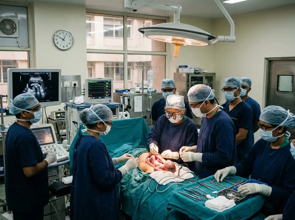
Doctors at AIIMS New Delhi have achieved a historic milestone by performing India's first open prenatal myelomeningocele repair on a 25-week unborn fetus. This landmark multidisciplinary procedure provides vital neuroprotective benefits, lowers hydrocephalus risks, and transforms maternal-fetal medicine nationwide.
Today
A randomized crossover trial reveals that daily fresh watermelon consumption significantly reduces baseline TMAO levels and favorably modulates lipid profiles, lowering triglycerides and LDL cholesterol in adults with overweight and obesity.
Today
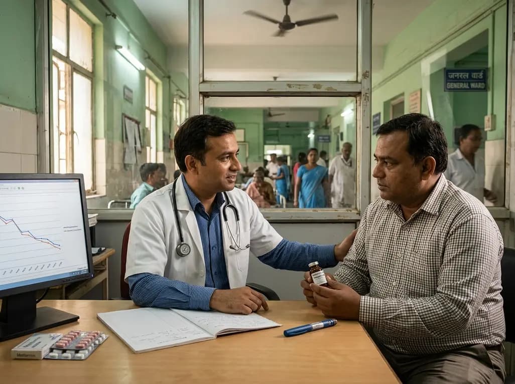
A large-scale retrospective cohort study demonstrates that GLP-1 receptor agonists significantly reduce all-cause hospitalizations, alcohol-related admissions, major cardiovascular events, and mortality in patients with overweight or obesity and alcohol use disorder, providing profound clinical insights.
Today
