

Loading, please wait...
Loading, please wait...

Comprehensive pediatric intracranial aneurysm management demands specialized neurovascular expertise because cerebrovascular lesions in children differ fundamentally from those in adults. Although intracranial aneurysms occur rarely in pediatric cohorts, their rupture causes devastating neurological morbidity. Clinicians must balance immediate procedural risks against the necessity for durable vascular occlusion. Consequently, understanding the comparative strengths of microsurgical clipping and endovascular coiling remains essential for multidisciplinary teams managing these neurovascular conditions.
Pediatric intracranial aneurysms represent fewer than five percent of all documented cerebral aneurysms. However, their biological behavior differs substantially from adult lesions. Adult aneurysms typically stem from acquired hemodynamic stress, atherosclerosis, and long-standing hypertension. In contrast, pediatric aneurysms frequently arise from congenital vessel wall defects, connective tissue disorders, direct trauma, or localized vascular inflammation.
Furthermore, pediatric lesions often present with unique morphological patterns. Clinicians frequently encounter giant, fusiform, or dissecting aneurysms rather than typical saccular forms. In addition, these lesions frequently involve the posterior circulation or the internal carotid artery terminus. Because children have pliable skull anatomy and resilient cerebral parenchymal adaptation, unruptured aneurysms may expand dramatically before manifesting focal mass effect symptoms.
When acute rupture does occur, the resulting subarachnoid hemorrhage presents with severe headache, intractable vomiting, meningismus, or rapid neurological deterioration. Consequently, emergency neuroimaging with computed tomography angiography or catheter-based digital subtraction angiography remains mandatory for precise diagnostic evaluation. Clinicians must promptly distinguish dissecting lesions from infectious or saccular varieties to chart an effective treatment plan.
Historically, open microsurgical clipping served as the gold standard for obliterating intracranial aneurysms across all age groups. In recent decades, however, endovascular techniques have gained tremendous popularity due to their minimally invasive profile. Endovascular coil embolization provides immediate protection against catastrophic rebleeding while avoiding craniotomy, brain retraction, and prolonged critical care stays.
Nevertheless, neurovascular teams encounter notable limitations when applying endovascular strategies to growing children. Pediatric cerebral vessels continue to mature, undergo hemodynamic remodeling, and increase in caliber over time. As a result, endovascularly coiled aneurysms exhibit higher rates of coil compaction, neck remnant expansion, and aneurysm recanalization compared to surgically clipped aneurysms.
Systematic literature evaluations indicate that microsurgical clipping achieves durable obliteration rates exceeding ninety percent, whereas endovascular approaches achieve complete initial occlusion in roughly seventy-five to eighty percent of cases. Furthermore, children treated endovascularly face a fourfold higher likelihood of requiring subsequent reintervention. Therefore, neurosurgeons must weigh the low acute invasiveness of coiling against the superior long-term permanence delivered by microsurgical vessel reconstruction.
A comprehensive literature review spanning over two decades evaluated 868 pediatric patients across 32 published clinical studies. Within this extensive international cohort, 410 children underwent microsurgical interventions, 334 underwent endovascular therapy, and the remaining patients received conservative surveillance. Notably, reintervention rates diverged significantly between both therapeutic modalities.
While only nine surgically treated patients required secondary procedures, twenty-two endovascularly managed patients experienced lesion recurrence necessitating repeat interventions. More importantly, over half of these endovascular failures—twelve patients—ultimately transitioned to open microsurgical salvage. This clinical reality underscores the recurring challenge of coil compaction and recanalization in young vessels.
Because children possess an extended life expectancy, incomplete occlusion poses an enduring risk of delayed subarachnoid rehemorrhage. In addition, juvenile arterial walls exhibit active growth, which often promotes late vascular remodeling around inert metal implants. Consequently, evidence confirms that while endovascular coiling provides critical acute stabilization, microsurgical clipping remains the ultimate benchmark for permanent exclusion. Multidisciplinary teams must actively incorporate these findings into primary procedural selection.
When endovascular coil embolization fails, rescue microsurgical clipping provides definitive exclusion, though it introduces formidable technical hurdles. Recurrent aneurysms often present with dense compaction of previous coils into the fundus, accompanied by significant neck regrowth. Furthermore, extensive inflammatory fibrosis frequently encases the parent artery and adjacent perforating branches.
A representative case illustrates an adolescent presenting with an acute rupture of an internal carotid artery terminus aneurysm. Although interventionalists achieved initial hemostasis with coil embolization, follow-up angiography revealed progressive coil compaction and significant neck recanalization. The multidisciplinary neurosurgical team successfully performed rescue microsurgical clipping to achieve complete vascular reconstruction.
During such rescue operations, surgeons encounter stiff coil masses that obstruct conventional clip blade closure across the aneurysm neck. Therefore, microdissection requires meticulous mobilization of adherent perforating vessels and parent arterial segments. In selected cases, neurosurgeons perform intraoperative coil extraction before clip application. Alternatively, surgeons apply specialized fenestrated or booster clips around the coil ball to avoid compromising parent flow. Intraoperative indocyanine green angiography remains indispensable to verify complete exclusion.
Given children's extended life expectancy, neurovascular management does not terminate at hospital discharge. On the contrary, pediatric patients exhibit a well-documented propensity for de novo aneurysm formation, with literature reporting annual rates between two and eight percent. Underlying structural vasculopathies, inherited connective tissue fragility, and altered hemodynamics from vessel occlusion drive this ongoing vulnerability.
Consequently, structured radiological surveillance remains mandatory throughout adolescence and into adulthood. While initial assessments utilize magnetic resonance angiography to reduce cumulative ionizing radiation exposure, clinicians should employ catheter angiography whenever imaging suggests recanalization or ambiguous remnants. Moreover, modern flow-diverting stents offer promising alternatives for complex lesions that resist direct clipping, although long-term antiplatelet therapy poses distinct pediatric risks.
Ultimately, neurovascular specialists must tailor therapy to the individual child. When anatomical features permit direct clipping, primary microsurgery offers unmatched durability and frees young patients from repeated retreatments. When clinicians choose endovascular therapy for high-risk surgical corridors, they must maintain rigorous longitudinal surveillance and prepare prompt rescue strategies if recanalization emerges.
Pediatric intracranial aneurysms recur more often after coiling because growing cerebral arteries experience active vascular wall remodeling and significant hemodynamic stress over time. Furthermore, coils placed within fragile juvenile lesions can compact into the fundus, reopening the aneurysm neck. Incomplete thrombosis allows persistent arterial inflow, which gradually re-expands the vascular defect. Consequently, pediatric patients require durable obliteration techniques to avoid lifelong structural failure and hazardous secondary hemorrhages.
Neurosurgeons favor rescue microsurgical clipping when recurrent aneurysms present with broad-based neck regrowth or repeated coil compaction that precludes stable endovascular packaging. Additionally, if the lesion produces progressive cranial nerve compression or significant parenchymal mass effect, open surgery permits mass reduction and local decompression. Microsurgical rescue provides definitive mechanical closure around the coil mass. Therefore, transitioning to open surgery eliminates recurring failure cycles and safeguards children across their remaining lifespan.
Pediatric patients require prolonged, often lifelong radiological surveillance because their growing cerebral vasculature remains susceptible to both late recanalization and de novo aneurysm development. Clinicians typically schedule vascular imaging at six months, one year, and then every two to three years thereafter. Magnetic resonance angiography serves as an ideal non-invasive modality to monitor vascular integrity without repeated radiation. However, any structural change or clinical suspicion warrants prompt catheter-based digital subtraction angiography.
Disclaimer: This content is for informational and educational purposes only... Refer to the latest local and national guidelines for clinical practice.
References

Read summarized clinical updates, watch expert medical content, and earn CME certifications right from your smartphone.
Open your smartphone camera to scan and install.





Pediatric intracranial aneurysms present unique management challenges due to high recurrence rates after endovascular coiling and extended life expectancy. A comprehensive literature review emphasizes durable obliteration and highlights microsurgical rescue clipping when endovascular recanalization occurs.
Today

The DOMINO study protocol presents a multidisciplinary implementation mapping approach to overcome provider, patient, and payer barriers to antiobesity medications, offering vital insights for Indian clinicians managing the growing diabesity burden.
Today

An intracranial EWSR1::CREM fusion sarcoma in a 31-year-old female manifested as a paraneoplastic inflammatory syndrome with anemia and secondary infertility. The dural mass mimicked meningioma, but gross-total resection resolved her systemic inflammation and restored natural fertility within one month.
Today

A real-world cohort study of 6,537 patients from the FOUNTAIN platform demonstrates that initiating finerenone significantly reduces albuminuria by 40.7% with minimal eGFR decline and manageable hyperkalemia risk, confirming its practical effectiveness and safety in diabetic kidney disease.
Yesterday

A comprehensive analysis of MAP therapy resistance in osteosarcoma, examining canonical and non-canonical mechanisms across damage avoidance, enhanced DNA repair, and replication stress tolerance to identify novel vulnerabilities.
Today
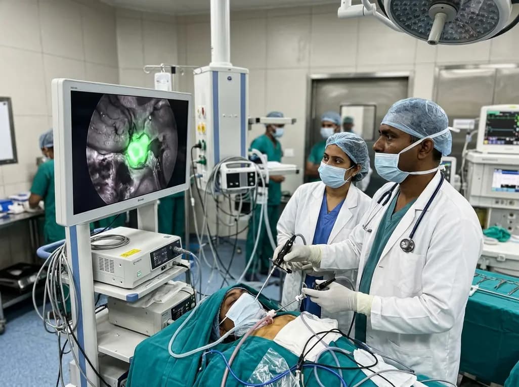
Near-infrared autofluorescence provides real-time label-free optical guidance during endoscopic transsphenoidal pituitary surgery. By highlighting stark signal contrasts between normal pituitary gland and adenomas, this technique aids gross-total tumor resection while safeguarding critical endocrine function.
Today
