.webp&w=384&q=75)

Loading, please wait...
Loading, please wait...
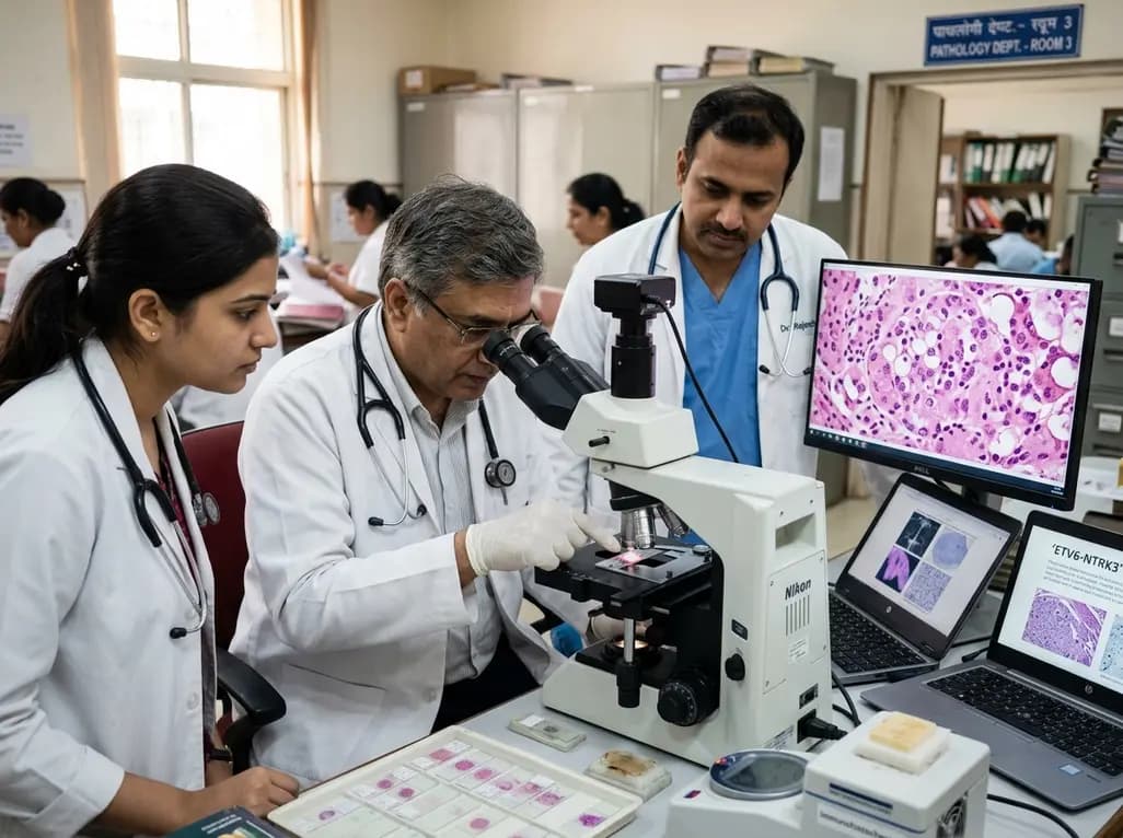
Salivary gland neoplasms present diverse morphological patterns that frequently complicate clinical and pathological evaluation. Among these unusual lesions, mammary analog secretory carcinoma represents a recently recognized malignant entity that predominantly affects the major salivary glands. Clinicians frequently encounter diagnostic dilemmas because these tumors mimic benign conditions both clinically and radiologically. Consequently, comprehensive histological and molecular assessments remain essential to guide definitive clinical management.
Mammary analog secretory carcinoma typically arises within the parotid gland, although minor salivary glands can occasionally host the lesion. Most patients present with a solitary, slow-growing, and painless swelling in the preauricular or infra-auricular region. Because these clinical features closely resemble benign parotid neoplasms, physicians frequently suspect pleomorphic adenoma during initial physical examinations. Clinicians often observe that lesions measure between two and three centimeters upon clinical evaluation, presenting as firm, mobile nodules without overlying cutaneous fixation. In addition, the tumor exhibits an equal distribution between sexes or a slight male predominance, commonly affecting young to middle-aged adults.
Furthermore, radiological investigations such as ultrasonography, computed tomography, and magnetic resonance imaging typically reveal well-circumscribed, lobulated masses. These imaging modalities often display homogeneous tissue characteristics with occasional cystic components, thereby reinforcing the initial benign impression. However, clinicians must maintain a high index of suspicion whenever evaluating persistent parotid lesions. Routine preoperative fine-needle aspiration cytology may assist in triage, but cytological overlaps frequently yield indeterminate classifications. Therefore, definitive diagnosis invariably relies on thorough postoperative histopathological assessment.
Histopathologically, secretory carcinoma displays an intricate microcystic, tubular, solid, or papillary-cystic architecture. The tumor cells appear uniform and bland, possessing round vesicular nuclei, distinct central nucleoli, and abundant pale eosinophilic or vacuolated cytoplasm. Moreover, the presence of characteristic intraluminal and extracellular colloid-like eosinophilic secretions strongly mirrors secretory carcinoma of the breast. These secretions typically test positive for periodic acid-Schiff staining and demonstrate resistance to diastase digestion.
However, differentiating this neoplasm from other salivary malignancies poses substantial diagnostic hurdles for pathologists. Historically, clinicians routinely misclassified these neoplasms as acinic cell carcinoma or low-grade mucoepidermoid carcinoma. In contrast to acinic cell carcinoma, secretory carcinoma lacks true serous acinar differentiation and cytoplasmic zymogen granules. Furthermore, mucicarmine staining frequently highlights abundant intraluminal mucin, which can easily mislead clinicians toward a diagnosis of low-grade mucoepidermoid carcinoma. Similarly, it lacks the prominent squamous differentiation and intermediate cells characteristic of mucoepidermoid carcinoma. Consequently, standard light microscopy alone often fails to establish a definitive diagnosis, necessitating ancillary immunohistochemical confirmation.
Immunohistochemistry serves as an indispensable tool for distinguishing this neoplasm from morphological mimickers. Lesional cells demonstrate strong and diffuse cytoplasmic and nuclear positivity for S100 protein and SOX10. Furthermore, the tumor cells consistently express mammaglobin, cytokeratin 7, and vimentin. In contrast, DOG1 and p63 staining remain uniformly negative, which reliably excludes conventional acinic cell carcinoma and pleomorphic adenoma. Thus, this distinct immunophenotype provides robust preliminary evidence supporting the diagnosis.
Nevertheless, molecular genetics provides the gold standard for unequivocal confirmation. The defining molecular hallmark of this entity is the balanced chromosomal translocation t(12;15)(p13;q25). This specific cytogenetic rearrangement fuses the ETV6 gene on chromosome 12 with the NTRK3 gene on chromosome 15. The resulting fusion transcript encodes a constitutively active chimeric tyrosine kinase that drives oncogenic intracellular signaling. Clinicians utilize fluorescence in situ hybridization or reverse transcription-polymerase chain reaction to detect this rearrangement. Additionally, pan-TRK immunohistochemistry offers a highly sensitive surrogate marker in routine laboratory practice.
Primary surgical resection represents the cornerstone of management for localized disease. Surgeons determine the operative extent based on tumor location, clinical stage, and proximity to crucial anatomical structures. For tumors confined to the superficial parotid lobe, superficial parotidectomy or extracapsular dissection provides excellent local disease control with low recurrence rates. However, surgeons must achieve clear surgical margins, as positive resection margins correlate with an increased risk of local failure. Postoperative histopathological margin assessment provides essential guidance regarding whether the patient requires adjuvant radiotherapy or re-excision.
Throughout surgical procedures, preservation of the facial nerve remains a paramount clinical objective. Therefore, surgical teams routinely employ intraoperative facial nerve monitoring to minimize traction injury and inadvertent nerve transection. Although temporary neuropraxia may occur following extensive parotid gland mobilization, most patients achieve complete functional recovery within several months. In addition, surgeons generally reserve elective neck dissection for cases demonstrating high-grade transformation, deep-lobe invasion, or clinically apparent regional lymphadenopathy. Overall, meticulous surgical technique balances oncological clearance with optimal functional and cosmetic outcomes.
Most cases of secretory carcinoma demonstrate low-grade biological behavior and carry a favorable clinical prognosis. Nevertheless, high-grade transformation, local recurrence, and distant metastatic spread to regional lymph nodes or lungs can occasionally develop. For patients who present with advanced, unresectable, or recurrent disease, conventional cytotoxic chemotherapy offers limited therapeutic benefit. Fortunately, the presence of the pathognomonic ETV6-NTRK3 gene fusion creates a valuable therapeutic target.
Recently, targeted small-molecule TRK inhibitors, including larotrectinib and entrectinib, have revolutionized the management of advanced NTRK-rearranged malignancies. Clinical trials demonstrate remarkable and durable objective response rates with manageable toxicity profiles in patients receiving these targeted agents. Consequently, molecular testing has become critical not only for diagnostic validation but also for therapeutic selection. Furthermore, clinicians must maintain rigorous, long-term clinical and radiological surveillance, as recurrences may arise several years following primary surgical intervention.
Mammary analog secretory carcinoma arises primarily from a recurrent genetic translocation, designated as t(12;15)(p13;q25). This chromosomal rearrangement fuses the ETV6 gene on chromosome 12 with the NTRK3 gene on chromosome 15. Consequently, the chimeric fusion gene produces an abnormal, constitutively active tyrosine kinase enzyme. This abnormal enzyme stimulates downstream oncogenic signaling pathways, driving uncontrolled cellular proliferation and tumor development within major and minor salivary tissues.
Pathologists distinguish secretory carcinoma from acinic cell carcinoma using detailed morphologic evaluation and targeted immunohistochemical panels. Secretory carcinomas consistently exhibit intense immunoreactivity for S100 protein, mammaglobin, and SOX10, while lacking DOG1 expression. In contrast, acinic cell carcinomas show characteristic serous acinar cells containing cytoplasmic zymogen granules and demonstrate positive DOG1 staining alongside absent mammaglobin expression. Molecular confirmation of the ETV6 rearrangement definitively resolves diagnostic uncertainty in challenging cases.
Targeted therapy plays a transformative role in managing advanced, unresectable, or metastatic secretory carcinoma harboring NTRK fusions. First-generation selective TRK inhibitors, such as larotrectinib and entrectinib, directly inhibit the aberrant chimeric tyrosine kinase activity driven by the fusion oncogene. These orally administered agents yield durable disease control, substantial tumor regression, and improved survival outcomes. Consequently, targeted therapy provides a highly effective alternative to traditional cytotoxic chemotherapy in advanced clinical settings.
Disclaimer: This content is for informational and educational purposes only... Refer to the latest local and national guidelines for clinical practice.
References

Read summarized clinical updates, watch expert medical content, and earn CME certifications right from your smartphone.
Open your smartphone camera to scan and install.





Mammary analog secretory carcinoma (MASC) is a rare salivary gland malignancy that often mimics benign lesions or acinic cell carcinoma. Accurate identification requires immunohistochemistry and molecular testing for ETV6-NTRK3 fusions, guiding precise surgical resection and novel targeted therapy.
Today

Ceftriaxone therapy can lead to biliary sludge and gallstone formation due to calcium-ceftriaxone precipitation. A recent cohort study shows that the noninvasive Fibrosis-4 (FIB-4) index, alongside treatment duration, independently predicts lithogenesis, enabling proactive risk stratification.
Today

A multiparametric MRI approach combining quantitative T2 shading, apparent diffusion coefficient (ADC) measurements, and morphological signs provides superior diagnostic accuracy over subjective evaluation to differentiate ovarian endometriomas from benign hemorrhagic cysts.
Today

A landmark case report reveals a donor heart beating for a century in a 63-year-old recipient, challenging conventional age limits in cardiac transplantation and offering critical lessons on organ longevity, donor selection, and long-term graft survival.
Today
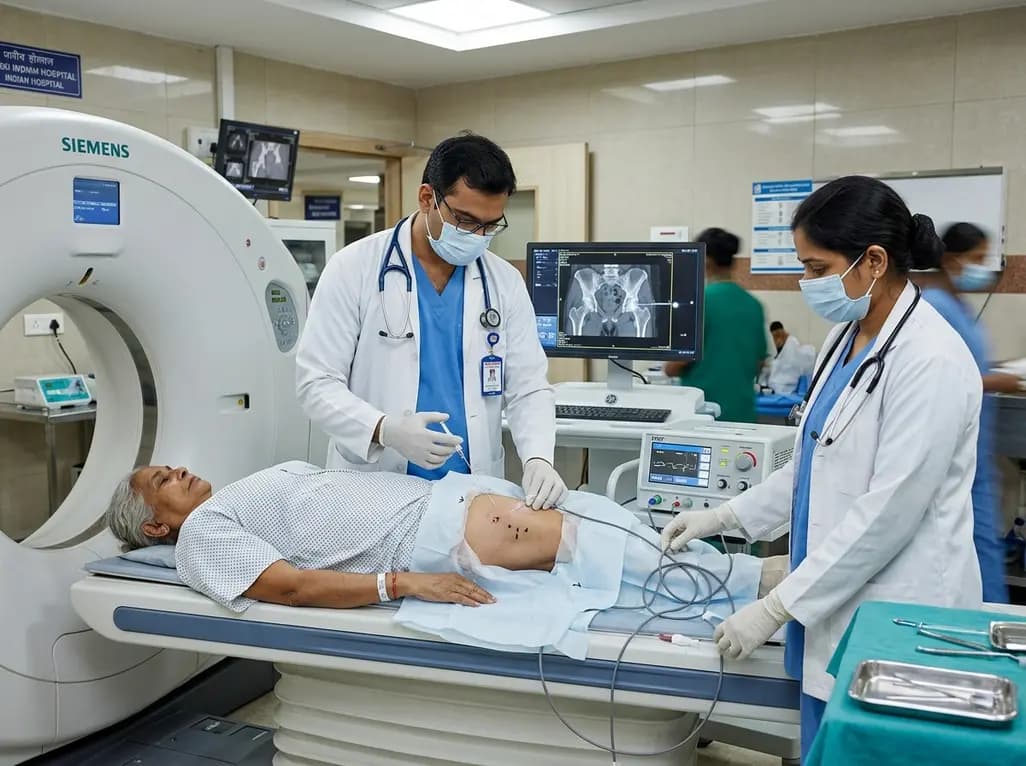
CT-guided pulsed radiofrequency ablation of the obturator nerve delivers durable pain relief up to 12 months in mild-to-moderate hip osteoarthritis, while offering a safe short-term palliative bridge for severe grade 4 disease without procedural complications.
Today

New research reveals that M1 macrophage-derived exosomes aggravate diabetic nephropathy by transferring WTAP to stabilize S1PR2 mRNA. Silencing WTAP in these vesicles attenuates endothelial injury and renal fibrosis, pointing toward innovative nanomedicine therapies.
Yesterday
