.webp&w=384&q=75)

Loading, please wait...
Loading, please wait...

Managing parasagittal meningiomas remains one of the most demanding tasks in modern neurosurgery. These benign neoplasms originate near the superior sagittal sinus and often involve crucial bridging veins. Historically, neurosurgeons prioritized radical extirpation to minimize recurrence risks. However, aggressive manipulation of the venous sinus can cause catastrophic venous infarction, severe brain edema, and permanent neurologic deficits. World Health Organization (WHO) grade I tumors generally carry a favorable histopathological prognosis. Yet, recurrence rates remain surprisingly high when sinus invasion precludes complete excision. Recently, Park and colleagues published a pivotal 20-year cohort study evaluating 115 patients treated between 2005 and 2025. Their findings provide nuanced insight into long-term tumor control and perioperative safety. Specifically, the authors investigated how clinical variables, histopathological markers, and technical resections influence patient outcomes. In neurosurgical centers across India, balancing complete tumor clearance against functional preservation is vital. Therefore, clinicians must understand which specific risk factors predict relapse and surgical morbidity. This article reviews the clinical evidence, dissects critical prognostic variables, and outlines refined surgical paradigms.
Parasagittal meningiomas present unique anatomical hazards because of their intimate proximity to the superior sagittal sinus. The superior sagittal sinus drains blood from both cerebral hemispheres. Consequently, any inadvertent injury or acute thrombosis produces devastating venous hypertension. The classic surgical dilemma centers on whether to pursue radical resection or accept subtotal debulking. In this recent 20-year cohort, investigators classified resections using the traditional Simpson grading system. Simpson grade I resections accounted for 24.3 percent of cases, whereas grade II resections comprised 60 percent. Grade III resections occurred in 15.7 percent of the patient cohort. Overall, surgical morbidities developed in 10.4 percent of individuals. The statistical analysis identified three primary predictors of perioperative complications: anatomical tumor localization, extent of surgical resection, and newly developed sinus occlusion. In particular, lesions located along the middle third of the sinus generated greater morbidity because of dense rolandic venous tributaries. Furthermore, aggressive attempts to achieve gross total resection significantly heightened the risk of acute sinus thrombosis. When a previously patent sinus becomes acutely occluded during surgery, severe cortical swelling usually follows. Therefore, neurosurgeons must exercise extreme restraint when handling partially patent venous structures.
Although WHO grade I lesions possess benign histology, tumor recurrence remains a substantial clinical challenge. In the study cohort, recurrence developed in 13.9 percent of patients over a mean follow-up of 67 months. The mean progression-free survival reached 50.1 months, though intervals ranged from 10.7 to 153.2 months. The authors identified four statistically significant predictors of tumor recurrence: biological sex, extent of sinus invasion, extent of resection, and the Ki-67 proliferation index. Specifically, male sex correlated significantly with earlier tumor recurrence (p = 0.001). Additionally, the extent of sinus invasion directly determined the risk of future relapse (p = 0.001). Tumors with extensive invasion into the sinus lumen often retain microscopic remnants after surgery. Moreover, the extent of surgical resection powerfully predicted recurrence (p = 0.001). Patients who underwent incomplete debulking naturally experienced higher relapse rates. Histopathologically, an elevated Ki-67 proliferation index demonstrated a strong correlation with shortened progression-free survival (p = 0.010). Even within histologically benign grade I cohorts, biological aggressiveness varies widely. Consequently, clinicians must integrate proliferation markers with anatomical features during postoperative planning.
Surgical morbidity occurred in 10.4 percent of treated patients, highlighting the hazardous nature of sinus manipulation. Tumor localization served as a paramount risk factor for postoperative neurological worsening (p = 0.005). The middle third of the superior sagittal sinus represents an unforgiving territory. In this region, rolandic bridging veins deliver vital cortical venous outflow from the motor cortex. Sacrificing these vessels frequently produces intractable intracranial hypertension or dense hemiplegia. Furthermore, the extent of resection directly correlated with perioperative morbidity rates (p = 0.001). Overzealous attempts to achieve clear margins often compromise surrounding dural collateral channels. Most importantly, newly developed superior sagittal sinus occlusion strongly predicted adverse outcomes (p = 0.010). When tumor invasion gradually obliterates the sinus preoperatively, cerebral collateral circulation matures over many months. In contrast, intraoperative injury or acute packing can cause sudden vascular occlusion without compensatory collaterals. As a result, massive venous congestion, venous infarction, and secondary hemorrhage can rapidly ensue. Therefore, neurosurgeons should avoid unnecessary aggressive resection along patent sinus walls. Protecting intact venous architecture must remain a higher priority than achieving radiographic radicality.
The study generated a practical stratification scheme linking recurrence risk directly to resection grade and tumor biology. Interestingly, low-grade tumors that underwent Simpson grade II resection demonstrated the lowest incidence of disease recurrence. In contrast, higher-grade tumors undergoing Simpson grade III resection exhibited the highest recurrence rates. This finding emphasizes that leaving gross residual disease inside the sinus sets the stage for inevitable progression. However, the study provided a crucial technical caveat regarding complete resection. The authors concluded that Simpson grade I resection becomes entirely feasible when preoperative angiography confirms complete sinus occlusion. If the sinus lumen is already fully occluded, robust venous collaterals have developed. Therefore, the neurosurgeon can safely resect the involved sinus segment and affected dural margins without precipitating venous hypertension. Conversely, when the sinus remains patent, attempting Simpson grade I resection carries unjustifiable neurological risks. In such cases, surgeons should remove the extra-sinusal tumor and coagulate the outer dural surface. Leaving a small intrasinus remnant preserves venous outflow and maintains patient safety.
In neurosurgical centers throughout India, managing these tumors demands an individualized, multimodal framework. Advanced neuroimaging, including MR venography and digital subtraction angiography, must guide all preoperative decisions. By assessing sinus patency and collateral venous anatomy, surgical teams can tailor their operative goals. Furthermore, microsurgery alone is no longer the sole therapeutic weapon for challenging lesions. Modern adjuvant modalities, such as stereotactic radiosurgery, offer excellent local control for residual intrasinus tumor tissue. Therefore, surgeons should not imperil neurological function to achieve radical excision in patent sinus segments. Instead, maximal safe resection followed by vigilant serial neuroimaging represents the optimal strategy. When residual tissue exhibits an elevated Ki-67 proliferation index, clinicians should consider early stereotactic radiosurgery. In resource-diverse environments across India, routine follow-up protocols must incorporate regular MRI surveillance for at least ten years. Because recurrences can manifest more than a decade after initial surgery, long-term vigilance remains paramount. Multidisciplinary neuro-oncology boards should evaluate complex cases collaboratively to balance curative ambition against functional preservation. Ultimately, understanding individual tumor biology and venous hemodynamics ensures the safest clinical outcomes.
The most significant factors predicting recurrence include male sex, extent of sinus invasion, extent of surgical resection, and the Ki-67 proliferation index. Higher proliferation markers and incomplete resection notably reduce progression-free survival. Consequently, neurosurgeons must track these specific clinical and pathological features closely during postoperative surveillance.
Aggressive Simpson grade I resection is justified primarily when preoperative angiography confirms complete superior sagittal sinus occlusion. Under these specific conditions, mature collateral venous pathways already protect the brain from venous infarction. Conversely, surgeons should avoid radical sinus resection when the sinus remains patent to prevent catastrophic venous hypertension and stroke.
Clinicians should monitor patients using contrast-enhanced magnetic resonance imaging at scheduled postoperative intervals over a minimum of ten years. Furthermore, if residual intrasinus tumor exhibits an elevated Ki-67 proliferation index, multidisciplinary teams should evaluate the patient for adjuvant stereotactic radiosurgery rather than attempting high-risk secondary microsurgical debulking.
Disclaimer: This content is for informational and educational purposes only. It is not intended to be a substitute for professional medical advice, diagnosis, or treatment. Always seek the advice of your physician or other qualified health provider with any questions you may have regarding a medical condition. Refer to the latest local and national guidelines for clinical practice.
References

Read summarized clinical updates, watch expert medical content, and earn CME certifications right from your smartphone.
Open your smartphone camera to scan and install.





A retrospective 20-year cohort study analyzes surgical recurrence and morbidity in WHO grade I parasagittal meningiomas, identifying critical risk factors including sinus invasion, resection extent, Ki-67 index, and venous occlusion to optimize surgical strategies.
Today
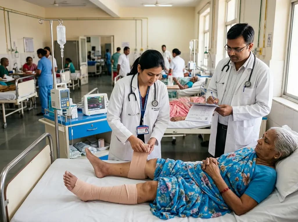
A Mayo Clinic study reveals that applying lower extremity elastic bandages in acute decompensated heart failure significantly improves diuretic efficiency by 340 mL per 40-mg furosemide equivalent. This mechanical therapy reduces required diuretic doses while safely maintaining necessary overall daily fluid clearance.
Today
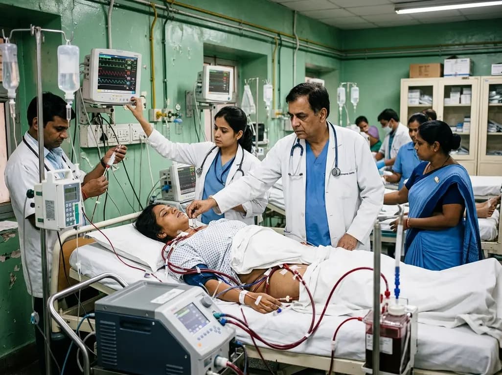
A 34-year-old postpartum woman suffered a pheochromocytoma crisis resulting in cardiac arrest and respiratory failure. Intensivists successfully deployed veno-arteriovenous ECMO to stabilize refractory arrhythmias and differential hypoxemia, bridging her safely to complete tumor resection and full clinical recovery.
Today

A study shows that the aggregate index of systemic inflammation (AISI) significantly associates with elevated myocardial injury markers, including cardiac troponin T and CK-MB, in patients with coronary heart disease, highlighting its potential utility for cardiovascular risk stratification.
Today

A narrative review clarifies key nuances of the AO Spine Primary Infection Classification, including canal boundaries dorsal to the PLL, Type 1 versus Type 2 criteria, neural compression thresholds, and modifier application in primary spinal infections.
Today
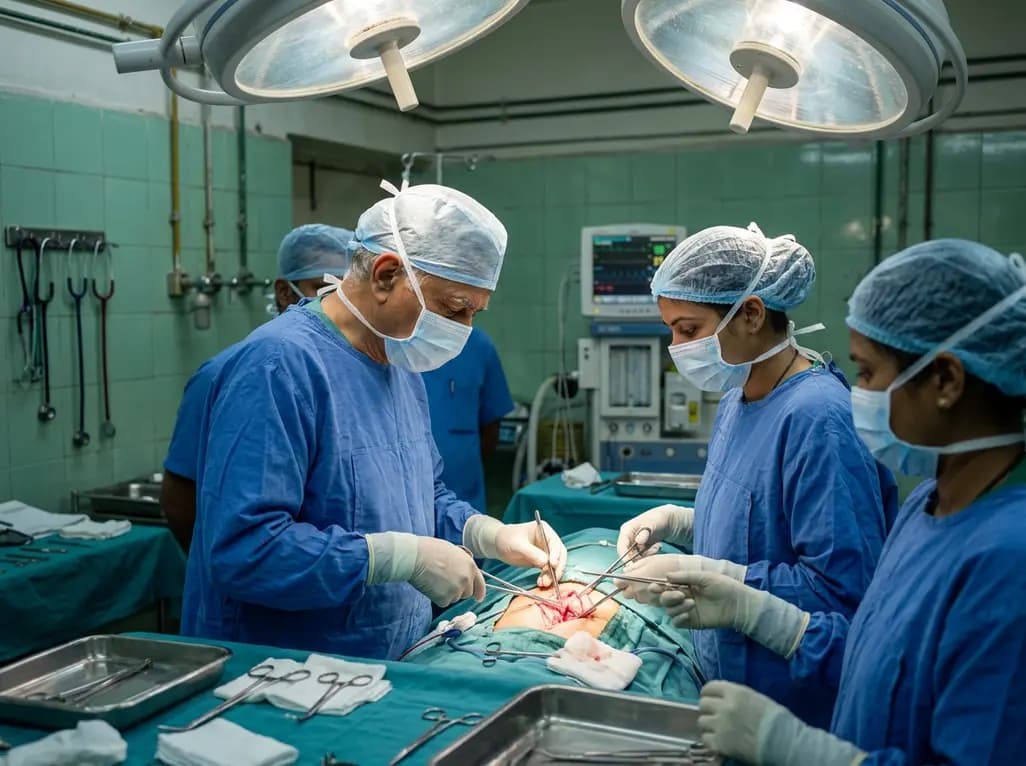
Calcimimetic therapy for primary hyperparathyroidism can induce profound tumor fibrosis, necrosis, and reduced vascularity. These changes alter ultrasound and 4D-CT imaging and cause dense recurrent laryngeal nerve adhesions, requiring heightened surgical vigilance during definitive parathyroidectomy.
Today
