.webp&w=384&q=75)

Loading, please wait...
Loading, please wait...

Primary spinal infections represent complex clinical conditions that can lead to rapid neurological deterioration and structural instability. Clinicians encounter these life-threatening presentations across emergency, orthopedic, and neurosurgical departments. To standardize global communication, researchers developed the AO Spine infection classification system. This structured framework categorizes infection morphology, neurological impairment, and patient-specific clinical modifiers. However, initial international validation trials revealed recurring ambiguities among practicing clinicians worldwide. Borderline cases and conflicting interpretations frequently complicated consistent application in routine practice. Therefore, expert authors published critical clarifications to refine clinical comprehension. Consequently, surgeons can now apply uniform definitions across varied healthcare environments. In regions with high infection burdens, such as India, these objective standards provide vital diagnostic clarity. Spine specialists must clearly understand how subtle morphological shifts influence urgent operative decision-making. Moreover, spinal infections often exhibit subtle initial presentations that mimic degenerative lumbar disease. Because delayed intervention causes permanent paralysis, systematic clinical terminology remains paramount. Standardized reporting prevents catastrophic diagnostic delays and ensures timely medical or surgical therapy.
The primary morphological tier of the classification scheme differentiates anatomical compartments based on infection spread. Type 0 infections represent purely extraspinal disease without osseous or intradural extension. For example, isolated psoas abscesses and paraspinal muscular collections fall strictly within this preliminary group. In contrast, clinicians categorize true spinal infection as Type 1 when osteomyelitis or spondylodiscitis occurs without canal involvement. Pathologists frequently observe vertebral body destruction or intervertebral disc erosion in this cohort. Nevertheless, the infectious process remains confined anteriorly and does not breach the spinal canal.
Furthermore, recognizing this distinction directly guides antimicrobial therapy and interventional procedural choices. Type 0 collections often resolve with targeted percutaneous drainage and systemic broad-spectrum antibiotics. Conversely, Type 1 infections demand strict radiographic monitoring for mechanical collapse and kyphotic progression. Clinicians must confirm the complete absence of epidural spread using high-resolution contrast magnetic resonance imaging. As a result, accurate baseline staging prevents premature surgical intervention while protecting spinal stability. In addition, routine inflammatory markers assist in tracking therapeutic response over subsequent weeks.
Clinicians frequently encounter diagnostic confusion when defining true spinal canal involvement in borderline presentations. According to the clarified consensus, the spinal canal compartment begins strictly dorsal to the posterior longitudinal ligament. Therefore, inflammatory tissue or phlegmon located ventral to this strong ligament does not constitute canal extension. Even when a retrovertebral collection bows the ligament backward, surgeons should not classify the lesion as Type 2. Instead, true Type 2 infections require documented inflammatory extension into the epidural space dorsal to the ligament.
Additionally, clinicians often debate whether cerebrospinal fluid effacement dictates category transitions. The updated guidance clarifies that any confirmed epidural infection qualifies as Type 2, regardless of subarachnoid effacement. However, the patient must show no objective evidence of neural compression centrally or peripherally. Thus, epidural phlegmon without mechanical thecal distortion remains firmly within the Type 2 category. Furthermore, eliminating subjective assessments of fluid effacement enhances inter-observer reliability among radiologists and surgeons. Consequently, clinical teams avoid unnecessary overtreatment in neurologically stable presentations.
Neurological compromise immediately escalates any presentation to a Type 3 infection. The expert consensus defines neural compression as any observable mechanical distortion of vital neural structures. Specifically, this includes thecal sac flattening centrally, cauda equina crowding, or exit foraminal nerve impingement. Clinicians often overlooked isolated foraminal involvement during early international validation exercises. Consequently, the authors emphasize that lateral canal compromise causing nerve root distortion constitutes true neural compression.
Moreover, clinicians must carefully evaluate superimposed infection in patients with pre-existing degenerative spinal stenosis. When acute epidural inflammation exacerbates baseline canal narrowing, distinguishing etiology becomes challenging. Nevertheless, if the active infection causes measurable mechanical deformity of neural elements, surgeons must assign Type 3. This designation alerts multidisciplinary teams to heightened risks of irreversible ischemic neuropathy. Rapid surgical decompression often becomes mandatory when neural compression combines with evolving neurological deficits. Therefore, early identification of Type 3 pathology prevents permanent motor and sensory impairment. In contrast, conservative antimicrobial therapy remains viable for stable non-compressive lesions.
Non-contiguous or extensive longitudinal infections introduce significant grading complexity for practicing spine surgeons. When infection spans cervicothoracic, thoracolumbar, or lumbosacral junctions, clinicians must document the junctional designation accurately. Furthermore, surgeons should classify each distinct level individually while prioritizing the highest morphological grade. In addition, the classification integrates patient-specific modifiers to capture holistic surgical risk. The system uses the plus sign (+) to denote substantial bony destruction. Meanwhile, neurological status grades range hierarchically from intact neurology (N0) to complete cord injury (N4). Clinical modifiers address intractable pain (M1), severe medical comorbidities (M2), and suspicion of atypical organisms (M3).
In India, applying this classification holds exceptional clinical relevance due to the high burden of spinal tuberculosis. Because tubercular spondylodiscitis frequently causes insidious multi-level destruction and cold abscesses, utilizing the M3 modifier remains vital. Similarly, adopting this standardized system promotes seamless inter-hospital transfers between community health centers and tertiary surgical institutes. Consequently, Indian spine specialists can streamline triage protocols and optimize operative decision-making across varied practice settings.
The fundamental distinction rests upon anatomical violation of the spinal canal dorsal to the posterior longitudinal ligament. Type 1 infections involve vertebral osteomyelitis or discitis without canal invasion. Conversely, Type 2 infections require documented inflammatory extension into the epidural space. Subarachnoid fluid effacement alone does not alter this category unless verifiable epidural tissue exists. Importantly, Type 2 lesions exhibit no mechanical compression of central thecal or peripheral foraminal neural structures.
Not automatically, because the classification demands definitive evidence of active mechanical neural compression caused or compounded by infection. If pre-existing degenerative stenosis exists but active epidural phlegmon does not deform the thecal sac or exiting nerve roots, it remains Type 2. However, if the infectious mass contributes to acute neural impingement centrally or within the exit foramen, clinicians must classify the condition as Type 3 to reflect heightened neurological risk.
Clinical modifiers capture critical patient-specific variables that morphological grading alone cannot convey. For example, the M1 modifier identifies severe mechanical instability and intractable pain preventing mobilization. Furthermore, the M2 modifier highlights serious systemic comorbidities like renal failure or immunosuppression that influence surgical safety. Finally, the M3 modifier flags suspected atypical pathogens such as tuberculosis or fungal infections. Consequently, these modifiers ensure tailored antimicrobial therapy and precise surgical risk stratification for every patient.
Disclaimer: This content is for informational and educational purposes only and should not be considered medical advice. Always consult a qualified healthcare professional regarding any medical condition or treatment. Refer to the latest local and national guidelines for clinical practice.
References

Read summarized clinical updates, watch expert medical content, and earn CME certifications right from your smartphone.
Open your smartphone camera to scan and install.





A narrative review clarifies key nuances of the AO Spine Primary Infection Classification, including canal boundaries dorsal to the PLL, Type 1 versus Type 2 criteria, neural compression thresholds, and modifier application in primary spinal infections.
Today
A Mayo Clinic study reveals that applying lower extremity elastic bandages in acute decompensated heart failure significantly improves diuretic efficiency by 340 mL per 40-mg furosemide equivalent. This mechanical therapy reduces required diuretic doses while safely maintaining necessary overall daily fluid clearance.
Today

A qualitative study examines reproductive autonomy and family planning among rural Muslim women, identifying cultural barriers, administrative obstacles, and covert coercion. Healthcare providers must adopt culturally sensitive counseling models to foster equitable maternal and reproductive health outcomes.
Today

A study shows that the aggregate index of systemic inflammation (AISI) significantly associates with elevated myocardial injury markers, including cardiac troponin T and CK-MB, in patients with coronary heart disease, highlighting its potential utility for cardiovascular risk stratification.
Today
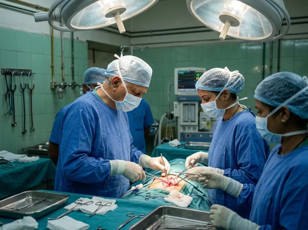
Calcimimetic therapy for primary hyperparathyroidism can induce profound tumor fibrosis, necrosis, and reduced vascularity. These changes alter ultrasound and 4D-CT imaging and cause dense recurrent laryngeal nerve adhesions, requiring heightened surgical vigilance during definitive parathyroidectomy.
Today
