.webp&w=384&q=75)

Loading, please wait...
Loading, please wait...
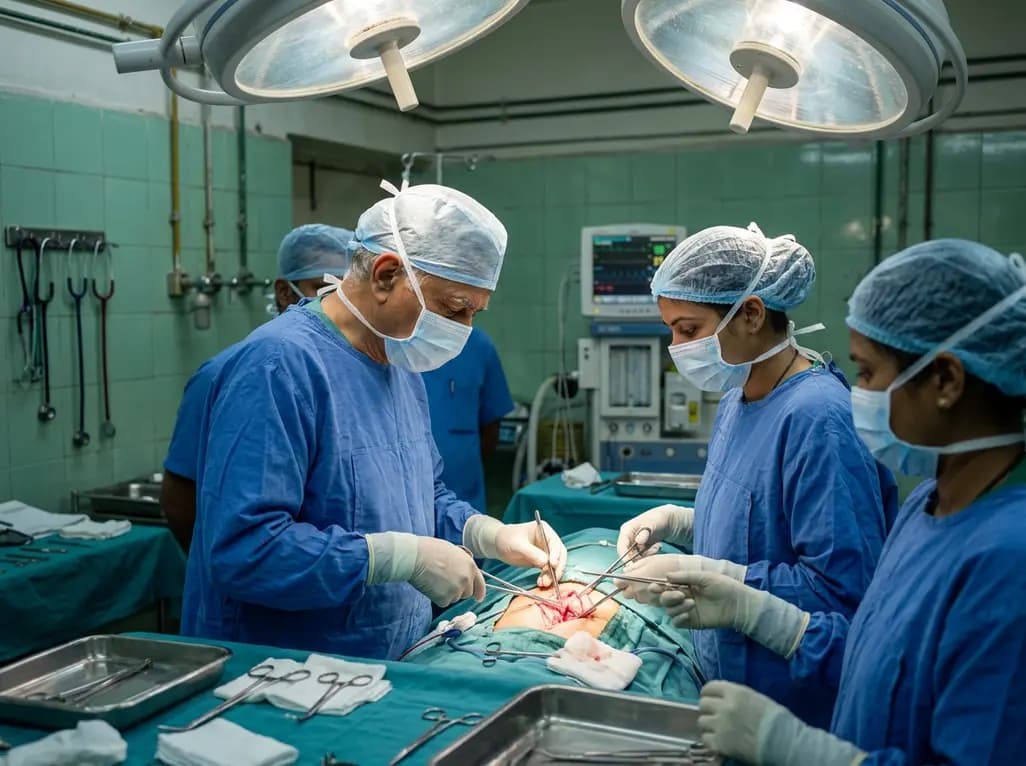
Primary hyperparathyroidism often requires targeted medical stabilization before definitive surgical excision. Clinicians frequently encounter unexpected diagnostic and operative complexities when managing a calcimimetic parathyroid adenoma. Recent clinical evidence demonstrates that calcimimetic administration substantially alters tumor vascularity, tissue planes, and histological architecture, demanding elevated clinical vigilance.
Clinicians prescribe calcimimetic agents like cinacalcet and evocalcet to control hypercalcemia by allosterically activating the calcium-sensing receptor. Consequently, these medications suppress parathyroid hormone secretion and rapidly decrease circulating serum calcium concentrations. However, prolonged or intensive pharmacological stimulation triggers profound biological changes within parathyroid tissues. Calcium-sensing receptor activation alters cellular signaling pathways, which ultimately inhibits cell proliferation and promotes programmed cell death.
Furthermore, pharmacotherapy can induce acute vascular shifts within hyperfunctioning parathyroid tissue. When parathyroid cellular turnover declines sharply, focal ischemia frequently develops across the adenoma parenchymal bed. As a result, cellular apoptosis transitions into coagulative necrosis and secondary degeneration. Clinicians must recognize that these pharmacological events do not merely restore biochemical equilibrium. Instead, they instigate an active local inflammatory response within the glandular capsule and surrounding soft tissues. Macrophages rapidly migrate into the necrotic areas, consuming cellular debris and initiating tissue repair cascades. Consequently, robust fibroblastic proliferation replaces healthy endocrine parenchyma with dense collagenous connective tissue. Therefore, medical therapies fundamentally reshape the biological landscape of parathyroid adenomas before surgical intervention.
Preoperative localization is critical for planning minimally invasive parathyroidectomy. Typically, classical parathyroid adenomas present as hypervascular, well-circumscribed hypoechoic lesions on neck ultrasonography with prominent peripheral vascular rimming. Furthermore, four-dimensional computed tomography routinely displays rapid peak arterial enhancement followed by characteristic delayed contrast washout. However, calcimimetic therapy dramatically disrupts these classic radiological hallmarks.
Notably, post-treatment sonographic evaluation often reveals markedly reduced or absent color Doppler flow within the lesion. Extensive tissue fibrosis and parenchymal infarction significantly diminish intra-tumoral microvascular networks. Similarly, 4D-CT imaging can exhibit weak, sluggish, or heterogeneous enhancement throughout the arterial and venous contrast phases. Clinicians who interpret these attenuated enhancement profiles may mistakenly classify the lesion as a cystic thyroid nodule, an atypical lymph node, or an inactive developmental cyst. Consequently, diagnostic sensitivity decreases, which may lead to inappropriate investigations or surgical delays. Therefore, radiologists and endocrinologists must obtain a meticulous medication history when assessing atypical parathyroid imaging. Recognizing these therapy-induced vascular changes prevents localization errors and ensures accurate preoperative mapping.
Endocrine surgeons increasingly rely on clear anatomical cleavage planes to perform focused parathyroidectomies safely. However, calcimimetic-induced inflammation frequently obliterates normal tissue planes around the parathyroid capsule. As local necrosis triggers capsular rupture and pericapsular fibrogenesis, the parathyroid tumor adheres tenaciously to adjacent vital cervical structures. Specifically, surgeons frequently encounter dense, woody scarring surrounding the tracheoesophageal groove and thyroid capsule.
Most importantly, this aggressive fibrosis often tethers the recurrent laryngeal nerve directly to the adenoma capsule. Consequently, blunt dissection becomes hazardous and technically demanding. Excessive traction on fibrotic bands can cause nerve neuropraxia or permanent vocal fold paralysis. Therefore, surgeons must abandon rapid dissection techniques in favor of meticulous, micro-surgical sharp dissection under magnified vision. Furthermore, surgical teams should routinely integrate continuous intraoperative neuromonitoring to track nerve integrity throughout difficult tissue mobilization. Additionally, dense pericapsular vascular proliferation can generate unexpected intraoperative capillary oozing, compromising surgical visibility. Thus, early awareness of prior calcimimetic exposure prepares operative teams to allocate adequate operating room time and assemble appropriate microsurgical instruments.
Pathologists examining parathyroidectomy specimens after calcimimetic treatment frequently observe striking histomorphological deviations. Classic parathyroid adenomas consist of uniform sheets, cords, or follicles of chief cells with minimal intervening stroma. In stark contrast, post-calcimimetic specimens display profound cellular pleomorphism, extensive coagulative necrosis, and broad bands of dense collagenous fibrosis. Furthermore, pronounced cystic degeneration often replaces large sections of the endocrine parenchyma.
Additionally, microscopic analysis reveals extensive chronic inflammatory infiltrates dominated by foamy, lipid-laden macrophages and lymphocytes. These scavenger cells congregate around resolving necrotic foci and areas of past interstitial hemorrhage. Notably, reactive cellular atypia and nuclear enlargement frequently appear near these fibrotic areas, reflecting cellular stress rather than malignancy. However, inexperienced pathologists might mistake broad fibrous bands, cellular spindling, and capsular adherence for parathyroid carcinoma. To avoid misinterpretation, pathologists must actively search for signs of genuine malignancy, such as unequivocal capsular penetration, vascular invasion, and atypical mitotic figures. Therefore, detailed clinical documentation regarding preoperative drug exposure provides crucial context that prevents overdiagnosis and ensures accurate, definitive pathological classification.
Managing primary hyperparathyroidism requires close coordination among endocrinologists, radiologists, endocrine surgeons, and pathologists. When clinicians prescribe calcimimetics as bridging therapy before surgery, they must establish rigorous biochemical surveillance protocols. In particular, clinicians should track serum calcium levels frequently to prevent symptomatic hypocalcemia. If overt biochemical oversuppression occurs, clinicians must titrate or discontinue the drug promptly.
Moreover, multidisciplinary communication remains vital for surgical success. Endocrinologists must explicitly alert the operative team whenever a patient receives calcimimetics preoperatively. This crucial communication allows surgeons to anticipate tough, adherent anatomy and plan surgical approaches accordingly. Similarly, surgeons should inform pathologists about the patient's pharmacological history before specimen examination. This collaborative workflow prevents diagnostic pitfalls and optimizes operative outcomes. Additionally, tertiary centers should consider standardized imaging protocols that correlate functional scintigraphy with anatomical imaging when pharmacotherapy obscures contrast enhancement. Consequently, institutions can maintain high operative success rates while minimizing patient morbidity. Through comprehensive interdisciplinary planning, surgical teams can successfully navigate the anatomical, radiological, and pathological challenges created by medical parathyroid therapies.
Calcimimetics activate calcium-sensing receptors, precipitating rapid parathyroid cellular changes and focal tissue infarction. Consequently, these agents induce marked microvascular regression and intratumoral fibrosis within the adenoma parenchyma. On neck ultrasonography, lesions frequently demonstrate a pronounced loss of internal color Doppler flow. Furthermore, four-dimensional computed tomography displays blunted, atypical contrast enhancement across arterial and venous phases. Therefore, clinicians must carefully differentiate these altered lesions from hypovascular thyroid nodules or cystic structures during diagnostic evaluations.
Preoperative calcimimetic exposure often triggers significant local necrosis, which incites an aggressive inflammatory repair cascade. As fibroblasts proliferate, dense collagenous scar tissue forms around the parathyroid capsule. Consequently, normal anatomical tissue planes become completely obliterated, firmly tethering the adenoma to adjacent cervical structures. Most critically, this dense fibrosis frequently encases the recurrent laryngeal nerve. Therefore, surgeons must perform meticulous, sharp microsurgical dissection while using continuous intraoperative neuromonitoring to prevent accidental traction injury and vocal cord palsy.
Yes, calcimimetic-induced architectural changes can closely simulate malignant parathyroid tumors. The therapy induces broad fibrotic bands, extensive coagulative necrosis, cystic degeneration, and reactive cellular atypia. These dramatic architectural alterations may mislead pathologists into suspecting parathyroid carcinoma. However, benign treated adenomas lack true capsular penetration, neurovascular invasion, and atypical mitotic figures. Therefore, surgical teams must inform pathologists about preoperative calcimimetic use to prevent diagnostic errors and avoid unnecessary, extensive secondary oncology interventions.
Disclaimer: This content is for informational and educational purposes only. It should not be used as a substitute for professional medical advice, diagnosis, or treatment. Refer to the latest local and national guidelines for clinical practice.
References

Read summarized clinical updates, watch expert medical content, and earn CME certifications right from your smartphone.
Open your smartphone camera to scan and install.





Calcimimetic therapy for primary hyperparathyroidism can induce profound tumor fibrosis, necrosis, and reduced vascularity. These changes alter ultrasound and 4D-CT imaging and cause dense recurrent laryngeal nerve adhesions, requiring heightened surgical vigilance during definitive parathyroidectomy.
Today

A randomized controlled trial demonstrates that a 12-week Tai Chi program significantly reduces post-transplant fatigue, enhances sleep quality, and increases 6-minute walk distance in renal transplant recipients, providing a safe and effective non-pharmacological rehabilitative exercise therapy.
Today

New clinical research demonstrates that adverse physical environments and housing instability strongly increase the risk of postpartum opioid readmissions within 180 days after delivery, highlighting the urgent need for comprehensive screening and supportive maternal community interventions.
Today

A study shows that the aggregate index of systemic inflammation (AISI) significantly associates with elevated myocardial injury markers, including cardiac troponin T and CK-MB, in patients with coronary heart disease, highlighting its potential utility for cardiovascular risk stratification.
Today

The state government is deploying helicopter emergency medical services across the Mumbai-Pune Expressway, Samruddhi Mahamarg, and Mumbai-Goa Highway. This initiative establishes unified standard operating procedures to enable rapid triage, advanced prehospital resuscitation, and seamless trauma care transfers.
Today
