.webp&w=384&q=75)

Loading, please wait...
Loading, please wait...

Locally advanced pancreatic cancer presents formidable therapeutic challenges due to complex retroperitoneal vascular encasement. Clinicians increasingly evaluate pancreatic irreversible electroporation as an effective non-thermal ablation modality for unresectable disease. Because the pancreas lies close to critical arteries and veins, operators require flawless spatial orientation during probe deployment. A groundbreaking single-center study has now evaluated a standardized intraprocedural vascular mapping workflow combining selective arteriography and portography. This innovative approach guides clustered electrode positioning, maximizing oncologic coverage while mitigating perioperative vascular damage.
Managing locally advanced pancreatic carcinoma demands meticulous intervention because tumors frequently encase major peripancreatic vessels. Traditional thermal ablation platforms often fail in this setting due to convective heat-sink effects and thermal injury risks to mesenteric vessels. In contrast, non-thermal electroporation delivers high-voltage electrical pulses that preserve underlying collagenous architecture and vascular elasticity. However, accurate electrode positioning remains exceptionally demanding. Operators must deploy multiple electrodes in strictly parallel configurations to achieve uniform electric field distribution across the entire target lesion. When needle electrodes deviate even slightly, the resulting electric geometry generates undertreated tumor margins. Furthermore, blind percutaneous punctures risk catastrophic vascular laceration or pseudoaneurysm formation. Consequently, interventional oncologists require high-resolution, real-time vascular roadmaps during needle manipulation. Preprocedural cross-sectional imaging provides substantial anatomical information, yet patient repositioning and respiratory excursions often alter tissue relationships. Therefore, intraprocedural guidance systems must definitively reveal both arterial and portal venous branches during needle insertion. By resolving these spatial uncertainties, proceduralists can safely target formerly inaccessible perivascular tumor margins without causing irreversible structural trauma to vital visceral conduits.
To overcome intraprocedural visualization barriers, investigators established an integrated vascular mapping workflow. The clinical cohort comprised forty-eight patients with locally advanced pancreatic cancer treated between August 2024 and December 2025. During these procedures, interventionalists completed selective arteriography in forty-seven patients, achieving a 97.9% application rate. Additionally, operators successfully performed indirect or direct portography in thirty-six patients, representing 75.0% of the cohort. By catheterizing the celiac trunk, superior mesenteric artery, and portal venous system, clinicians obtained continuous fluoroscopic reference points. These dynamic angiographic roadmaps allowed the team to pinpoint exact tumor-vessel boundaries throughout electrode advancement. Furthermore, the protocol introduced clustered electrode arrays, utilizing supplementary configurations in forty-four patients. This adaptable modular setup enabled operators to calibrate probe geometry according to unique perivascular tumor shapes. Only three patients required electrode repositioning, demonstrating the high initial precision of the angiographic technique. Most importantly, the entire cohort achieved a 100% technical success rate for completing the planned mapping and electrode deployment workflow. Consequently, intraoperative angiographic visualization proved entirely feasible, establishing reliable guidance in complex retroperitoneal anatomy where distorted landmarks frequently baffle standard imaging.
Securing complete electrical field coverage represents the primary technical determinant of long-term oncologic control in electroporation procedures. In this study, clinicians systematically calculated the ablation zone-to-tumor size ratio using immediate post-procedure contrast-enhanced computed tomography. Remarkably, every single patient in the forty-eight patient cohort achieved an ablation zone-to-tumor ratio greater than or equal to 1.0. The median ablation ratio reached 1.29, spanning a favorable overall range from 1.12 to 1.69. These metrics confirm that the clustered electrode geometry consistently encompassed the entire gross tumor volume with protective margins. In addition, evaluators assessed early imaging responses on follow-up computed tomography scans at a median interval of eighty-five days. Among the forty evaluable patients, thirty-eight individuals maintained stable disease, representing an impressive 95.0% disease control rate. Only two patients exhibited disease progression during this initial evaluation period. Therefore, the standardized vascular mapping protocol directly supported reproducible, full-thickness necrosis around encased retroperitoneal arteries and veins. These robust early radiological findings suggest that intraprocedural angiographic roadmaps translate directly into measurable technical and local tumor control advantages for challenging pancreatic malignancies.
Although pancreatic electroporation preserves tissue architecture, aggressive perivascular instrumentation still carries distinct procedural risks. In this series, immediate intraprocedural complications occurred in only two of the forty-eight patients, establishing a low 4.2% acute complication rate. Specifically, both adverse events involved localized bleeding episodes that the clinical team successfully managed without emergent surgical exploration. In addition, severe vascular encasement required prophylactic or therapeutic portal vein stenting in eleven patients, accounting for 22.9% of the cohort. Among these stented individuals, two patients developed stent-related complications during follow-up. One patient experienced systemic sepsis requiring targeted antimicrobial therapy, while another developed portal vein thrombosis with intrahepatic extension that mandated anticoagulant therapy. Fortunately, no intraprocedural deaths occurred across the entire patient group. These clinical observations indicate that intraprocedural arteriography and portography provide vital real-time visual warnings that limit severe parenchymal and arterial laceration. Nevertheless, clinicians must maintain heightened vigilance regarding thrombotic and infectious risks whenever endovascular venous stents accompany extensive pancreatic ablation protocols, ensuring prompt intervention should ischemic or septic signs arise.
The successful execution of this vascular mapping protocol offers transformative implications for interventional oncology and surgical practice. Historically, locally advanced tumors involving the celiac axis or superior mesenteric vessels precluded definitive local interventions. However, standardized angiographic mapping converts hazardous percutaneous procedures into controlled, reproducible therapeutic interventions. Multidisciplinary teams, including interventional radiologists, medical oncologists, and hepatobiliary surgeons, can now incorporate clustered electrode ablation into multimodal paradigms. Because systemic chemotherapy often achieves disease stabilization without tumor downstaging, adding definitive local ablation may prevent debilitating local complications like duodenal obstruction or vascular occlusion. Furthermore, this dual-modality workflow provides a clear structural framework for prospective randomized trials evaluating overall survival and progression-free survival. Interventional teams adopting this strategy should prioritize comprehensive training in simultaneous catheter angiography and cross-sectional computed tomography guidance. As clinical centers refine these advanced techniques, image-guided non-thermal ablation will assume a prominent role alongside systemic therapies, expanding viable treatment avenues for patients with historically intractable retroperitoneal disease.
Intraprocedural vascular mapping utilizes selective arteriography and portography to illuminate peripancreatic blood vessels under continuous fluoroscopic visualization. Because locally advanced tumors encase crucial mesenteric and celiac vessels, real-time angiographic roadmaps allow interventionalists to steer clustered electrodes safely past arterial walls. Consequently, this dynamic guidance markedly reduces inadvertent vessel puncture, minimizes major hemorrhagic complications, and guarantees that electrical fields cover perivascular tumor margins without disrupting structural vascular integrity.
Clustered electrodes offer superior electric field uniformity and predictable ablation geometries compared to widely spaced single needles. When operators treat irregularly shaped tumors surrounding major abdominal vessels, clustered configurations deliver concentrated high-voltage pulses that reliably encompass target margins. Furthermore, they simplify spatial alignment during needle advancement, reduce procedural repositioning attempts, and consistently achieve ablation zone-to-tumor size ratios exceeding 1.0, ensuring complete microscopic coverage across difficult perivascular anatomical planes.
Clinicians assess therapeutic efficacy using contrast-enhanced computed tomography immediately after the procedure to measure the ablation zone-to-tumor size ratio. Subsequently, routine cross-sectional imaging occurs at approximately two to three months post-ablation to determine formal disease control. Radiologists evaluate non-enhancing hypodense ablation zones, vessel patency, and regional lymph nodes. In clinical trials, stable disease on serial computed tomography confirms successful local cytoreduction and guides ongoing maintenance chemotherapy protocols.
Disclaimer: This content is for informational and educational purposes only... Refer to the latest local and national guidelines for clinical practice.
References

Read summarized clinical updates, watch expert medical content, and earn CME certifications right from your smartphone.
Open your smartphone camera to scan and install.





A standardized vascular mapping workflow combining selective arteriography and portography facilitates accurate clustered electrode placement in pancreatic irreversible electroporation for locally advanced pancreatic cancer, achieving complete ablation coverage with favorable procedural safety.
Today
A systematic review evaluates the role of branched-chain amino acids (BCAAs) in depression, highlighting neurobiological mechanisms, clinical trial findings, and lifestyle synergies.
Today

Hormonal shifts throughout puberty, menstruation, fertility treatments, pregnancy, and menopause cause profound glucose excursions in women with diabetes. Understanding these endocrine transitions and utilizing advanced continuous glucose monitoring enables clinicians to optimize personalized diabetes care.
Today

A Bayesian network meta-analysis evaluates exercise training in HFpEF, demonstrating that combined aerobic and resistance training, HIIT, and moderate continuous training significantly enhance peak VO2, while functional electrical stimulation uniquely improves quality of life without altering cardiac remodeling.
Today

Cervi-AI achieves expert-level agreement in evaluating cervical spinal stenosis and spinal cord compression on MRI, while its automated imaging metrics reliably predict anterior versus posterior surgical approaches in multilevel degenerative cervical myelopathy.
Today
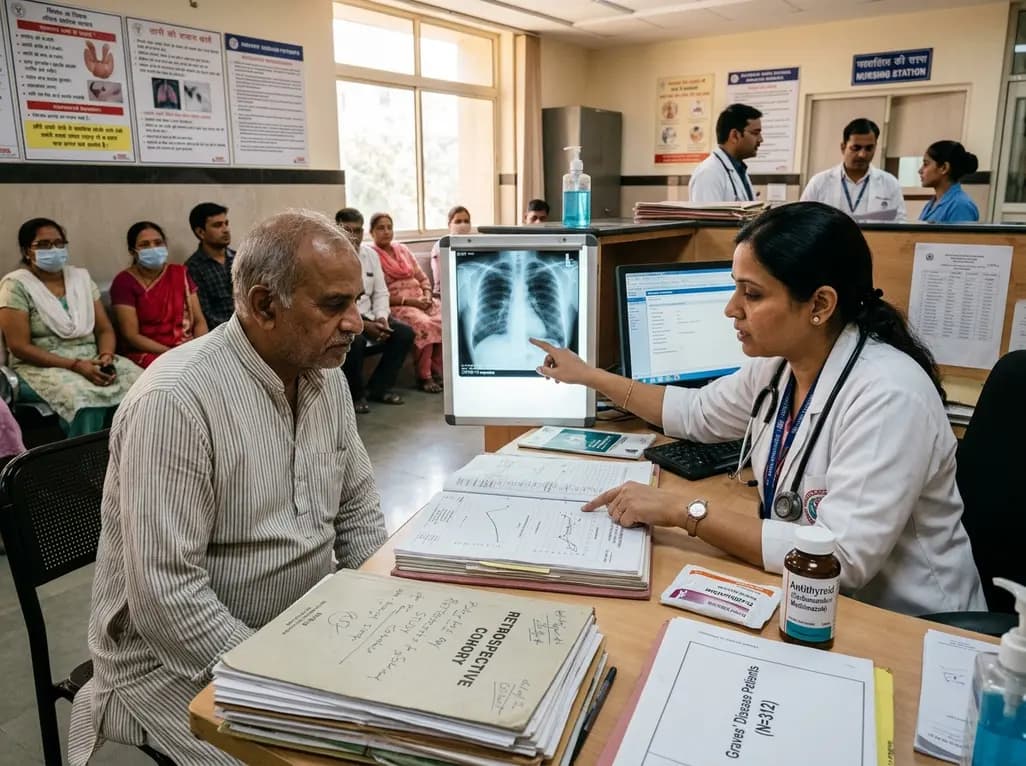
A retrospective cohort study evaluates the short- and long-term impacts of SARS-CoV-2 infection on patients with Graves' disease receiving antithyroid drug therapy, highlighting thyroid status destabilization, clinical symptom exacerbations, and post-viral sequelae.
Yesterday
