

Loading, please wait...
Loading, please wait...

Head and neck squamous cell carcinoma remains one of the most challenging malignancies managed with multimodal therapy. Unfortunately, osteoradionecrosis after radiation therapy represents an excruciating and intractable late toxicity that significantly impairs functional survival. Radiation oncologists and head and neck surgical teams frequently mandate preventive dental clearances to avert this complication. However, real-world clinical data demonstrate variable rates of efficacy across distinct patient subsets. A recent 24-year retrospective cohort study provides essential insights into disease-related severity factors and peri-treatment dental interventions that drive bone necrosis. Understanding these independent predictors enables clinicians to tailor multidisciplinary protocols, minimize invasive procedures post-irradiation, and safeguard long-term skeletal health.
Osteoradionecrosis describes an area of exposed, devitalized irradiated bone that fails to heal over several months without persistent malignancy. Radiation damages microvascular endothelial cells, which induces severe obliterative endarteritis, cellular depletion, and progressive tissue fibrosis. Consequently, bone remodeling capacity collapses under physiological stressors or physical trauma. To elucidate specific predictive factors, researchers evaluated 462 consecutive patients diagnosed with head and neck squamous cell carcinoma treated between March 2000 and January 2024 at a high-volume academic institution. The study team comprehensively gathered demographic profiles, primary tumor subsites, oncologic stages, reconstructive surgeries, radiation dosimetry, and dental clearance records. Overall, 47 individuals developed clinically confirmed osteoradionecrosis, reflecting a cumulative incidence rate of 10.2 percent across the 24-year study timeline. Notably, investigators stratified all participants based on preradiation dental clearance documentation and invasive postradiation dental treatments. This granular methodology allowed the authors to isolate mechanical dental triggers from baseline tumor severity. Therefore, the findings deliver actionable, real-world prognostic information for oncology clinicians, maxillofacial surgeons, and dental teams managing high-risk cancer survivors.
Surgical protocols have long mandated rigorous preradiation dental clearance to eliminate infected or structurally compromised dentition before radiotherapy initiation. In this long-term cohort, 182 patients (39.4 percent) underwent formally documented preradiation dental clearance before radiotherapy. Surprisingly, the incidence of osteoradionecrosis remained virtually identical between patients who completed clearance and those who did not. Specifically, 10.4 percent of patients receiving pre-treatment clearance developed necrosis, compared to 10.0 percent in the unmanaged group. This finding does not imply that pre-treatment dental assessments lack therapeutic clinical value. Instead, it suggests that patients selected for aggressive pre-radiation extractions often present with worse baseline oral disease, periodontitis, and high-dose bone exposure. Furthermore, inadequate healing intervals between tooth extractions and radiotherapy initiation can leave healing bone vulnerable to radiation-induced vascular death. Because oncologists cannot delay radiation without compromising locoregional cancer control, aggressive extractions right before radiotherapy may introduce additional alveolar socket vulnerability. Consequently, clinicians must weigh the immediate hazards of pre-treatment dental trauma against long-term infectious risks. A selective, conservative approach may preserve bone integrity better than universal wholesale extractions.
In stark contrast to pre-treatment interventions, postradiation dental treatment emerged as a potent driver of mandibular and maxillary necrosis. In the cohort, 88 patients (19.0 percent) underwent invasive dental procedures following radiation completion. Among these individuals, 23.9 percent suffered osteoradionecrosis, compared to only 7.0 percent of patients who avoided post-radiation dental trauma. The difference achieved substantial statistical significance, confirming post-irradiation invasive dental surgery as a catastrophic trigger. Irradiated bone exists in a chronic state of hypoxic, hypocellular, and hypovascular insufficiency. When a dental provider extracts a tooth or traumatizes irradiated periosteum years later, the compromised tissue cannot mount an adequate wound-healing response. As a result, microorganisms invade the unhealed socket, triggering persistent non-healing cortical exposure and osteomyelitis. Furthermore, patients with persistent xerostomia and radiation-induced rampant decay often seek urgent dental interventions after therapy. If general dentists remain unaware of prior radiation dosages, routine exodontia can rapidly precipitate severe osteonecrosis. Therefore, preventing subsequent oral trauma represents the single most important clinical strategy to reduce late bone damage in cancer survivors.
Beyond dental interventions, underlying disease severity and radiation targets heavily dictate skeletal vulnerability. The multivariable logistic regression analysis demonstrated that advanced T-category, primary oral cavity and oropharyngeal subsites, and cumulative radiation doses correlate strongly with osteoradionecrosis. Specifically, tumors situated directly adjacent to the mandible expose the bone to high radiation fields exceeding 60 to 70 Gray. Furthermore, concurrent chemotherapy elevates mucosal toxicity, prolongs local healing times, and exacerbates systemic inflammatory stress. When primary tumor extirpation requires extensive periosteal stripping or segmental bone resection, the residual vascular network suffers additional ischemic insults. Tobacco abuse and chronic alcohol consumption further amplify these vascular detriments by impairing microcirculatory perfusion and blunting native immune defenses. Notably, the study revealed that patients combining advanced disease severity with postradiation invasive dental manipulation faced the highest cumulative danger. Consequently, multidisciplinary cancer teams must identify these vulnerable patients during initial staging and contour radiation fields meticulously to spare mandibular structures. By systematically calculating high-dose bone volumes, radiation oncologists can proactively shield vulnerable osseous anatomy.
Preventing osteoradionecrosis requires strict, seamless collaboration among surgical oncologists, radiation oncologists, restorative dentists, and oral maxillofacial specialists. Before starting radiotherapy, dental teams must evaluate patients immediately upon cancer diagnosis to allow adequate wound healing after necessary extractions. Evidence suggests that dental extractions require a healing period of at least two to three weeks before radiotherapy begins. If time does not allow full mucosal closure, endodontic therapy and coronectomy represent safer conservative alternatives. Following radiation completion, oncologists must educate patients regarding lifelong risks and aggressive daily oral hygiene regimens. For example, prescription-strength neutral sodium fluoride gels and remineralizing varnishes protect compromised enamel against severe radiation caries. If tooth loss becomes unavoidable in irradiated jaws, oral surgeons should employ minimally traumatic extraction techniques, primary mucosal closure, and prophylactic systemic therapies where indicated. In addition, close clinical coordination between community dentists and cancer centers prevents accidental extractions within high-dose radiation target volumes. Ultimately, lifelong surveillance and conservative preventive dental care remain indispensable to eliminate the devastating physical and financial burdens of necrosis.
Radiation therapy permanently damages the microvasculature supplying the mandible and maxilla, causing chronic tissue hypoxia and cell depletion. When invasive dental procedures like extractions occur after radiation, the hypovascular bone cannot generate a normal inflammatory and healing response. Consequently, the extraction socket fails to heal, oral bacteria penetrate the bone, and progressive necrosis develops. Thus, postradiation surgical trauma directly provokes severe, chronic osseous breakdown.
Patients who receive preradiation dental clearance frequently present with significantly worse baseline dentition, advanced periodontal infections, and heavier radiation fields. In addition, extractions performed immediately before radiotherapy may not have adequate healing time before cytotoxic radiation begins. Therefore, the residual surgical wounds suffer immediate vascular injury, neutralizing the theoretical protective benefits of clearance. A conservative, timely approach to pre-treatment extractions is critical.
Clinicians often prefer conservative endodontic therapies, coronectomy, or non-surgical restorative stabilization over surgical exodontia in heavily irradiated bone. Removing the coronal pulp and smoothing the tooth roots below the gingival margin maintains mucosal coverage and avoids alveolar bone trauma. However, if extraction proves mandatory, surgeons must employ atraumatic techniques, preserve the periosteum, achieve tension-free primary closure, and maintain close postoperative surveillance.
Disclaimer: This content is for informational and educational purposes only and does not constitute medical advice. Treatment decisions should always be made in consultation with a qualified healthcare provider. Refer to the latest local and national guidelines for clinical practice.
References

Read summarized clinical updates, watch expert medical content, and earn CME certifications right from your smartphone.
Open your smartphone camera to scan and install.





A 24-year cohort study reveals key predictors of osteoradionecrosis in head and neck cancer. While preradiation dental clearance showed comparable necrosis rates, postradiation dental treatment tripled the risk, underscoring the urgent need for conservative long-term oral care.
Today
An intracranial EWSR1::CREM fusion sarcoma in a 31-year-old female manifested as a paraneoplastic inflammatory syndrome with anemia and secondary infertility. The dural mass mimicked meningioma, but gross-total resection resolved her systemic inflammation and restored natural fertility within one month.
Today
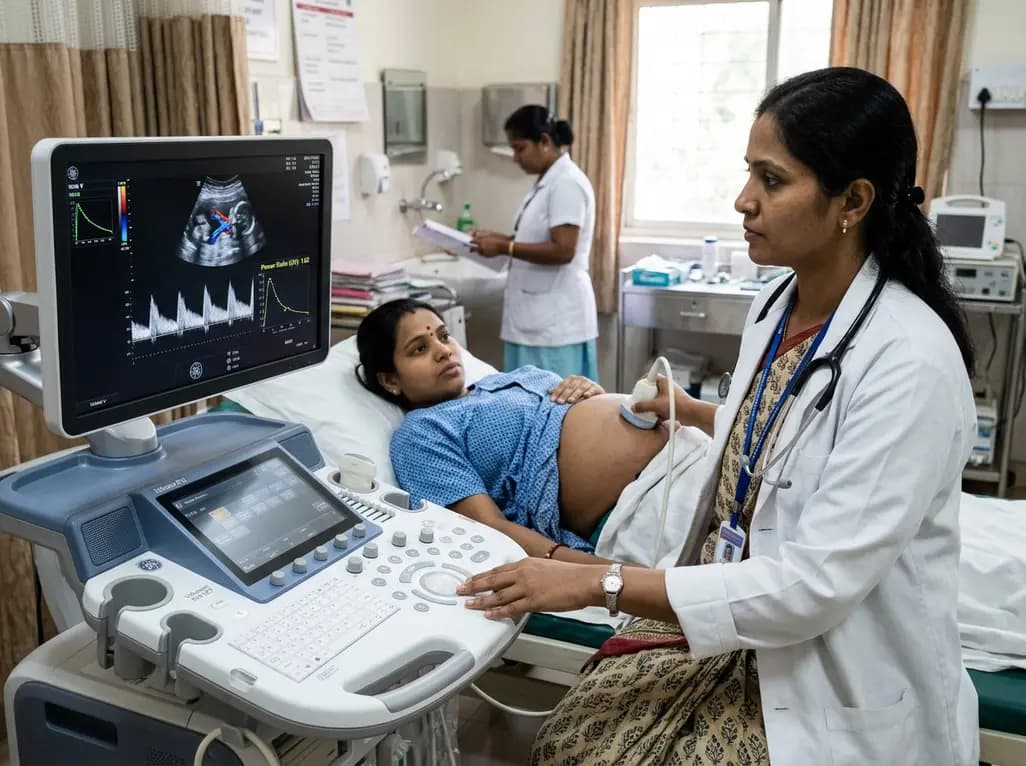
Adaptive spectral analysis of umbilical venous Doppler recordings reveals that an elevated second-trimester maternal-fetal power ratio can distinguish pregnancies at risk of preeclampsia, offering a novel biophysical approach to enhance early vascular risk stratification.
Today

A real-world cohort study of 6,537 patients from the FOUNTAIN platform demonstrates that initiating finerenone significantly reduces albuminuria by 40.7% with minimal eGFR decline and manageable hyperkalemia risk, confirming its practical effectiveness and safety in diabetic kidney disease.
Yesterday

A comprehensive analysis of MAP therapy resistance in osteosarcoma, examining canonical and non-canonical mechanisms across damage avoidance, enhanced DNA repair, and replication stress tolerance to identify novel vulnerabilities.
Today
Near-infrared autofluorescence provides real-time label-free optical guidance during endoscopic transsphenoidal pituitary surgery. By highlighting stark signal contrasts between normal pituitary gland and adenomas, this technique aids gross-total tumor resection while safeguarding critical endocrine function.
Today
