

Loading, please wait...
Loading, please wait...

Down syndrome, or trisomy 21, presents complex craniofacial and dental challenges requiring specialized interdisciplinary care. Among these presentations, Class III malocclusion occurs frequently due to severe midfacial hypoplasia and altered growth dynamics. Furthermore, congenital anomalies like dental agenesis and canine transpositions compound existing spatial deficiencies. While orthognathic surgery resolves severe skeletal discrepancies, systemic risks often favor non-surgical orthodontic camouflage to restore functional occlusion.
Individuals diagnosed with Down syndrome exhibit distinct craniofacial patterns that fundamentally influence dental development. Specifically, deficient cartilaginous growth at the cranial base impairs forward projection of the nasomaxillary complex. This growth restriction causes pronounced midfacial retrusion, creating a relative mandibular prognathism. Therefore, an Angle Class III dental relationship frequently develops during mixed dentition. Furthermore, transverse maxillary hypoplasia produces severe unilateral or bilateral crossbites. Muscular hypotonia and a low resting tongue posture exert outward mechanical force on mandibular teeth, worsening anterior discrepancies. As a result, affected individuals experience impaired mastication, speech articulation challenges, and compromised airways. Concurrently, congenital dental anomalies occur in over half of these patients. In particular, tooth agenesis commonly targets maxillary lateral incisors and mandibular premolars. Additionally, ectopic eruption pathways frequently precipitate canine transpositions within the narrow upper arch. Consequently, treating clinicians must identify these skeletal and dental disharmonies through early radiographic and clinical evaluations. Recognizing these interrelated anomalies early enables timely intervention, preventing progressive dentoalveolar compensation and irreversible functional decline.
Comprehensive diagnostic evaluation remains vital when managing complex syndromic malocclusions. In this representative case, radiographic imaging confirmed bilateral maxillary lateral incisor agenesis alongside a missing mandibular incisor. Simultaneously, panoramic views revealed a complete transposition between the maxillary canines and adjacent first premolars. Managing transposed teeth requires prudent mechanical judgment. Orthodontists must evaluate whether moving roots past each other presents unacceptable biological risks. Forcing root movement across dense cortical bone often triggers root resorption, loss of vitality, and alveolar dehiscence. Furthermore, correcting transposition drastically increases treatment duration, stressing patient cooperation and periodontal health. Therefore, clinicians elected to maintain the transposed order, adopting a canine-premolar substitution strategy. Cephalometric assessment simultaneously highlighted a severe anterior crossbite, negative overjet, and reduced lower facial height. Moreover, bilateral posterior crossbites reflected significant transverse constriction of the basal maxilla. Clinicians synthesized these findings to formulate realistic therapeutic objectives. Prioritizing non-surgical compensation over rigid anatomical ideals preserved fragile periodontal tissues while addressing the patient's primary functional and aesthetic deficits.
Executing orthodontic camouflage requires precise biomechanical planning to resolve sagittal and vertical disharmonies. Clinicians utilized fixed preadjusted edgewise appliances combined with continuous Class III intermaxillary elastics. These mechanics promoted controlled proclination of maxillary incisors while retroclining mandibular anterior teeth. Consequently, the team corrected the anterior crossbite and established positive overjet and overbite. Concurrently, mechanics induced a slight clockwise rotation of the mandible. This rotation beneficially increased the lower anterior facial height, significantly improving the patient's soft tissue profile. Orthodontists achieved space closure by drawing posterior teeth forward into missing incisor sites. Maintaining transposed canines avoided complex root uprighting and shortened overall chair time. Throughout active therapy, practitioners used light, continuous forces through flexible nickel-titanium and stainless steel archwires. In addition, the dental team monitored periodontal parameters diligently at every activation visit. Because patients with Down syndrome experience rapid periodontal breakdown, meticulous biofilm control proved vital. This conservative biomechanical approach avoided surgical morbidity while fulfilling comprehensive functional objectives.
Orthodontic space closure in the presence of agenesis and transposition requires close restorative partnership. Following fixed appliance therapy, practitioners initiated restorative recontouring to establish dental harmony. Specifically, canines positioned in lateral incisor sites received conservative enamel reduction and composite reshaping. Adding composite resin modified the pointed canine morphology into natural lateral incisor anatomy. Similarly, clinicians modified the first premolars located in canine sites. By grinding palatal cusps and augmenting buccal contours, dentists established adequate canine guidance and cosmetic symmetry. This restorative intervention also prevented traumatic occlusal interferences during lateral jaw movements. In the mandibular arch, restorative dentists conservatively recontoured anterior teeth, compensating for the missing lateral unit. Direct composite resin provided an ideal, minimally invasive restorative medium. Furthermore, direct bonding avoided extensive enamel removal, preserving pulpal health and reducing financial strain. Ultimately, this seamless interface between orthodontics and restorative dentistry delivered balanced occlusion, protected periodontal tissues, and substantially enhanced smile aesthetics.
Long-term retention presents unique hurdles following syndromic malocclusion correction. Persistent hypotonia and an anterior tongue thrust continuously threaten incisal position. Consequently, relying on removable retainers frequently fails due to variable patient compliance. Clinicians routinely place fixed multistrand stainless steel retainers on the lingual surfaces of anterior teeth. Bonded wires provide continuous stabilization against incisor relapse without demanding patient cooperation. In the maxillary arch, combining fixed retainers with nocturnal vacuum-formed retainers preserves transverse expansion. Furthermore, establishing positive overbite and overjet mechanically locks the corrected occlusion, resisting sagittal relapse. Correcting the crossbite also improves lip competence, swallowing mechanics, and speech clarity. Additionally, widening the maxillary perimeter enhances intraoral airway space, reducing habitual mouth breathing. Clinicians must establish structured maintenance schedules, scheduling professional prophylaxis every three to four months. Regular evaluations safeguard gingival margins and verify retainer bonding integrity. Through diligent interdisciplinary retention protocols, dental teams maintain hard-earned functional and aesthetic results throughout adult life.
Orthognathic surgery presents significant perioperative risks for patients with Down syndrome. Specifically, these individuals frequently present with congenital cardiac defects, generalized ligamentous laxity, and narrow upper airways that elevate anesthetic hazards. In contrast, orthodontic camouflage provides a reliable, non-surgical alternative that repositions teeth to compensate for underlying skeletal discrepancies. Consequently, camouflage treatment substantially improves chewing efficiency, eliminates traumatic crossbites, and enhances facial aesthetics without subjecting medically fragile patients to major invasive surgical interventions.
Clinicians typically choose between active biomechanical correction and maintaining the transposed dental alignment. In cases involving anatomic constraints or syndromic conditions, maintaining the transposition offers significant advantages. Specifically, forcing tooth roots past each other through dense cortical plates risks severe root resorption, alveolar dehiscence, and prolonged treatment. Therefore, maintaining the transposition preserves periodontal structures. Restorative clinicians subsequently reshape the canine into a lateral incisor and recontour the adjacent first premolar to replicate normal canine aesthetics and guidance.
Retaining corrected occlusion in patients with Down syndrome demands individualized retention protocols. Because muscle hypotonia and low resting tongue posture exert forward pressure on the anterior dentition, relapse risks remain exceptionally high. Consequently, practitioners prefer fixed bonded lingual retainers over removable appliances to eliminate compliance dependence. In addition, combining maxillary bonded wires with clear overlay retainers stabilizes transverse expansion. Routine recall examinations ensure continuous bracket and wire integrity, reinforced oral hygiene, and lasting functional occlusal stability.
Disclaimer: This content is for informational and educational purposes only... Refer to the latest local and national guidelines for clinical practice.
References

Read summarized clinical updates, watch expert medical content, and earn CME certifications right from your smartphone.
Open your smartphone camera to scan and install.





A case report demonstrates successful orthodontic camouflage in a patient with Down syndrome, addressing Class III malocclusion, crossbite, dental agenesis, and transposition without orthognathic surgery to achieve stable occlusion and improved aesthetics.
Today
A multicenter study introduces BAT-AKI, a biomarker-aware transformer that achieves superior acute kidney injury prediction and post-onset prognostic modeling across diverse hospital electronic health record systems.
Today
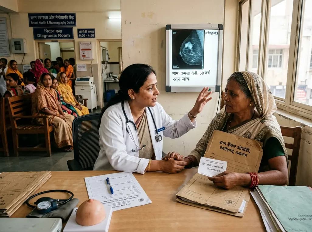
A KGMU study reveals over half of women with breast abnormalities fail to undergo diagnostic confirmation, leading to advanced-stage detection. Discover the socio-cultural barriers, clinical risks of incomplete resection, and community-level solutions needed to bridge the critical screening-to-diagnosis gap in India.
Yesterday
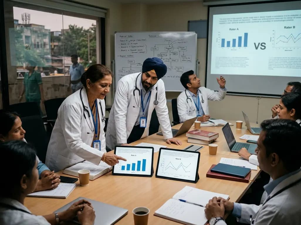
A controlled evaluation reveals that AI raters in clinical LLM benchmarking disagree significantly due to divergent scoring models, challenging automated healthcare AI validation.
Today
A phase I/II trial evaluated E. coli-derived rhBMP-2 combined with beta-tricalcium phosphate for long-bone nonunion. The combination demonstrated high radiographic union, functional limb recovery, undetectable systemic absorption, and transient antibody formation without clinical adverse events.
Today

A meta-analysis of 18 studies reveals that bariatric surgery does not significantly alter circulating homocysteine levels, regardless of follow-up duration. Clinicians must maintain vigilance over post-surgical micronutrient supplementation to prevent hyperhomocysteinemia.
Today
