.webp&w=384&q=75)

Loading, please wait...
Loading, please wait...

Oral leukoplakia (OL) remains a significant concern in clinical practice as it is the most common oral potentially malignant disorder. While clinicians often identify these white patches during routine examinations, predicting their progression to malignancy is challenging. A recent study has investigated oral leukoplakia salivary cytokines to better understand the local and systemic immunological changes involved in this condition.
The study utilized a comprehensive panel of 13 cytokines to compare 30 patients with oral leukoplakia against 30 healthy controls. Researchers focused on both saliva and plasma to capture local and systemic inflammatory profiles. Consequently, they found that certain biomarkers, such as IL-6 and IL-8/CXCL8, show significant variation based on the severity of the lesion. Furthermore, the researchers observed that these levels could distinguish between different stages of neoplasia.
The classification of Oral Intraepithelial Neoplasia (OIN) provides a structured way to assess these changes. Specifically, higher grades of OIN were associated with distinct cytokine elevations. Therefore, these proteins may serve as non-invasive tools for monitoring disease progression. However, while salivary levels showed strong correlations with local tissue changes, plasma levels offered a broader view of systemic inflammation.
Using oral leukoplakia salivary cytokines as biomarkers could revolutionize early detection strategies. Traditionally, clinicians rely on invasive biopsies to assess the risk of malignant transformation. In contrast, salivary analysis is non-invasive and easy to repeat. This approach allows for closer monitoring of high-risk patients without the discomfort of frequent surgical interventions. Additionally, identifying specific cytokine profiles helps in understanding the underlying pathophysiology of oral disorders.
Further research is necessary to validate these findings across larger, more diverse populations. Nevertheless, the current data suggests that the integration of immunological profiling into clinical workflows is promising. By identifying patients with high transformation risk early, medical professionals can implement more aggressive preventive measures and improve long-term outcomes.
Salivary biomarkers provide a non-invasive method to monitor the local immune environment. They can help identify which lesions are more likely to progress to oral cancer.
The OIN (Oral Intraepithelial Neoplasia) classification categorizes the severity of dysplasia in oral tissues. This helps clinicians determine the risk level and appropriate treatment plan for leukoplakia.
Disclaimer: This content is for informational and educational purposes only. It does not constitute professional medical advice, diagnosis, or treatment. Always seek the advice of your physician or other qualified health provider with any questions you may have regarding a medical condition. Refer to the latest local and national guidelines for clinical practice.
References
Yülek H et al. Immunological Biomarkers in Oral Leukoplakia According to Oral Intraepithelial Neoplasia (OIN) Classification: A Comparative Analysis of Salivary and Plasma Cytokines. J Oral Pathol Med. 2026 Mar 10. doi: 10.1111/jop.70131. PMID: 41807270.
Brailo V et al. Salivary and serum interleukin 1 beta, interleukin 6 and tumor necrosis factor alpha in patients with leukoplakia and oral cancer. Oral Oncol. 2006.
Deepthi G et al. Salivary Tumour Necrosis Factor-α as a Biomarker in Oral Leukoplakia and Oral Squamous Cell Carcinoma. Asian Pac J Cancer Prev. 2019.

Read summarized clinical updates, watch expert medical content, and earn CME certifications right from your smartphone.
Open your smartphone camera to scan and install.





Research identifies salivary and plasma cytokines as potential biomarkers for assessing oral leukoplakia severity and progression risk using OIN classificat...
7 months ago

An observational study from the NIH All of Us Research Program reveals that adding a multi-ancestry polygenic risk score to machine learning models provides only modest improvements in predicting adult weight gain, underscoring the enduring significance of routine clinical and lifestyle factors.
Today

A study in JAMIA Open shows lightweight open-source reasoning LLMs achieve up to 95% sensitivity and accelerate evidence synthesis screening by nearly twentyfold, presenting a cost-effective, scalable AI solution for biomedical research and systematic reviews.
Today
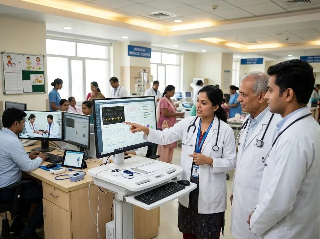
A landmark study details how an enterprise pediatric remote patient management model expanded from a single cardiology pilot to 16 diverse specialty programs by combining centralized digital operations with specialty clinical leadership.
Today
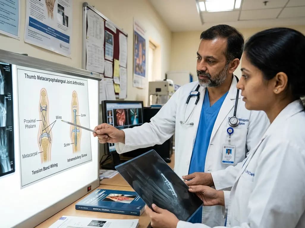
A comparative review of tension band wiring and intramedullary screw fixation in thumb metacarpophalangeal arthrodesis, evaluating union rates, hardware complications, and clinical outcomes.
Yesterday
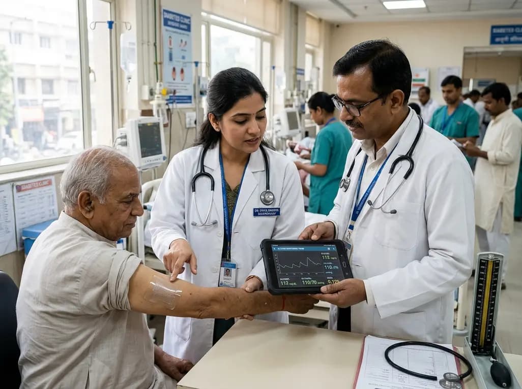
A breakthrough ultrathin multimodal sensor decouples physical hemodynamic monitoring from disposable electrochemical calibration, achieving long-term wearable glucose prediction with 81.36% accuracy and a 9.12% MARD. This offers a robust, drift-free solution for outpatient metabolic surveillance.
Yesterday
