

Loading, please wait...
Loading, please wait...
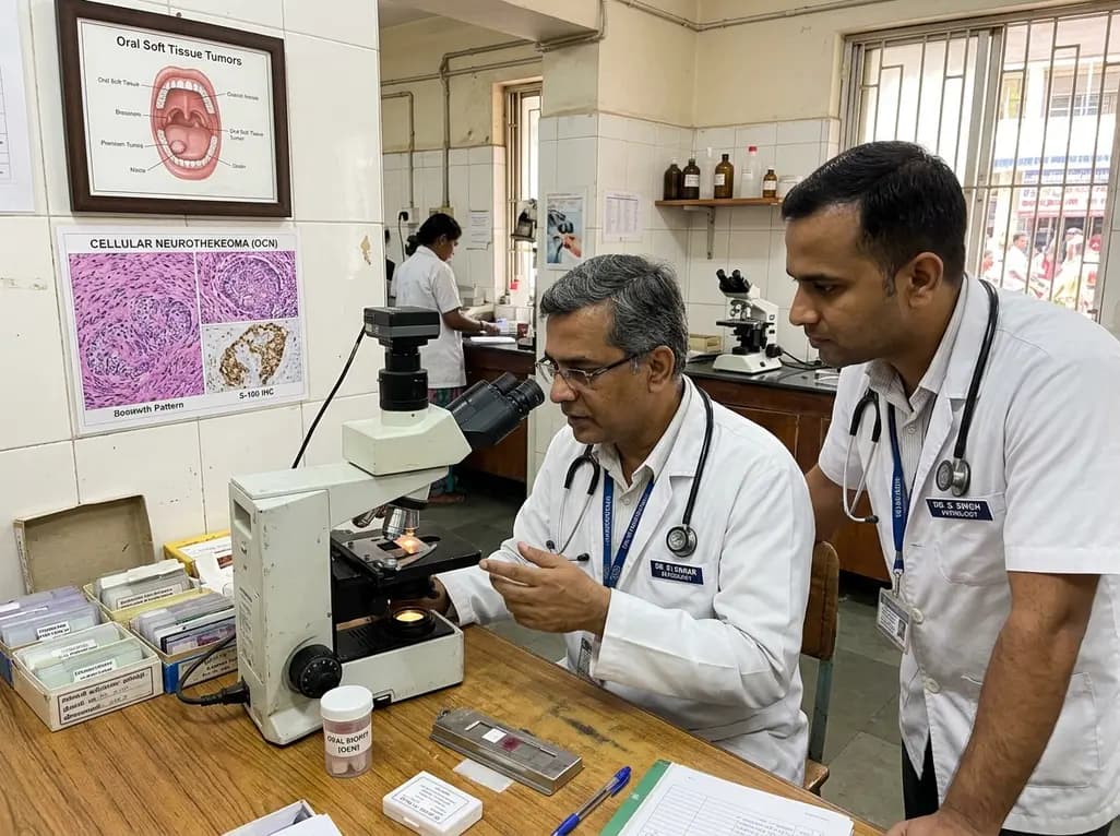
A mucosal cellular neurothekeoma represents an exceptionally unusual clinical entity within oral pathology. Clinicians routinely encounter various soft tissue nodules in the gingival and buccal mucosa. However, distinguishing rare benign mesenchymal neoplasms from reactive proliferations remains challenging. In particular, cellular neurothekeoma displays distinctive architectural features, yet it frequently causes diagnostic confusion. Because lesions mimic common reactive processes, clinicians and pathologists must maintain heightened awareness to ensure precise diagnostic evaluation.
Historically, pathologists categorized cellular neurothekeoma alongside nerve sheath myxoma due to overlapping lobular growth patterns. However, modern molecular and immunohistochemical studies demonstrate that cellular neurothekeoma is a distinct mesenchymal neoplasm. Researchers initially debated whether this tumor arose from peripheral nerve sheath elements or primitive neuroectodermal cells. Currently, significant scientific consensus suggests a fibrohistiocytic or myofibroblastic lineage rather than true neural derivation. Furthermore, traditional nerve sheath myxomas consistently express S100 protein and display clear peripheral nerve differentiation. In contrast, cellular neurothekeoma remains characteristically negative for S100 protein and SOX10. Consequently, modern classifications separate it entirely from benign peripheral nerve tumors. Dermal presentations typically emerge on the face, neck, and upper extremities of young adult females. Nevertheless, intraoral mucosal involvement occurs in exceedingly rare instances. Therefore, pathologists must carefully evaluate mucosal spindle cell proliferations to avoid diagnostic errors. Moreover, modern diagnostic criteria emphasize distinguishing true cellular neurothekeomas from myxoid variants to avoid misinterpreting benign lesions as sarcomatous processes. Ultimately, recognizing this unique histogenesis helps clinicians understand the tumor's benign biological behavior despite its high cellularity.
Intraoral presentations of this tumor remain exceptionally unusual in clinical dental practice. For instance, a recent report highlighted a twenty-two-year-old woman presenting with a painful, enlarging mass. The lesion involved her left buccal and labial gingiva over a five-month timeframe. Because the mass progressively expanded, it caused significant mechanical difficulty with mastication and speech. Clinical examination revealed a firm submucosal mass without superficial mucosal ulceration. Additionally, radiographic imaging confirmed an ill-defined soft tissue lesion lacking underlying cortical bone erosion. Most cutaneous neurothekeomas present as painless, slow-growing, asymptomatic nodules. In contrast, oral mucosal tumors frequently cause pain or tenderness due to persistent masticatory trauma. Consequently, clinicians often mistake these gingival swellings for common reactive lesions such as pyogenic granulomas. Therefore, dental surgeons must gather comprehensive clinical details and obtain representative biopsy tissue. Furthermore, clinicians should perform careful palpation and radiographic assessment to evaluate any cortical disruption or deeper tissue involvement before intervention. Moreover, early clinical recognition prevents unnecessary delays in delivering definitive care.
Histopathologic assessment of oral spindle cell lesions often presents notable diagnostic dilemmas. For example, the patient's initial excisional biopsy was interpreted as nodular fasciitis. Nodular fasciitis represents a benign myofibroblastic proliferation that frequently mimics sarcomas due to rapid initial growth. Furthermore, both entities display spindle-shaped cells, varying degrees of myxoid stroma, and inflammatory infiltrates. However, cellular neurothekeoma exhibits a distinct nested and micronodular architecture separated by dense collagen bands. In contrast, nodular fasciitis typically features a feathery, tissue-culture-like growth pattern without organized cellular nests. Moreover, clinical recurrence of the lesion six months later prompted critical diagnostic re-evaluation. Nodular fasciitis almost never recurs after surgical removal, whereas incomplete excision of neurothekeoma leads to recurrence. Consequently, clinicians should regard post-surgical recurrence as a red flag challenging an initial diagnosis of nodular fasciitis. In addition, reviewing multiple histological sections prevents sampling errors that frequently arise from superficial incisional biopsies of heterogeneous submucosal masses. Therefore, pathologists must carefully scrutinize architectural lobulation and cytologic features before concluding a final diagnosis.
Immunohistochemistry serves as an indispensable tool for resolving complex soft tissue differentials. In this notable case, definitive evaluation revealed a submucosal neoplasm containing spindle to epithelioid cells in fascicular arrangements. Furthermore, the neoplastic cells demonstrated complete negativity for S100 protein, cytokeratin AE1/AE3, desmin, CD34, EMA, and ALK. In contrast, the tumor cells exhibited focal immunoreactivity for smooth muscle actin and PGP9.5. Additionally, the Ki-67 proliferative index remained characteristically low, confirming an indolent neoplastic process. Absence of S100 protein definitively excludes schwannoma, neurofibroma, and conventional nerve sheath myxoma. Similarly, negativity for cytokeratin and desmin rules out spindle cell carcinoma, leiomyoma, and rhabdomyosarcoma. The lack of CD34 excludes solitary fibrous tumor, while negative ALK staining eliminates inflammatory myofibroblastic tumor. Although focal smooth muscle actin positivity occurs, the absence of diffuse myofibroblastic markers differentiates it from myofibroma. Furthermore, evaluating protein gene product 9.5 and smooth muscle markers provides useful diagnostic support when morphological features remain ambiguous. Therefore, an extensive immunohistochemical panel ensures diagnostic precision and eliminates malignant mimics.
Conservative surgical resection represents the primary therapeutic strategy for managing intraoral cellular neurothekeoma. In the featured case, conservative re-excision achieved clear surgical margins without causing extensive tissue destruction. Because this tumor behaves in a benign manner, radical resection or extensive margin clearance remains entirely unnecessary. However, complete excision is essential because positive surgical margins significantly increase the probability of local recurrence. Subsequently, the clinical team implemented structured postoperative surveillance to ensure long-term stability. While cellular neurothekeoma carries virtually no metastatic potential, incomplete removal frequently leads to local persistence. Furthermore, younger patients require diligent surveillance due to higher recurrence rates documented in published series. Clinicians should maintain regular clinical oral evaluations every three to six months during the first two postoperative years. In addition, patient counseling regarding recurrence symptoms empowers individuals to seek early clinical evaluation if mucosal swelling reappears. Consequently, routine follow-up examinations enable prompt identification of recurrent nodules while preserving normal oral function. Therefore, coordinated management between oral surgeons and surgical pathologists provides the optimal framework for favorable patient outcomes.
Although historically linked, cellular neurothekeoma is biologically distinct from nerve sheath myxoma. Classic nerve sheath myxoma exhibits strong, diffuse immunoreactivity for S100 and SOX10, confirming Schwann cell origin. In contrast, cellular neurothekeoma is uniformly negative for S100 and SOX10. Furthermore, cellular neurothekeoma features dense nested or fascicular arrangements of epithelioid cells with limited myxoid stroma, reflecting a putative fibrohistiocytic lineage rather than true peripheral nerve differentiation.
Both nodular fasciitis and cellular neurothekeoma present as rapidly growing spindle cell lesions with variable myxoid changes and inflammatory cells. Consequently, superficial biopsies can easily lead to diagnostic confusion. However, nodular fasciitis displays a loose, tissue-culture-like growth pattern and rarely recurs after excision. In contrast, cellular neurothekeoma forms distinct lobules separated by dense collagenous septa and recurs locally if surgical margins remain incomplete.
Complete conservative surgical excision with histologically negative margins represents the definitive treatment. Because cellular neurothekeoma is completely benign, wide radical resections are unnecessary. The overall prognosis is excellent, and metastases do not occur. However, incomplete excision can lead to local recurrence, as demonstrated in this case. Therefore, clinicians must ensure clear operative margins and maintain regular clinical follow-up for at least one to two years.
Disclaimer: This content is for informational and educational purposes only... Refer to the latest local and national guidelines for clinical practice.
References
Martínez SC et al. Oral Cellular Neurothekeoma: Case Report and Brief Review of the Literature. Am J Dermatopathol. 2026 Nov 16. doi: 10.1097/DAD.0000000000003475. PMID: 43037609.
Hornick JL, Fletcher CD. Cellular neurothekeoma: detailed characterization in a series of 133 cases. Am J Surg Pathol. 2007;31(3):329-340.
Vered M, Fridman E, Carpenter WM, Buchner A. Classic neurothekeoma (nerve sheath myxoma) and cellular neurothekeoma of the oral mucosa: immunohistochemical profiles. J Oral Pathol Med. 2011;40(2):174-180.
Fetsch JF, Laskin WB, Hallman JR, Lupton GP, Miettinen M. Neurothekeoma: an analysis of 178 tumors with detailed immunohistochemical data and long-term patient follow-up information. Am J Surg Pathol. 2007;31(7):1103-1114.

Read summarized clinical updates, watch expert medical content, and earn CME certifications right from your smartphone.
Open your smartphone camera to scan and install.





Oral cellular neurothekeoma is an exceedingly rare benign mesenchymal tumor that can mimic reactive or malignant lesions. Accurate diagnosis requires histopathology and immunohistochemistry to guide conservative surgical excision.
Today

The randomized EARLY-RESYNC trial reveals that early left bundle branch pacing combined with guideline-directed medical therapy significantly improves LVEF and reduces LV end-systolic volume compared to medical therapy alone in patients with complete LBBB and mildly reduced ejection fraction (36%-50%).
Today

A comparative evaluation of INNO-LiPA, Cobas 4800, and Amplicor highlights significant assay-dependent variation in high-risk HPV detection, emphasizing the clinical distinction between analytical sensitivity and screening specificity.
Today

Recent data from PGIMER reveals that young adults diagnosed with type 2 diabetes face an aggressive disease trajectory. They encounter severe cardiovascular and microvascular damage within five years, occurring at higher rates than in type 1 diabetes despite lower mean blood glucose levels.
Today

Discover the six core principles of responsible AI in orthopaedics, emphasizing algorithmic fairness, transparency, clinical reliability, and specialist oversight to safeguard patient trust and outcomes.
Today

A novel smartphone-assisted biosensor utilizing molecularly imprinted polymers and catalytic nanozymes offers antibody-free, enzyme-free urinary microalbumin testing, enabling rapid, low-cost point-of-care screening for early chronic kidney disease.
Today
