.webp&w=384&q=75)

Loading, please wait...
Loading, please wait...

Oral cavity severe dysplasia represents a complex clinical challenge for head and neck specialists, surgical oncologists, and dental practitioners. Characterized by advanced cytological and architectural atypia confined to the oral epithelium, severe dysplasia and carcinoma in situ present a high risk of malignant progression to invasive oral squamous cell carcinoma. Despite its clinical significance, the natural history of this entity remains heterogenous, leading to widespread variability in management approaches across clinical centers. Clinicians often grapple with choosing among surgical resection, thermal or laser ablation, or close watchful surveillance. Recent population-based registry analyses have sought to clarify the epidemiological trends, treatment patterns, and survival outcomes associated with oral cavity severe dysplasia to inform evidence-based practice guidelines.
Managing oral cavity severe dysplasia effectively requires understanding the baseline risk profile and the limitations of current diagnostic modalities. Histopathological grading serves as the primary determinant of risk, yet interobserver variability among pathologists can complicate decision-making. Furthermore, field cancerization—where broad areas of mucosal tissue exhibit molecular alterations despite normal or mildly dysplastic macroscopic appearances—increases the likelihood of recurrent or metachronous lesions. Consequently, establishing standard therapeutic protocols is essential to mitigate overall mortality while minimizing surgical morbidity in affected patient populations.
Epidemiological evaluation of long-term cancer registry data from 2004 through 2021 provides crucial insights into how therapeutic modalities for non-invasive oral mucosal lesions have evolved over nearly two decades. Analysis of thousands of TisN0 cases reveals a pronounced reliance on surgical interventions compared to nonsurgical or observational pathways. Surgical resection remains the predominant definitive treatment modality selected by oncologists and head and neck surgeons across diverse institutional settings. This preference reflects the primary goal of achieving pathologically clear margins, which provides both diagnostic confirmation and therapeutic local control.
However, temporal trend analyses demonstrate shifting paradigms in the application of alternative treatment modalities. While formal surgical extirpation maintains its dominant role, local ablative techniques such as carbon dioxide laser ablation and electrocautery have gained traction in select clinical scenarios, particularly for superficial or widespread mucosal lesions where extensive resection might cause significant functional impairment. Concurrently, primary surveillance strategies are increasingly reserved for patients with severe medical comorbidities or those refusing invasive procedures. Understanding these shifting practice patterns helps clinicians evaluate how institutional practices align with broader national standards and highlights the necessity of standardized treatment frameworks for premalignant oral mucosal disease.
Survival outcomes in oral mucosal neoplasia vary significantly according to disease stage and anatomical extent. When evaluating overall survival across classifications ranging from carcinoma in situ (Tis) to locally advanced invasive carcinomas (T1 through T4), a clear biological gradient emerges. Univariate and multivariable Cox regression analyses demonstrate that higher T-classifications correlate directly with poorer long-term overall survival outcomes. Over a ten-year observational window, patients diagnosed with Tis lesions demonstrate significantly superior overall survival rates compared to those diagnosed with invasive T1, T2, T3, or T4 oral cavity carcinomas.
This survival disparity highlights the critical prognostic threshold between non-invasive epithelial dysplasia and invasive squamous cell carcinoma. Even early-stage invasive tumors, such as T1 lesion cases, carry a higher mortality risk than severe dysplasia, underscoring the protective benefit of intercepting disease at the pre-invasive stage. Multivariable modeling confirms that T-classification remains an independent prognostic predictor after adjusting for patient demographics, age, socioeconomic status, and treatment facility type. Consequently, accurate clinical staging and prompt therapeutic intervention for non-invasive mucosal lesions are vital steps in preventing disease progression and optimizing long-term patient survival outcomes in clinical oncology practice.
Selecting the optimal therapeutic strategy for oral cavity severe dysplasia requires balancing oncologic efficacy against functional morbidity. Surgical resection offers the definitive advantage of complete histopathological evaluation of the excised tissue specimen, allowing pathologists to verify surgical margins and rule out occult microinvasive carcinoma. Multivariable survival analyses demonstrate that definitive surgical resection is associated with improved overall survival compared to non-resective options, reinforcing surgery as the primary standard of care for resectable severe dysplastic lesions in suitable candidates.
In contrast, laser ablation and thermal destruction techniques preserve mucosal architecture and minimize postoperative functional deficits, such as speech and swallowing impairment. However, ablative procedures do not yield tissue specimens for histological margin verification, potentially leaving undetected invasive foci untreated. Surveillance alone, while avoiding procedural risks, carries the inherent danger of undetected disease progression to invasive carcinoma. Therefore, while ablation or surveillance may be appropriate for select high-risk surgical candidates or extensive, field-changed mucosa, complete surgical excision remains the benchmark intervention for achieving optimal oncologic control and long-term overall survival in patients suffering from severe mucosal dysplasia.
The clinical management of oral cavity severe dysplasia necessitates a coordinated, multidisciplinary strategy involving head and neck surgeons, oral maxillofacial specialists, pathologists, and radiation oncologists. Given the demonstrated survival benefit associated with active surgical intervention over observation, multidisciplinary tumor boards play a central role in formulating individualized management plans. Rigorous histological evaluation of biopsy specimens is essential to accurately distinguish severe dysplasia from invasive disease, ensuring that patients receive appropriately aggressive initial therapy while avoiding unnecessarily morbid radical resections.
Furthermore, long-term post-treatment surveillance is vital for all patients diagnosed with severe dysplasia, regardless of the primary treatment modality utilized. Due to the biological phenomenon of field cancerization, patients remain at ongoing risk for disease recurrence or second primary mucosal neoplasms throughout their lifetime. Clinicians must implement strict follow-up schedules incorporating comprehensive visual and tactile oral examinations, complemented by prompt re-biopsy of any suspicious mucosal changes. Patient education regarding tobacco cessation, alcohol reduction, and early symptom recognition further enhances long-term management success, ultimately improving survival outcomes and quality of life for individuals affected by high-risk oral mucosal lesions.
Definitive surgical resection is widely considered the standard treatment recommendation for oral cavity severe dysplasia. Surgical excision allows for complete histopathological examination of the specimen to confirm margin status and exclude occult microinvasive carcinoma. Comparative clinical survival studies demonstrate that active surgical intervention is associated with superior overall survival outcomes compared to watchful waiting or non-resective ablative strategies in eligible surgical candidates.
Carcinoma in situ represents severe dysplasia confined to the oral epithelium without invasion through the basement membrane into underlying stroma. Prognostically, patients with carcinoma in situ experience significantly higher ten-year overall survival rates compared to those with invasive oral squamous cell carcinoma, even stage T1 lesions. Intercepting and treating disease at the in situ stage prevents regional nodal spread and improves long-term survival outcomes.
Long-term follow-up is critical after treating severe oral dysplasia because of the biological concept of field cancerization. The surrounding oral mucosa remains at significant long-term risk for developing recurrent dysplasia, metachronous lesions, or frank invasive squamous cell carcinoma. Regular clinical examinations allow for early detection and prompt intervention, thereby safeguarding overall patient survival and minimizing the need for radical secondary surgical procedures.
Disclaimer: This content is for informational and educational purposes only and does not constitute medical advice. Refer to the latest local and national guidelines for clinical practice.
References
Ho SM et al. Management trends and mortality risk of oral cavity severe dysplasia/carcinoma in situ. Oral Oncol. 2026 Aug 10. doi: undefined. PMID: 42574789.

Read summarized clinical updates, watch expert medical content, and earn CME certifications right from your smartphone.
Open your smartphone camera to scan and install.





An analysis of U.S. registry data highlights management trends, survival differences, and the impact of surgical resection versus non-resective approaches in patients diagnosed with oral cavity severe dysplasia and carcinoma in situ.
last month

The Food Safety and Standards Authority of India has issued draft regulations prohibiting the sale and manufacture of non-dairy imitation substitutes as paneer. This nationwide regulatory action aims to eliminate consumer deception, protect dietary protein quality, and curtail lipid-related metabolic risks.
Today
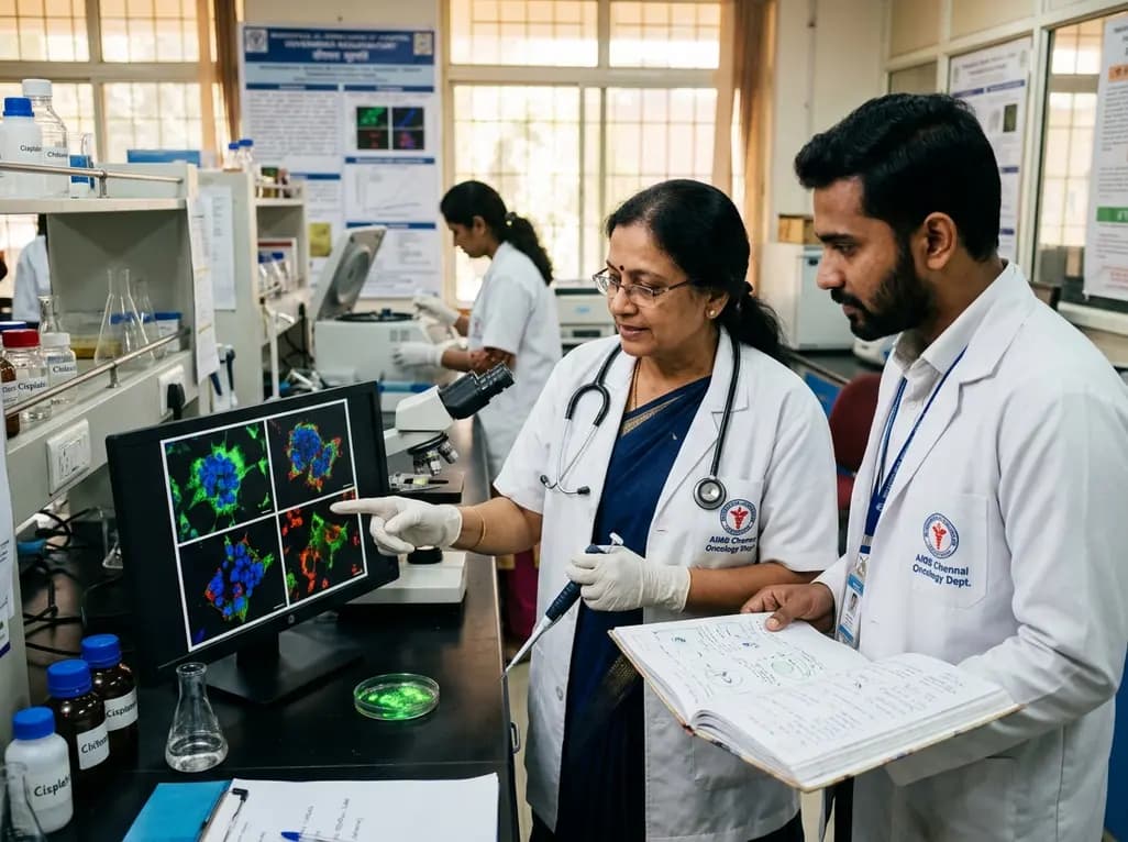
A novel green-synthesized nanocarrier uses ellagic acid to crosslink chitosan for localized cisplatin delivery in cervical cancer. Preclinical results demonstrate extended vaginal retention, potent cytotoxicity, ROS-driven apoptosis, and superior tumor regression compared with traditional crosslinkers.
Today

Managing refractory hypoxemia after cardiothoracic surgery becomes perilous when prolonged air leak limits positive-pressure ventilation. This case-based review details how high-flow nasal cannula, paired with awake rehabilitation, enabled successful extubation and avoided invasive re-intubation.
Today

A longitudinal study reveals that patients undergoing staged bilateral hip arthroscopy experience progressive contralateral labral deterioration during surgical intervals. However, structural degradation does not compromise 2-year postoperative clinical outcomes or patient-reported satisfaction scores.
Today

SWEDEPAD-1 trial insights show paclitaxel-coated devices do not improve long-term limb salvage in patients with chronic limb-threatening ischemia and tissue loss. While one-year reinterventions decreased, the devices were linked to a higher risk of major amputation at three months.
Today
