.webp&w=384&q=75)

Loading, please wait...
Loading, please wait...

Diagnosing inner ear disorders like Ménière's disease requires robust visualization of labyrinthine architecture. Delayed contrast-enhanced magnetic resonance imaging provides direct insight into fluid space distension. Consequently, protocols for endolymphatic hydrops 3D FLAIR imaging have become indispensable tools for modern neuro-otologists and head-and-neck radiologists. However, conventional sequences often demand prolonged acquisition times that expose scans to severe patient motion artifacts. Furthermore, achieving an optimal signal-to-noise ratio in tiny labyrinthine structures remains technically challenging. Recent advancements in artificial intelligence now offer substantial workflow improvements. Deep learning reconstruction algorithms specifically target raw k-space noise without sacrificing subtle anatomic margins. Therefore, assessing how deep learning algorithms perform across varying scan speeds represents a crucial milestone for inner ear diagnostics.
Evaluating hydropic ear disease requires precise discrimination between the endolymphatic and perilymphatic fluid compartments. Following intratympanic or delayed intravenous gadolinium administration, perilymph enhances brightly whereas endolymph shows no enhancement. Consequently, radiologists can delineate abnormal saccular expansion, utricular distension, and cochlear duct displacement. However, capturing these microscopic compartments within the temporal bone demands high spatial resolution. Traditional non-accelerated scans frequently exceed seven minutes per ear or sequence acquisition. As a result, patient fatigue, swallowing, and involuntary head tremors introduce substantial phase-encoding artifacts. Such blurring obscures the fine vestibular membranes and undermines diagnostic confidence. In addition, lengthy scanning protocols decrease patient throughput in busy imaging centers across India. Clinicians therefore need rapid acquisition strategies that preserve contrast boundaries. Deep learning reconstruction addresses these technical bottlenecks by filtering underlying noise directly from raw data. Furthermore, the algorithm preserves sharp transitions between perilymph and the adjacent non-enhancing endolymphatic space. Thus, optimizing these imaging sequences facilitates accurate staging while improving overall patient compliance.
To investigate reconstruction capabilities, researchers designed a prospective trial enrolling fifty-three consecutive patients with suspected hydropic disorders. Each participant underwent specialized inner ear imaging assessing four distinct three-dimensional sequences. Specifically, investigators acquired conventional 3D FLAIR taking seven minutes and fourteen seconds alongside a high-resolution sequence taking six minutes and fifty-four seconds. In addition, the team reconstructed high-resolution data using deep learning reconstruction software. Finally, they evaluated an accelerated deep learning FLAIR sequence requiring only five minutes and thirty-four seconds. Two independent radiologists rigorously scored each dataset for noise levels, edge sharpness, and overall diagnostic quality. Moreover, the investigators used conventional sequences as the established reference standard to evaluate hydrops grading accuracy. Interobserver agreement metrics validated the clinical reliability across both readers. Consequently, this study framework permitted direct side-by-side comparisons of physical signal metrics and qualitative diagnostic efficacy. The team thereby proved how artificial intelligence algorithms process fine structural data within restricted scan times.
The objective findings demonstrated outstanding quantitative improvements across all reconstructed sequences. Most notably, deep learning reconstruction significantly increased signal-to-noise ratio compared to non-DLR acquisitions with high statistical significance. Furthermore, qualitative analysis demonstrated superior edge sharpness and noticeable suppression of high-frequency background noise. Consequently, readers identified fine anatomic borders between endolymphatic and perilymphatic spaces with enhanced clarity. Moreover, the accelerated sequence demonstrated complete diagnostic concordance with conventional reference scans. Both independent radiologists achieved excellent interobserver agreement when grading vestibular and cochlear hydrops. Therefore, shortening the acquisition by nearly twenty-five percent did not compromise disease detection. Nevertheless, subtle differences in texture smoothing require careful scrutiny during initial deployment. Some reconstructed images exhibit minor synthetic smoothing across soft tissue boundaries if denoising strength is excessive. Overall, however, deep learning preservation of contrast boundaries delivers diagnostic reliability that matches or exceeds conventional baseline imaging.
Temporal bone imaging remains exceptionally sensitive to minor patient movement during prolonged magnetic resonance scanning. Because involuntary swallowing and micro-movements generate significant image blur, conventional seven-minute sequences often require repeated acquisitions. Consequently, patient restlessness leads to wasted scanner slots and diagnostic ambiguity. In contrast, reducing scan duration to five and a half minutes substantially diminishes the likelihood of motion-induced degradation. Furthermore, deep learning filters suppress ghosting artifacts caused by slight phase instabilities. This acceleration directly enhances operational throughput for diagnostic facilities handling heavy clinical caseloads. Moreover, geriatric individuals and patients experiencing acute vertigo tolerate shorter examination periods far better. As a result, departmental cancellation rates drop while patient comfort increases noticeably. Clinicians also benefit from consistent image quality that facilitates longitudinal monitoring of therapy. Therefore, accelerated deep learning sequences transform inner ear MRI from an exhausting ordeal into an efficient, routine clinical investigation.
Implementing deep learning reconstructions in routine neuro-otology practice offers substantial advantages for multidisciplinary patient management. Otolaryngologists, neurologists, and head-and-neck radiologists can collaborate more effectively when inner ear hydrops visualizations remain crisp and reproducible. In addition, rapid scanning protocols simplify the coordination of delayed post-contrast imaging sessions. For instance, diagnostic centers can comfortably schedule patients four hours after intravenous gadolinium without extending scanner table times excessively. Furthermore, standardizing automated deep learning post-processing reduces inter-operator variability across different MRI suites. However, clinicians must establish careful validation protocols before completely replacing standard sequences. Radiologists should initially review parallel reconstructions to confirm that algorithmic denoising does not conceal subtle blood-labyrinth barrier breakdowns. Similarly, institutional protocols must calibrate spatial matrix settings to maintain accurate volumetric hydrops measurements. Ultimately, embracing deep learning reconstruction allows diagnostic centers to optimize resource utilization while delivering superior anatomical detail for complex vertigo evaluations.
Delayed contrast-enhanced 3D FLAIR MRI directly visualizes endolymphatic hydrops by demonstrating non-enhancing dilated endolymphatic compartments against brightly enhancing perilymphatic fluid. This objective imaging technique confirms Ménière's disease pathology in patients presenting with fluctuating hearing loss, tinnitus, and episodic vertigo. Consequently, clinicians can accurately differentiate hydropic ear disease from other vestibular conditions, guide targeted intratympanic therapy, and monitor disease progression over time with high diagnostic confidence.
Deep learning reconstruction utilizes neural network architectures trained on high-quality datasets to identify and remove noise from raw k-space data. Consequently, this technology significantly boosts signal-to-noise ratios while preserving crisp anatomical boundaries within delicate inner ear structures. Furthermore, it enables substantial scan time acceleration without sacrificing diagnostic resolution, thereby decreasing patient motion artifacts and improving clinical feasibility for individuals suffering from severe vertigo or claustrophobia.
Yes, recent clinical studies indicate that accelerated deep learning 3D FLAIR sequences can safely replace traditional longer acquisitions. Specifically, accelerated protocols achieve nearly identical diagnostic accuracy and excellent interobserver agreement for grading vestibular and cochlear hydrops. Moreover, by reducing table time by approximately twenty-five percent, these sequences substantially limit involuntary motion artifacts while delivering sharper margins and superior overall diagnostic efficiency in everyday clinical practice.
Disclaimer: This content is for informational and educational purposes only. Refer to the latest local and national guidelines for clinical practice.
References

Read summarized clinical updates, watch expert medical content, and earn CME certifications right from your smartphone.
Open your smartphone camera to scan and install.





Deep learning reconstruction significantly optimizes 3D FLAIR MRI for endolymphatic hydrops evaluation. The technology enhances signal-to-noise ratio, sharpens anatomical edges, and reduces acquisition time by nearly 25%, offering reliable hydrops grading while minimizing patient motion artifacts.
Today

The Food Safety and Standards Authority of India has issued draft regulations prohibiting the sale and manufacture of non-dairy imitation substitutes as paneer. This nationwide regulatory action aims to eliminate consumer deception, protect dietary protein quality, and curtail lipid-related metabolic risks.
Today
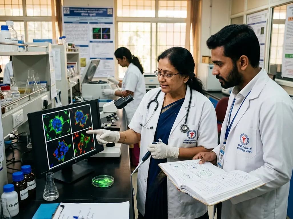
A novel green-synthesized nanocarrier uses ellagic acid to crosslink chitosan for localized cisplatin delivery in cervical cancer. Preclinical results demonstrate extended vaginal retention, potent cytotoxicity, ROS-driven apoptosis, and superior tumor regression compared with traditional crosslinkers.
Today

Managing refractory hypoxemia after cardiothoracic surgery becomes perilous when prolonged air leak limits positive-pressure ventilation. This case-based review details how high-flow nasal cannula, paired with awake rehabilitation, enabled successful extubation and avoided invasive re-intubation.
Today

A longitudinal study reveals that patients undergoing staged bilateral hip arthroscopy experience progressive contralateral labral deterioration during surgical intervals. However, structural degradation does not compromise 2-year postoperative clinical outcomes or patient-reported satisfaction scores.
Today

SWEDEPAD-1 trial insights show paclitaxel-coated devices do not improve long-term limb salvage in patients with chronic limb-threatening ischemia and tissue loss. While one-year reinterventions decreased, the devices were linked to a higher risk of major amputation at three months.
Today
