

Loading, please wait...
Loading, please wait...

Endovascular therapy has revolutionized the management of cerebrovascular lesions, yet microscopic assessment of vascular healing remains difficult. Implementing optical coherence tomography aneurysm evaluation offers unprecedented micron-scale resolution inside intracranial vessels. While digital subtraction angiography provides excellent macroscopic visualization of vessel architecture, it cannot depict real-time stent strut apposition, localized thrombus formation, or fine neointimal coverage. Consequently, interventionalists often face challenges in predicting delayed ischemic complications or incomplete neck occlusion. High-resolution intravascular imaging bridges this critical diagnostic gap by providing cross-sectional anatomical detail directly at the vessel wall interface.
Historically, neurovascular interventionists have relied on conventional angiography and flat-panel computed tomography to evaluate device deployment. However, these traditional imaging modalities lack the spatial resolution required to identify microscopic malapposition or delicate platelet aggregation. Because incomplete device apposition strongly correlates with delayed aneurysm occlusion and thromboembolic events, high-precision imaging remains essential. Optical coherence tomography delivers spatial resolution between 10 and 15 micrometers, which far surpasses intravascular ultrasound. Therefore, this near-infrared light technology allows clinicians to inspect the lumen-device boundary with remarkable fidelity.
Furthermore, navigating coronary imaging catheters into tortuous intracranial anatomy previously presented significant procedural challenges. Recent technical refinements now enable safer navigation across the internal carotid artery and vertebrobasilar systems. As interventionalists gain experience with these tools, they can systematically inspect stent-assisted coiling and flow diversion procedures. Thus, endovascular operators can detect subtle structural abnormalities that conventional angiography regularly misses.
In a landmark clinical study, researchers evaluated the intravascular characteristics of flow diverter devices and neurovascular stents in female patients undergoing endovascular aneurysm reconstruction. The investigators performed nine optical coherence tomography procedures across five complex aneurysms in both anterior and posterior circulations. Notably, all patients received standard dual antiplatelet therapy consisting of aspirin and clopidogrel to prevent acute construct thrombosis.
The procedural findings demonstrated complete technical success and perfect device wall apposition in every patient. Importantly, the real-time optical coherence tomography aneurysm imaging confirmed that no patients developed acute intrastent thrombosis. Instead, the imaging revealed intrasaccular thrombosis alongside localized fibrin meshes forming along the outer margins of the constructs. Consequently, all treated aneurysms achieved successful neck occlusion during serial follow-up. In addition, every vital side branch originating adjacent to the target reconstruction maintained robust patency, confirming therapeutic efficacy without compromised perfusion.
Biological healing after flow diversion depends entirely on progressive neoendothelialization across the parent vessel defect. Optical coherence tomography uniquely enables longitudinal monitoring of this vital reparative cascade. By reflecting infrared light off native tissue, the imaging displays progressive neointimal growth spanning across the implanted metallic struts. Therefore, clinicians can clearly differentiate between incomplete healing, healthy tissue incorporation, and excessive neointimal hyperplasia.
Moreover, tracking this biological integration helps specialists assess long-term arterial remodeling with objective measurements. When endothelial cells fail to cover exposed metal struts, patients face a prolonged risk of late in-stent thrombosis. Conversely, excessive tissue proliferation can induce in-stent restenosis and downstream cerebral ischemia. Through serial intraluminal evaluations, practitioners can accurately establish the precise timeline of vessel endothelization. Consequently, these direct observations guide personalized pharmacotherapy and optimize long-term clinical surveillance.
Optimizing dual antiplatelet therapy remains one of the most controversial aspects of modern neurointerventional practice. Clinicians often struggle to determine the ideal duration of antiplatelet regimens following flow diversion. If an operator stops platelet inhibition prematurely, naked metal struts can trigger catastrophic thromboembolism. However, maintaining aggressive antiplatelet therapy indefinitely exposes vulnerable patients to systemic and intracranial bleeding risks.
Because optical coherence tomography directly visualizes strut coverage, it provides objective criteria for titrating antiplatelet agents. For instance, detecting complete neoendothelialization might allow safe de-escalation from dual therapy to monotherapy. Conversely, identifying persistent strut malapposition or lingering thrombus suggests that the patient requires intensified platelet inhibition. Furthermore, identifying mechanical malapposition intraoperatively allows immediate corrective action, such as post-dilation balloon angioplasty. Therefore, optical coherence tomography functions as an invaluable decision-making instrument that reduces avoidable adverse events.
Although current results are highly promising, broader clinical adoption requires dedicated neurovascular optical coherence tomography hardware. Most current procedures employ modified coronary imaging catheters, which feature stiffer delivery shafts and require rapid blood clearance via contrast flush. In tortuous intracranial anatomies, these coronary designs can increase vasospasm risks or procedural friction. Therefore, medical engineers must prioritize ultra-flexible microcatheters tailored specifically for delicate cerebral vasculature.
Additionally, automated artificial intelligence algorithms will soon assist operators by instantly calculating malapposition volumes and endothelial coverage percentages. Such objective computational metrics will eliminate inter-observer variability during high-stakes interventions. As ongoing clinical trials validate safety and diagnostic yield across larger cohorts, intravascular imaging may become the standard of care for complex aneurysm repair. Ultimately, precision intravascular imaging will empower clinicians to deliver safer, customized endovascular care for every patient.
Optical coherence tomography is an intravascular imaging technique utilizing near-infrared light. It provides microscopic, cross-sectional views of cerebral blood vessels at ten-micrometer resolution. Consequently, neurointerventionalists use it to assess stent apposition, detect thrombus formation, and monitor endothelial healing following endovascular aneurysm repair.
Flow diverters require complete wall contact to promote parent vessel healing and prevent ischemic complications. Intraluminal imaging accurately detects microscopic strut malapposition and platelet aggregation that angiography misses. Thus, clinicians can immediately perform balloon angioplasty or adjust antiplatelet therapy to prevent ischemic strokes.
Yes, high-resolution optical coherence tomography clearly visualizes covered arterial branch ostia. The imaging confirms whether collateral perforators and side branches remain widely patent after stent deployment. Therefore, operators can confidently treat complex bifurcations and wide-neck aneurysms without risking downstream territory infarction.
Disclaimer: This content is for informational and educational purposes only and does not constitute medical advice, diagnosis, or treatment. Healthcare professionals should exercise their independent clinical judgment. Refer to the latest local and national guidelines for clinical practice.
References
Gutiérrez-Baños JJ et al. Early experience using optical coherence tomography for intraluminal evaluation of vascular healing associated with endovascular treatment of intracranial aneurysms. J Neurosurg. 2025 Sep 01. doi: 10.3171/2025.1.JNS242458. PMID: 40344764.
Li L, Gong P, Li M, Song Y, Fan C, Yuan F, et al. Evaluation of the apposition in unruptured aneurysms treated with flow diverters by optical coherence tomography: Preliminary clinical experience. Front Neurol. 2022;13:1015638.
Gounis MJ, Ughi GJ, Marosfoi M, et al. Intravascular optical coherence tomography for neurointerventional surgery. Stroke. 2019;50(1):218-223.

Read summarized clinical updates, watch expert medical content, and earn CME certifications right from your smartphone.
Open your smartphone camera to scan and install.





Optical coherence tomography offers unprecedented microscopic resolution to assess stent apposition, neoendothelialization, and thrombus formation during endovascular treatment of intracranial aneurysms, providing vital insights for neurointerventionalists.
Today

The DOMINO study protocol presents a multidisciplinary implementation mapping approach to overcome provider, patient, and payer barriers to antiobesity medications, offering vital insights for Indian clinicians managing the growing diabesity burden.
Today

An intracranial EWSR1::CREM fusion sarcoma in a 31-year-old female manifested as a paraneoplastic inflammatory syndrome with anemia and secondary infertility. The dural mass mimicked meningioma, but gross-total resection resolved her systemic inflammation and restored natural fertility within one month.
Today

A real-world cohort study of 6,537 patients from the FOUNTAIN platform demonstrates that initiating finerenone significantly reduces albuminuria by 40.7% with minimal eGFR decline and manageable hyperkalemia risk, confirming its practical effectiveness and safety in diabetic kidney disease.
Yesterday

A comprehensive analysis of MAP therapy resistance in osteosarcoma, examining canonical and non-canonical mechanisms across damage avoidance, enhanced DNA repair, and replication stress tolerance to identify novel vulnerabilities.
Today
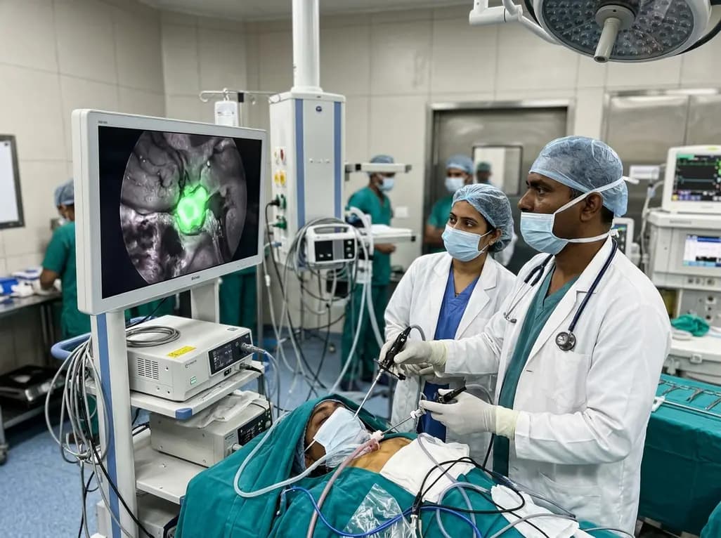
Near-infrared autofluorescence provides real-time label-free optical guidance during endoscopic transsphenoidal pituitary surgery. By highlighting stark signal contrasts between normal pituitary gland and adenomas, this technique aids gross-total tumor resection while safeguarding critical endocrine function.
Today
