.webp&w=384&q=75)

Loading, please wait...
Loading, please wait...

Vitreoretinal surgeons frequently encounter idiopathic epiretinal membranes in routine ophthalmic practice. Today, optical coherence tomography angiography provides remarkable noninvasive views of retinal microvasculature before and after epiretinal membrane surgery. However, interpreting postoperative changes in flow parameters has sparked continuous clinical debate. When quantitative scans show reduced central perfusion density, clinicians often worry about capillary loss or surgical trauma. A recent landmark study illuminates this paradox. The authors demonstrate that postoperative vascular alterations represent topographic displacement rather than genuine vascular closure. Consequently, vitreoretinal specialists can interpret postoperative scans with greater confidence and diagnostic precision.
Idiopathic epiretinal membrane formation exerts progressive tangential and centripetal traction on the underlying retina. As a result, the macula thickens, the foveal pit flattens, and surrounding retinal vessels migrate toward the foveal center. This pathological centripetal displacement crowds the macular capillaries into a smaller surface area. Consequently, baseline quantitative measurements often display artificially elevated central vessel density and an abnormally constricted foveal avascular zone.
Surgical intervention aims to relieve this mechanical traction completely. During vitrectomy, surgeons carefully peel the fibrocellular membrane and internal limiting membrane. Therefore, removing this physical constraint allows the distorted neurosensory retina to relax and restore its anatomical configuration. Following surgical release, the displaced retinal architecture gradually moves outward toward its native peripheral position. However, clinicians often misunderstand the resulting quantitative optical coherence tomography angiography parameters. When post-peeling scans display decreased foveal perfusion density, observers mistakenly suspect capillary nonperfusion. In reality, the surgical procedure relieves mechanical compaction. Thus, the apparent reduction in central microvascular metrics simply reflects physiological vessel redistribution across the re-expanded macula.
To elucidate these architectural shifts, researchers evaluated thirty-seven patients with unilateral idiopathic epiretinal membrane who underwent surgical repair. The investigative team followed each patient for at least six months postoperatively to capture steady-state vascular changes. They utilized a wide six-by-six millimeter volume scan on swept-source optical coherence tomography angiography. This advanced imaging modality provides enhanced visualization through edematous tissue. Furthermore, it reliably segments both the superficial capillary plexus and the deep capillary plexus.
The investigators assessed changes in the foveal avascular zone area and perfusion density within one-millimeter and three-millimeter concentric rings. Notably, both capillary networks demonstrated significant postoperative modifications. Within the innermost one-millimeter circle, perfusion density decreased significantly in both the superficial and deep vascular layers. Simultaneously, the foveal avascular zone expanded significantly after surgery. However, deeper analysis showed that these alterations were not uniform across wider retinal zones. Outside the foveal epicenter, the three-millimeter ring exhibited distinct regional redistributions. Therefore, the microvascular alterations mirrored structural relaxation rather than progressive microangiopathy or tissue death.
Differentiating true capillary dropout from geometric tissue shift is essential for clinical decision-making. In vascular occlusive diseases or diabetic retinopathy, capillary dropouts cause permanent nonperfusion, tissue hypoxia, and visual loss. In contrast, the microvascular changes observed after membrane peeling tell an entirely different physiological story. Specifically, the mechanical release allows capillaries that were bunched centrally to slide outward.
To verify this mechanism, the study evaluated the precise distance between the fovea and the major vascular arcades. Postoperative measurements confirmed a significant elongation of this fovea-to-arcade anatomical interval. Furthermore, the degree of arcade displacement correlated directly with the expansion of the foveal avascular zone. As the retinal tissue returns toward its native position, the microvasculature disperses centrifugally. Consequently, the concentration of blood vessels per square millimeter decreases centrally without any actual vascular destruction. Therefore, retinal blood flow remains robust despite lower numerical density values. Clinicians can reassure patients that these image metrics indicate successful mechanical restoration rather than iatrogenic ischemic damage.
The foveal avascular zone serves as a vital structural biomarker in vitreoretinal disease. In eyes with epiretinal membranes, centripetal contracture shrinks the foveal avascular zone into a tiny, irregular shape. Following successful peeling, the foveal avascular zone predictably enlarges toward normal anatomical dimensions. Notably, this study proved that postoperative enlargement correlates with the widening distance between the fovea and the retinal vascular arcades.
Moreover, functional visual outcomes align with this mechanical realignment. Patients in the cohort achieved meaningful improvements in best-corrected visual acuity over the six-month follow-up window. If reduced perfusion density represented true capillary occlusion, visual acuity would deteriorate or plateau prematurely. Instead, vision improved while central vessel density decreased. This inverse relationship confirms that expanding avascular zones and lower central densities reflect normalized retinal geometry. In addition, relieving macular contracture significantly reduces metamorphopsia and foveal distortion. Therefore, surgeons must interpret enlarging avascular contours as signs of anatomical recovery rather than capillary loss.
These optical coherence tomography angiography insights have direct practical applications for comprehensive ophthalmologists and vitreoretinal surgeons. First, clinicians should avoid misinterpreting reduced central perfusion metrics as microvascular ischemia or surgical phototoxicity. Understanding the distinction between topographic remodeling and capillary loss prevents unnecessary invasive testing, unwarranted medical therapies, and undue patient anxiety.
Second, surgical planning and postoperative counseling benefit tremendously from this biomechanical framework. When monitoring patients after surgery, clinicians should integrate en face vascular displacement data with traditional cross-sectional b-scans. Furthermore, evaluating fovea-to-arcade distances provides an objective benchmark for tissue relaxation. If a patient displays delayed visual recovery despite good membrane peeling, surgeons can assess whether persistent traction or incomplete centrifugal realignment exists. In addition, routine use of wide-field swept-source angiography enables precise tracking of perifoveal capillary return. Consequently, retina specialists can provide accurate prognostic counseling, reassuring patients that anatomical remodeling often continues for months after vitrectomy.
Perfusion density decreases centrally after surgery because releasing membrane traction allows compressed retinal tissue to relax centrifugally. Preoperatively, pathological traction pulls capillaries toward the foveal center, causing artificial vascular crowding. Once surgeons peel the membrane, vessels shift outward toward their natural anatomical locations. Consequently, the concentration of capillaries within the central millimeter declines. This numerical drop represents structural redistribution across an expanding retinal surface rather than ischemic capillary loss.
No, foveal avascular zone enlargement does not indicate ischemic damage in this setting. During active epiretinal membrane contraction, tangential forces squeeze the avascular zone into an unnaturally small, distorted boundary. Following vitrectomy and membrane peeling, the inward traction disappears, allowing the margins of the foveal pit to expand naturally. Therefore, avascular zone enlargement reflects anatomical decompression and restoration of normal foveal contour rather than progressive capillary dropout or pathological ischemia.
Clinicians should monitor patients using swept-source optical coherence tomography angiography combined with high-resolution cross-sectional b-scans. Rather than relying solely on automated vessel density numbers, ophthalmologists must assess qualitative capillary architecture and track the distance between the fovea and vascular arcades. Furthermore, clinicians should correlate vascular shifts with visual acuity recovery and metamorphopsia reduction. This comprehensive multimodal approach ensures accurate evaluation of anatomical healing without confusing physiological displacement with retinal disease.
Disclaimer: This content is for informational and educational purposes only... Refer to the latest local and national guidelines for clinical practice.
References
Park NH et al. Perfusion Density Changes After Epiretinal Membrane Surgery Predominantly Reflect Topographic Redistribution of Macular Vasculature. Retina. 2026 Sep 29. doi: 10.1097/IAE.0000000000005027. PMID: 42808983.
Lee SM, Park SW, Byon I. Topographic changes in macula and its association with visual outcomes in idiopathic epiretinal membrane surgery. PLoS One. 2025;20(1):e0316450.
Govetto A, Lalane CA, Sarraf D, et al. Insights into Epiretinal Membranes: Presence of Ectopic Inner Foveal Layers and a New Optical Coherence Tomography Staging System. Ophthalmology. 2017;124(10):1511-1521.
Mastropasqua R, D'Aloisio R, Di Antonio L, et al. Optical Coherence Tomography Angiography in Epiretinal Membrane Surgery: A Systematic Review. J Clin Med. 2021;10(19):4416.

Read summarized clinical updates, watch expert medical content, and earn CME certifications right from your smartphone.
Open your smartphone camera to scan and install.





A retrospective OCTA study demonstrates that macular perfusion density reductions and foveal avascular zone expansion after epiretinal membrane surgery represent mechanical topographic redistribution of retinal vessels rather than true capillary dropout, offering critical reassurance for vitreoretinal surgeons.
Today
A novel microgel platform presenting FasL and protease-resistant CXCL12 enables sustained allogeneic islet survival and glycemic control without systemic immunosuppression, enhancing local Treg recruitment and graft revascularization.
Today

A recent qualitative study evaluates institutional gaps across health, police, and justice sectors in supporting survivors of intimate partner violence. Healthcare providers must bridge procedural fragmentation with empathetic, trauma-informed care and cross-sector coordination to safeguard vulnerable patients.
Today
A study evaluates axillary arteriovenous fistula creation for resolving pulmonary arteriovenous malformations after bidirectional cavopulmonary connection. The technique achieved 75% clinical improvement and successfully bridged patients to total cavopulmonary connection.
Today
A cross-sectional study reveals a strong positive correlation between problematic internet use and kinesiophobia in adults aged 18-45 with non-specific low back pain, highlighting how digital overuse reinforces movement fear and complicates musculoskeletal recovery.
Today
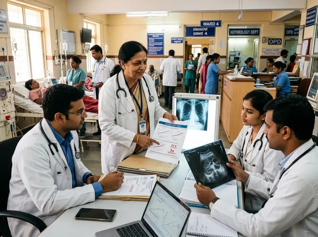
Recent data from Karnataka reveals a threefold surge in genitourinary and renal deaths over ten years. Driven by uncontrolled diabetes, hypertension, and emerging lifestyle hazards, this epidemiological shift highlights an urgent need for proactive screening and multidisciplinary nephrology management in primary care.
Today
