.webp&w=384&q=75)

Loading, please wait...
Loading, please wait...
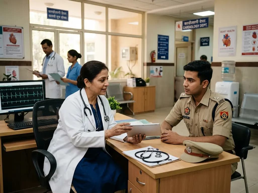
First responders regularly face potentially psychologically traumatic events during their daily duties. These critical incidents include direct or indirect exposure to severe injury, sudden violence, or life-threatening situations. Consequently, high-risk personnel experience an elevated risk of developing posttraumatic stress injuries. While mental health disorders represent a recognized consequence of operational trauma, cardiovascular health also deteriorates under chronic psychological strain. Recent medical literature indicates that psychological trauma accelerates subclinical cardiovascular disease through systemic pathways. Diastolic cardiac function appears particularly sensitive to chronic sympathetic hyperactivity, persistent inflammation, and altered autonomic tone. Therefore, clinicians must recognize that psychological trauma influences cardiovascular outcomes alongside traditional cardiac risk factors. Early detection of microvascular and ventricular changes remains essential to prevent long-term adverse events. Consequently, occupational health programs must expand beyond psychological support to safeguard cardiovascular wellness effectively.
Longitudinal data from police cohorts highlight alarming trends regarding early cardiovascular decline. Specifically, significant reductions in diastolic cardiac function emerge as early as the second year of active field service. Concurrently or shortly thereafter, measurable changes in systolic parameters develop, demonstrating progressive myocardial strain. This rapid onset suggests that subclinical ventricular impairment may directly constitute a physical manifestation of posttraumatic stress injuries. Traditionally, clinicians associated occupational heart disease with years of chronic exposure or advanced age. However, young officers with a mean age around thirty years display quantifiable cardiac changes. Early diastolic impairment reflects decreased ventricular compliance and impaired relaxation during the cardiac cycle. Consequently, filling pressures rise, predisposes individuals to future heart failure. Regular monitoring during initial career years can identify early physiological shifts before structural remodeling becomes permanent. Thus, healthcare providers must evaluate early-career personnel for subtle physiological impairments.
The biological pathways connecting traumatic stress to cardiac dysfunction involve complex neuroendocrine and vascular responses. First, exposure to traumatic incidents triggers sustained activation of the hypothalamic-pituitary-adrenal axis and sympathetic nervous system. As a result, elevated circulating catecholamines increase resting heart rate and arterial stiffness. Prolonged sympathetic surge leads to endothelial dysfunction, impaired coronary flow reserve, and persistent microvascular constriction. In addition, chronic stress elevates pro-inflammatory cytokines, including interleukin-6 and tumor necrosis factor-alpha, which promote myocardial fibrosis. Fibrotic changes directly impair myocardial relaxation, leading to measurable deficits in diastolic performance. Furthermore, altered autonomic regulation reduces heart rate variability, signaling impaired vagal withdrawal. Over time, these combined physiological stressors compromise left ventricular filling kinetics and increase myocardial workload. Therefore, clinicians should recognize posttraumatic stress injury as a systemic disorder with profound cardiovascular ramifications.
Detecting subtle cardiac changes requires reliable, non-invasive diagnostic tools suitable for routine longitudinal screening. Seismocardiography has emerged as a practical technology that measures chest wall vibrations produced by cardiac mechanical activity. Specifically, this modality records micro-vibrations corresponding to valve openings, valve closures, and ventricular contraction phases. Unlike standard electrocardiography, which records electrical activity, seismocardiography assesses mechanical performance and myocardial timing intervals. For instance, changes in isovolumic contraction and relaxation times provide precise insights into subtle systolic and diastolic shifts. Moreover, seismocardiography sensors are portable, user-friendly, and cost-effective, making them ideal for field-based occupational health assessments. Serial seismocardiographic evaluations allow clinicians to track individual baseline trends over time. Consequently, practitioners can identify early mechanical deterioration before significant clinical symptoms appear. Integrating this technology into routine health examinations provides a scalable solution for high-stress organizations.
Mitigating cardiovascular risk in traumatized personnel requires targeted lifestyle and exercise interventions. Standard physical conditioning may not adequately address stress-induced diastolic dysfunction or autonomic imbalance. Therefore, occupational health programs should incorporate best-practice cardiac rehabilitation exercise prescription guidelines. Structured aerobic exercise tailored to enhance myocardial relaxation improves ventricular compliance and microvascular perfusion. Additionally, exercise training modulates autonomic tone by enhancing vagal activity and reducing excessive sympathetic outflow. Consequently, structured physical activity restores heart rate variability and lowers resting arterial pressure. Furthermore, combining moderate-intensity aerobic conditioning with resistance training promotes favorable metabolic adaptations and reduces systemic inflammation. Clinicians should ensure exercise protocols are individualized, taking into account baseline fitness and trauma severity. Regular exercise participation also reduces anxiety and posttraumatic stress symptoms, producing synergistic psychological and cardiovascular benefits.
Healthcare providers and occupational medical teams must adopt comprehensive strategies to manage trauma-exposed populations. First, baseline cardiovascular risk assessments should occur during initial recruitment or training phases. Incorporating non-invasive diagnostic tools, such as seismocardiography, provides valuable reference parameters for long-term comparison. Second, periodic health evaluations must routinely screen for both psychological stress and early subclinical cardiac changes. When early diastolic or systolic declines are detected, clinicians should initiate prompt multidisciplinary care. This collaborative approach should involve cardiologists, mental health specialists, and exercise physiologists. In addition, organizational policies must encourage early reporting of traumatic exposure without fear of occupational stigma. Providing structured wellness programs, resilience training, and access to exercise facilities significantly mitigates long-term health risks. Consequently, proactive health management preserves operational readiness while reducing major adverse cardiac events.
Psychological trauma activates the sympathetic nervous system and hypothalamic-pituitary-adrenal axis, triggering a sustained release of stress hormones. Consequently, chronic elevation of cortisol and catecholamines induces systemic inflammation, arterial stiffness, and endothelial dysfunction. Over time, these biochemical changes impair myocardial relaxation during diastole and reduce coronary perfusion. As a result, affected individuals experience early deterioration in diastolic cardiac function and progressive systolic changes, even in young, physically active populations.
Seismocardiography is a non-invasive diagnostic technique that records chest wall micro-vibrations generated by cardiac mechanical activity. Unlike standard electrocardiography, which measures electrical signals, seismocardiography evaluates mechanical events, including valve movements and myocardial contraction phases. By measuring specific timing intervals such as isovolumic relaxation time, this portable technology detects subtle diastolic and systolic changes early. Consequently, it offers a practical, reliable screening tool for longitudinal health monitoring in high-stress occupational environments.
Yes, structured physical exercise plays a critical role in restoring cardiac function and autonomic balance. Tailored aerobic conditioning improves ventricular compliance, enhances endothelial function, and increases coronary blood flow reserve. Furthermore, regular exercise training increases parasympathetic vagal tone while suppressing excessive sympathetic activity. When integrated into occupational health programs alongside cardiac rehabilitation guidelines, structured exercise effectively counteracts trauma-induced diastolic deterioration and reduces the overall risk of major adverse cardiovascular events.
Disclaimer: This content is for informational and educational purposes only and does not constitute medical advice, diagnosis, or treatment. Healthcare professionals should rely on their clinical judgment and individualized patient evaluations. Refer to the latest local and national guidelines for clinical practice.
References

Read summarized clinical updates, watch expert medical content, and earn CME certifications right from your smartphone.
Open your smartphone camera to scan and install.





A longitudinal study reveals early deterioration of diastolic cardiac function in trauma-exposed police officers within three years of service. Non-invasive seismocardiography and targeted cardiac exercise offer crucial early detection and preventive interventions.
last month

A comparative analysis shows that older age, elevated blood glucose, and abdominal adiposity are primary drivers of hypertension. Comprehensive evaluation of cardiometabolic risk profiles enables clinicians to detect overlapping metabolic dysfunctions early and optimize targeted prevention strategies.
Today

A premature neonate developed upper limb compartment syndrome after uterine rupture extruded the arm through a scar defect. Conservative management with continuous monitoring yielded complete functional recovery and normal limb growth at 10-year follow-up, highlighting non-operative safety in selected cases.
Today
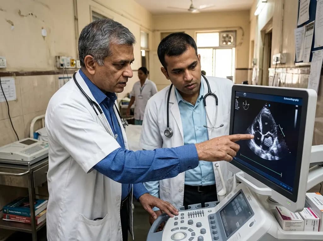
Left ventricular thrombus is a severe complication of acute decompensated heart failure. This clinical review explores the pathophysiology, imaging challenges, and pharmacotherapeutic considerations of laminated thrombi, evaluating the evolving role of direct oral anticoagulants versus traditional warfarin.
Today
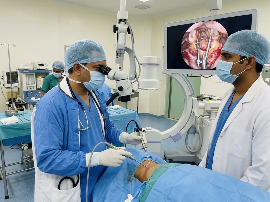
Endoscopic posterior cervical fusion combines minimally invasive decompression, joint preparation, and rigid screw-rod fixation for atlantoaxial pathologies. Early clinical findings demonstrate solid bony union, excellent symptom relief, and minimal soft-tissue morbidity without significant vascular compromise.
Yesterday

Atherosclerosis involves extensive glycometabolic reprogramming across immune and vascular cells. This review examines how glycolysis, the pentose phosphate pathway, and lactate-driven epigenetic shifts fuel plaque vulnerability, while highlighting novel therapeutic targets like PFKFB3 and LDHA.
Today
