.webp&w=384&q=75)

Loading, please wait...
Loading, please wait...
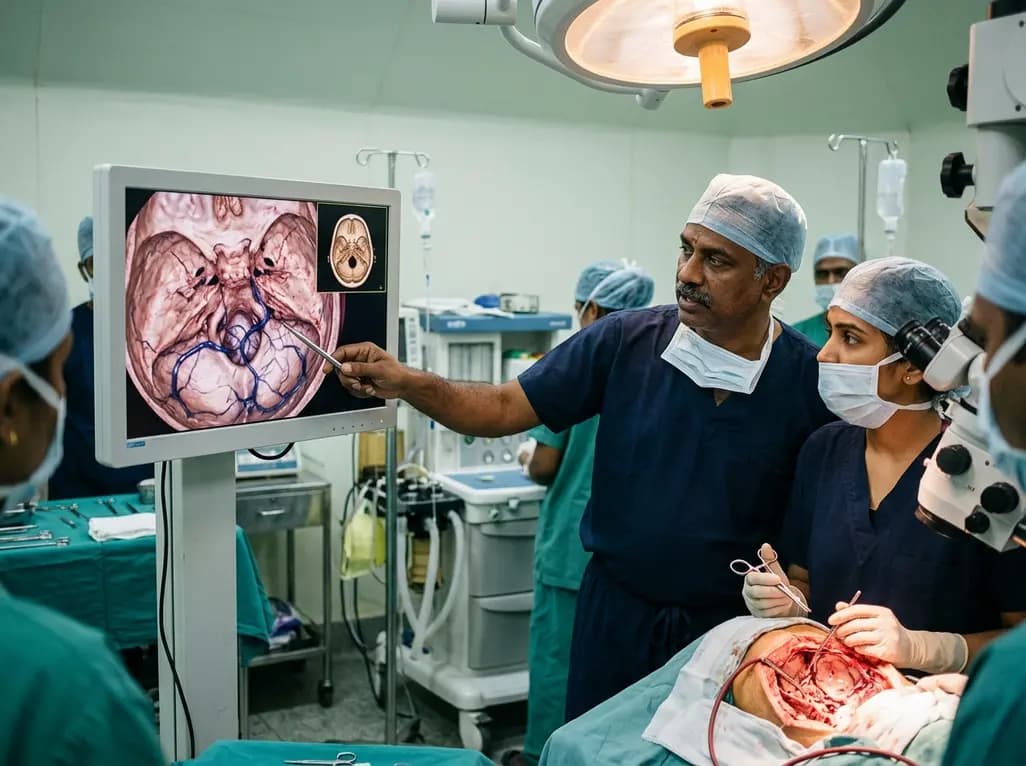
The occipital emissary vein represents a clinically critical yet frequently underappreciated venous channel traversing the posterior cranial vault. Skull base surgeons regularly operate in the suboccipital and retrosigmoid regions where venous anatomy dictates operative safety. Although mastoid emissary veins receive significant attention in neurosurgical literature, occipital emissary channels present complex anatomical variability. Consequently, unexpected injury to these valveless transosseous vessels can cause brisk intraoperative hemorrhage, bone bleeding, and dangerous air embolism. A rigorous anatomical investigation examining 250 adult dry skulls and 14 cadaveric specimens provides essential morphometric data to guide skull base procedures. This detailed analysis clarifies the true prevalence of external and internal occipital foramina while detailing their intradiploic pathways. Furthermore, these findings emphasize that surgeons cannot rely on superficial bony landmarks alone to predict deep venous anatomy. Therefore, mastering the nuances of this emissary system allows operative teams to anticipate venous variations, optimize craniotomy design, and avoid catastrophic dural complications.
Anatomical assessment reveals substantial variability in the presence and configuration of occipital foramina. In the landmark investigation of 250 adult human skulls, approximately 16% completely lacked any occipital foramina. External foramina were absent in 34% of skulls, whereas internal foramina were absent in 22% of the examined specimens. Bilateral external foramina occurred in only 8% of specimens, illustrating that unilateral presentations predominate. When present, external foramina typically clustered within 1 to 2 cm lateral to the midline. In contrast, internal foramina consistently aggregated near the internal occipital protuberance and the confluence of sinuses. Complete osseous conduits connecting an external opening to an internal opening occurred in only 10% on the left side and 8% on the right side. These patent canals demonstrated an oblique, descending trajectory from intracranial confluence to extracranial musculature. Moreover, isolated foramina frequently ended as blind bony pits within the diploic space rather than traversing both cortical tables. Thus, surgeons must recognize that a palpable or visible external foramen rarely indicates a straight, patent transosseous channel.
Dissection of latex-injected adult cadaveric specimens demonstrates that venous networks within the occipital bone exhibit complex compartmentalization. Researchers identified macroscopic internal or external emissary veins in 50% of latex-injected specimens. Additionally, two of these specimens exhibited a small companion artery traveling alongside the venous channel within the osseous canal. Histological analysis of bone segments confirmed that these vessels traverse distinct microvascular diploic channels rather than simple fibrous tracks. Cannulation and blue latex injection of dry skulls showed extensive intradiploic spread without direct, unobstructed through-passage between external and internal surfaces. Therefore, the occipital squama houses distinct internal and external diploic communication systems that interact through an intradiploic trabecular meshwork. This intricate arrangement explains why localized osteotomies often provoke generalized diploic oozing even when a continuous canal appears absent. Furthermore, these venous pathways communicate freely with the suboccipital venous plexus and the dural venous sinuses. Understanding this intradiploic architecture helps surgical teams appreciate how retrograde flow develops during acute elevations of intracranial or systemic venous pressure.
Posterior fossa surgery requires meticulous planning to avoid unpredicted venous bleeding during bone removal. The suboccipital and retrosigmoid approaches bring the surgeon into direct proximity with the transverse-sigmoid junction and torcular region. Because external foramina do not reliably align with internal dural entry points, external landmarks fail to predict deep venous danger zones. Consequently, raising a suboccipital bone flap can unexpectedly tear a large occipital emissary vein anchored firmly to the underlying dural sinuses. Internal emissary veins frequently exist without any corresponding external opening, remaining entirely hidden during subperiosteal soft tissue reflection. As a result, sudden tearing of these tethered channels can occur during mechanical dural elevation from the inner table. Such vascular avulsions provoke vigorous bleeding that obscures the operative field and increases the risk of paradoxically fatal air embolism, especially in sitting positions. Additionally, bone edges may bleed profusely from unroofed diploic channels. Surgeons must therefore maintain immediate access to bone wax, hemostatic matrix, and bipolar coagulation whenever dissecting near the internal occipital protuberance.
Preoperative neuroimaging plays an indispensable role in identifying high-risk venous variations before suboccipital craniotomy. High-resolution computed tomography of the temporal and occipital bones effectively delineates osseous foramina, canal trajectories, and intradiploic channels. Similarly, magnetic resonance venography and CT angiography visualize the caliber and drainage patterns of posterior cranial fossa emissary channels. Radiologists and neurosurgeons must actively evaluate these scans for prominent emissary veins, which often dilate under pathological conditions. For instance, chronic dural sinus thrombosis, intracranial hypertension, and skull base vascular malformations frequently enlarge collateral emissary pathways. In these circumstances, the occipital emissary vein serves as a vital outflow conduit that maintains venous drainage from the posterior fossa. Inadvertent surgical ligation or occlusion of an enlarged collateral emissary vein may precipitate venous infarction, cerebellar edema, or elevated intracranial pressure. Therefore, surgical teams should systematically incorporate three-dimensional vascular reconstructions into their preoperative workflow. Routine review of bone windows and venous phase reconstructions prevents intraoperative surprises and guides individualized placement of cranial burr holes.
Achieving rapid, definitive hemostasis when encountering emissary vein bleeding requires structured technical maneuvers. When an emissary vein tears at the osseous margin, immediate application of sterile bone wax directly into the bony canal halts bleeding effectively. Simultaneously, applying gentle pressure with cottonoids and oxidized regenerated cellulose controls venous oozing from the adjacent dural surface. If the tear occurs at the dural sinus junction, primary suture repair with fine monofilament or hemoclip placement may become necessary. Furthermore, maintaining strict patient hydration and positive end-expiratory pressure reduces the pressure gradient that favors venous air entry during posterior fossa procedures. Surgeons should avoid placing the primary burr hole directly over the inion or midline occipital crest where internal foramina cluster. Instead, initiating craniotomies laterally and gently dissecting the dura under direct magnified visualization minimizes the risk of abrupt vascular avulsion. By combining meticulous surgical technique with a thorough understanding of occipital venous anatomy, surgical teams significantly improve operative safety and patient outcomes.
The occipital emissary vein is an anatomical vascular channel traversing the squamous occipital bone. It connects the intracranial dural venous sinuses, particularly the torcula or transverse sinus, with extracranial veins in the suboccipital plexus. Because it lacks valves, blood can flow bidirectionally depending on physiological and intracranial pressure dynamics.
External occipital foramina are unreliable landmarks because their superficial positions do not predict deep intracranial venous entry points. Furthermore, complete through-and-through canals occur in less than 10% of cases. Many internal emissary veins exist without any external opening, creating unpredictable tethering risks during dural elevation.
Surgeons manage bleeding by applying sterile bone wax firmly into the transosseous canal to seal diploic flow. Concurrently, they apply hemostatic flowable gelatin matrix or cottonoid pressure to the dural margin. Suture ligatures or bipolar coagulation secure direct tears at the dural sinus junction while preventing venous air embolism.
Disclaimer: This content is for informational and educational purposes only. It does not constitute formal medical advice, surgical instruction, or institutional clinical guidance. Healthcare professionals must evaluate each patient individually and rely on their clinical judgment, multidisciplinary expertise, and verified diagnostic imaging. Refer to the latest local and national guidelines for clinical practice.
References
1. Campbell A et al. The enigmatic occipital emissary vein, foramina, and canals: anatomical study with application to skull base surgery. Neurosurg Rev. 2026 Jun 08. doi: 10.1007/s10143-026-04340-8. PMID: 42252332.
2. Singhal S, Ravindranath R. Occipital emissary foramina in South Indian modern human skulls. ISRN Anat. 2013;2013:727489. doi: 10.5402/2013/727489.
3. Maslarski I, Belenska-Todorova L. Variants of mastoid and occipital emissary foramens and their clinical correlation. J of IMAB. 2021;27(4):4056-4060. doi: 10.5272/jimab.2021274.4056.
4. Mortazavi MM, Schwab K, Loukas M, Tubbs RS. Anatomy and pathology of the cranial emissary veins: a review with surgical implications. Neurosurgery. 2012;70(5):1312-1319. doi: 10.1227/NEU.0b013e31824388f8.

Read summarized clinical updates, watch expert medical content, and earn CME certifications right from your smartphone.
Open your smartphone camera to scan and install.





A comprehensive anatomical study reveals significant variations in the occipital emissary vein and its bony foramina, emphasizing vital clinical implications for posterior fossa craniotomies and skull base procedures.
last month

With post-monsoon dengue surges threatening South Asian health systems, physicians must refine triage and management. This guide covers vital warning signs, plasma leakage detection, crystalloid titration, and vector containment strategies to prevent mortality during seasonal arboviral epidemics.
Today

Findings from Project Viva indicate that sustained postpartum weight retention, rather than excessive gestational weight gain alone, directly worsens women's mid-life cardiovascular health, degrading blood pressure and glycemic parameters decades after delivery.
Today

A large UK Biobank study reveals that plasma proteomic signatures capture preclinical organ damage and improve multiorgan risk prediction across early CKM syndrome stages 0 to 2 beyond traditional PREVENT clinical models.
Today

A retrospective CBCT study reveals that superior TMJ space asymmetry correlates moderately with occipital condyle-atlas-axis asymmetry in unilateral TMJ osteoarthritis, highlighting an imaging correlate between jaw biomechanics and upper cervical alignment rather than a standalone diagnostic marker.
Today

A Bayesian multilevel meta-analysis reveals that aerobic training combined with moderate carbohydrate restriction modestly lowers HbA1c in type 2 diabetes. However, sparse data and very low certainty leave incremental benefits over exercise or diet alone unproven, highlighting the need for individualized care.
Today
