.webp&w=384&q=75)

Loading, please wait...
Loading, please wait...

Surgical resection remains a gold-standard therapeutic intervention for drug-resistant left temporal lobe epilepsy, offering substantial rates of long-term seizure freedom. However, resective procedures involving the language-dominant temporal cortex carry an inherent risk of post-operative cognitive morbidity. Among these cognitive challenges, anomia—manifesting as a post-operative decline in object naming function—represents one of the most significant and distressing sequelae reported by surgical candidates. Although neurosurgeons and epileptologists frequently warn patients about immediate postoperative naming deficits, longitudinal data regarding long-term recovery trajectory and specific neuroanatomical risk factors have historically remained limited. Recent clinical investigations now provide vital insights into who is at highest risk and how naming function evolves over nearly a decade post-surgery.
Understanding which patients face the greatest vulnerability to visual naming loss requires careful analysis of baseline neuropsychological profiles and specific neuroanatomical landmarks. In patients undergoing resections for left temporal lobe epilepsy, baseline language performance and verbal memory scores serve as essential predictive biomarkers. Clinical research demonstrates that individuals presenting with lower preoperative verbal memory capabilities face a heightened statistical risk of suffering a marked postoperative naming decline. Furthermore, older age at seizure onset correlates strongly with a higher probability of persistent language dysfunction, likely reflecting reduced functional neuroplasticity within mature neural networks. Neuroanatomical extent of the surgical resection also plays a decisive role. Resections extending posteriorly—specifically measuring forty millimeters or more from the temporal pole—engraft a dramatically elevated risk of object naming deterioration. This anatomical boundary often intersects critical white matter pathways, including the inferior longitudinal fasciculus and arcuate fibers, which mediate fast lexical access and object recognition. Consequently, clinicians must meticulously integrate preoperative neuropsychological assessments with high-resolution magnetic resonance imaging. This integrative mapping allows surgical teams to balance optimal seizure control against language preservation.
During the early postoperative period, typically evaluated between six and twelve months following surgery, naming deficits become strikingly apparent in a substantial subset of patients. Longitudinal cohort evaluations reveal that approximately forty-two percent of individuals undergoing surgery for language-dominant left temporal lobe epilepsy demonstrate a clinically significant decline on standard confrontation naming assessments, such as the Boston Naming Test. In stark contrast, only a tiny fraction—approximately three percent—of control patients undergoing non-dominant right temporal resections exhibit comparable language deficits. This stark disparity underscores the specialized lateralization of semantic retrieval mechanisms in the dominant hemisphere. Patients experiencing early decline often describe profound frustration with word-finding difficulty, which adversely impacts their vocational productivity, social interactions, and overall health-related quality of life. Furthermore, the magnitude of initial decline varies across cohorts, ranging from subtle item dropouts to severe, debilitating dysnomia. Clinical teams should actively screen patients during early follow-up visits to quantify naming loss accurately. Early identification of naming deficits enables timely referral to speech-language pathologists, allowing structured cognitive rehabilitation strategies to begin during the critical window of early neuroplastic recovery.
While initial postoperative testing frequently paints a concerning picture of acute language impairment, long-term observation reveals a surprisingly robust capacity for neural recovery. Extended follow-up studies spanning nearly ten years demonstrate that roughly thirty-nine percent of patients who suffer early naming decline eventually achieve complete recovery of their pre-surgical naming baseline. This remarkable longitudinal trajectory suggests that the human brain retains dynamic functional reorganization capabilities well into adulthood. Interestingly, the degree of initial postoperative decline strongly predicts the probability and timeline of ultimate recovery. Patients demonstrating mild to moderate early naming loss—specifically dropping fewer than ten items on standard naming panels—exhibit the highest likelihood of achieving full functional restoration over time. Conversely, patients who experience severe immediate drops often contend with persistent, long-term naming deficits. This physiological recovery dynamic suggests that early deficits often reflect transient disruption from acute surgical edema, localized diaschisis, or temporary white matter tract traction rather than permanent destruction of essential cortical language nodes. Recognizing this favorable recovery trajectory provides profound reassurance to patients and families navigating the early, challenging months after epilepsy surgery.
Minimizing language disruption requires precise surgical planning that tailors the anterior temporal resection boundary to individual anatomical and functional maps. Traditional anterior temporal lobectomies historically utilized standardized measurement boundaries; however, modern epilepsy surgery emphasizes tailored resections. Surgical teams now utilize advanced neuroimaging tools, including diffusion tensor imaging tractography and intraoperative electrocorticography, to delineate vital white matter bundles. By carefully preserving posterior temporal structures beyond the critical forty-millimeter boundary, neurosurgeons can significantly reduce the incidence of severe postoperative anomia. Furthermore, selective surgical approaches, such as selective amygdalohippocampectomy or MRI-guided stereotactic laser interstitial thermal therapy, offer targeted lesionectomy while sparing neocortical language networks. Clinicians must weigh the trade-offs between aggressive cortical resection for complete seizure freedom and conservative tissue preservation to safeguard cognitive integrity. Interdisciplinary case conferences involving epileptologists, neurosurgeons, neuroradiologists, and neuropsychologists are vital for formulating personalized surgical strategies. These comprehensive pre-surgical evaluations help establish clear expectations for patients, identifying those who require tailored surgical boundaries or specialized post-operative speech therapy.
Postoperative management must extend beyond seizure monitoring to encompass targeted cognitive and speech rehabilitation. For patients who exhibit immediate naming decline following dominant temporal resection, structured speech-language therapy provides essential tools to accelerate semantic retrieval and compensate for lexical access gaps. Rehabilitation programs focus on semantic feature analysis, phonological cueing, and compensatory communication strategies that leverage intact right-hemispheric or frontal neural networks. Additionally, repeated neuropsychological re-evaluations at twelve months and subsequent multi-year intervals provide critical benchmarks to track cognitive trajectory and adjust therapeutic interventions. Family members and caregivers should also receive education on supportive communication techniques to reduce patient frustration during conversational pauses. Combining structured speech therapy with cognitive stimulation creates an environment that fosters long-term neuroplastic adaptation. As clinical evidence demonstrates that language recovery can continue for many years after surgery, ongoing rehabilitation and periodic neurocognitive reassessments remain essential components of comprehensive postoperative care in epilepsy centers worldwide.
Effective preoperative counseling empowers patients with drug-resistant epilepsy to make fully informed decisions regarding surgical intervention. Translating empirical risk data into understandable clinical conversations allows patients to weigh seizure reduction benefits against potential language risks accurately. Clinicians should explicitly explain that while approximately four out of ten dominant-hemisphere surgical candidates experience temporary or persistent naming drop, nearly forty percent of those affected demonstrate complete long-term recovery. Highlighting specific risk factors—such as older age at seizure onset, lower baseline verbal memory, and planned posterior extension of cortical resections—helps establish realistic postoperative expectations. Tailored risk communication reduces acute psychological distress when patients encounter early word-finding difficulties post-surgery. Furthermore, pre-surgical discussions should outline clear postoperative rehabilitation plans, offering patients proactive support mechanisms from the onset. By integrating empirical outcome data into patient-centered communication, surgical teams foster greater patient trust, improve treatment compliance, and optimize overall long-term satisfaction following dominant temporal lobe epilepsy surgery.
Approximately 42% of patients undergoing language-dominant left temporal lobe surgery experience a significant decline in object naming during the early follow-up period (6 to 12 months post-surgery). In comparison, only about 3% of patients undergoing non-dominant right temporal lobe surgery experience similar naming deficits.
Primary risk factors for postoperative naming decline include lower preoperative verbal memory performance, older age at seizure onset, and surgical resections extending posteriorly by 40 millimeters or more from the temporal pole. These factors reflect reduced cognitive reserve and potential disruption of critical language pathways.
Yes, significant recovery is possible over time. Research shows that approximately 39% of patients who experience early naming decline fully recover their naming abilities during late follow-up, averaging nearly ten years post-surgery. Patients with milder early declines demonstrate the highest rates of complete recovery.
Disclaimer: This content is for informational and educational purposes only and does not constitute medical advice, diagnosis, or treatment. Always seek the advice of a qualified healthcare provider with any questions you may have regarding a medical condition or treatment options. Refer to the latest local and national guidelines for clinical practice.
References

Read summarized clinical updates, watch expert medical content, and earn CME certifications right from your smartphone.
Open your smartphone camera to scan and install.





A study on object naming outcomes after left temporal lobe epilepsy surgery reveals that 42% of patients experience early naming decline, but nearly 39% achieve full recovery over long-term follow-up. Risk factors include lower preoperative memory, older age at onset, and posterior resections.
last month

The Food Safety and Standards Authority of India has issued draft regulations prohibiting the sale and manufacture of non-dairy imitation substitutes as paneer. This nationwide regulatory action aims to eliminate consumer deception, protect dietary protein quality, and curtail lipid-related metabolic risks.
Today
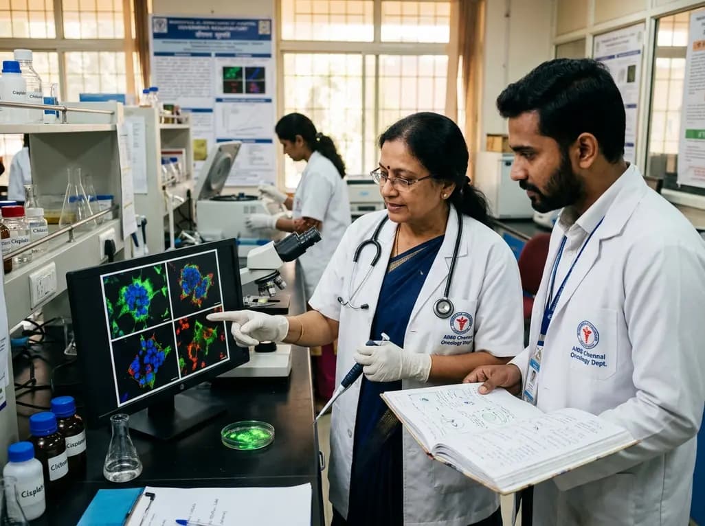
A novel green-synthesized nanocarrier uses ellagic acid to crosslink chitosan for localized cisplatin delivery in cervical cancer. Preclinical results demonstrate extended vaginal retention, potent cytotoxicity, ROS-driven apoptosis, and superior tumor regression compared with traditional crosslinkers.
Today
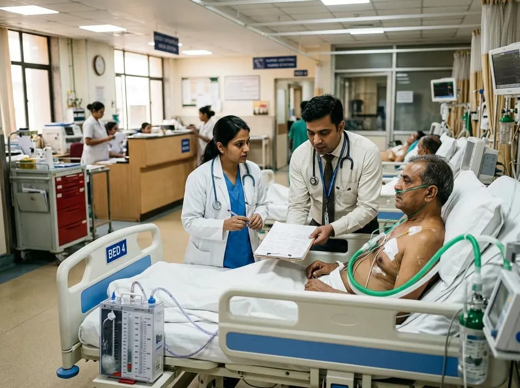
Managing refractory hypoxemia after cardiothoracic surgery becomes perilous when prolonged air leak limits positive-pressure ventilation. This case-based review details how high-flow nasal cannula, paired with awake rehabilitation, enabled successful extubation and avoided invasive re-intubation.
Today

A longitudinal study reveals that patients undergoing staged bilateral hip arthroscopy experience progressive contralateral labral deterioration during surgical intervals. However, structural degradation does not compromise 2-year postoperative clinical outcomes or patient-reported satisfaction scores.
Today

SWEDEPAD-1 trial insights show paclitaxel-coated devices do not improve long-term limb salvage in patients with chronic limb-threatening ischemia and tissue loss. While one-year reinterventions decreased, the devices were linked to a higher risk of major amputation at three months.
Today
