

Loading, please wait...
Loading, please wait...

Orbital trauma typically results from direct blunt impact, yet an orbital roof blowout fracture can occasionally develop without physical violence. When sudden periorbital swelling and vision changes follow forceful respiratory maneuvers, physicians must maintain high clinical suspicion. Such unexpected presentations challenge standard diagnostic intuition, demanding rapid multidisciplinary evaluation to preserve vision and prevent severe intracranial complications.
Typically, blunt facial trauma fractures the paper-thin medial wall or fragile orbital floor. In contrast, an orbital roof blowout fracture represents an exceedingly rare event because the frontal bone offers substantial protection. However, violent pneumatic pressure changes can overcome even sturdy craniofacial bone plates, especially in elderly adults with pre-existing bone demineralization.
When an individual blows their nose forcefully with compressed nostrils, intranasal air pressure spikes instantaneously. This rapid pressure pulse transmits directly across the ethmoidal labyrinth into the retrobulbar cone. Consequently, orbital hydrostatic pressure rises drastically within milliseconds. Although hydraulic blowout fractures traditionally decompress downward into maxillary cavities, pressurized air and soft tissue can displace superiorly. As a result, the structural partition separating the ocular orbit from the anterior cranial fossa fractures outward under sheer hydraulic force.
Furthermore, this disruption breaches the anatomical barrier isolating the intracranial vault. The resulting bone defect allows orbital fat and extraocular muscle tissue to enter the cranial space. Clinicians evaluating acute periorbital edema must recognize that pneumatic barotrauma alone can cause severe structural blowout injuries.
Exploring hydraulic mechanics clarifies how routine respiratory actions provoke significant bone compromise. Under normal circumstances, the paranasal sinuses equalize air pressure seamlessly through open ostia. However, pinching the nostrils during vigorous nose blowing causes nasopharyngeal pressure to surge dramatically. If mucosal edema or structural variations obstruct normal sinus outflow, this kinetic energy seeks alternative decompression pathways.
Consequently, pressurized air fractures the fragile lamina papyracea, forcing compressed air into the rigid retrobulbar space. Because the conical orbital cavity cannot expand easily, intraorbital volume loads escalate abruptly. Therefore, powerful hydraulic forces push directly against the orbital roof.
Moreover, if age-related bone thinning affects the frontal plate, the bone buckles toward the low-pressure intracranial compartment. This explosive hydraulic vector drives intraorbital fat and superior muscle fibers through lacerated periosteum and dura mater. Additionally, sharp bone fragments can lacerate local dural capillaries, causing inferior frontal lobe contusions or micro-hematomas. Thus, benign household barotrauma rapidly converts into a complex cranio-orbital emergency requiring prompt intervention.
Patients suffering nontraumatic superior blowout fractures present with recognizable clinical features. Immediately following forceful nose blowing, individuals experience periorbital edema, epistaxis, and severe orbital pain. In addition, subcutaneous emphysema frequently produces palpable crepitus across the upper eyelid.
However, neuro-ophthalmic symptoms provide the most decisive diagnostic clues. When the superior rectus muscle and orbital fat herniate upward, vertical ocular motility suffers immediately. Consequently, patients develop pronounced vertical diplopia and impaired downward gaze. Unlike standard floor blowouts that restrict upward movement, superior fractures impede downward travel due to superior tissue tethering.
Furthermore, clinicians must actively look for serious intracranial red flags. Because the anterior cranial floor is breached, watery nasal discharge may signal a cerebrospinal fluid leak. Patients may also exhibit severe frontal headaches, subtle cognitive changes, or neurological deficits from expanding frontal lobe hematomas. Therefore, emergency clinicians must never dismiss sudden periorbital swelling as simple allergy. Early identification of these warning signs ensures rapid neurosurgical referral before permanent damage occurs.
Accurate diagnosis depends on prompt multi-modality neuroimaging. High-resolution non-contrast computed tomography of the brain and orbits represents the essential frontline diagnostic modality. Coronal, sagittal, and axial CT reconstructions clearly delineate bony disruptions in the medial wall and orbital roof. Furthermore, CT rapidly identifies pneumocephalus, orbital emphysema, and displaced bone fragments protruding into the anterior cranial fossa.
Nevertheless, computed tomography cannot adequately visualize soft-tissue entrapment or dural injury. Magnetic resonance imaging provides vital complementary detail, especially through fat-suppressed coronal T2-weighted sequences. MRI precisely outlines soft-tissue displacement, confirming whether the superior rectus muscle or orbital fat extends into the cranial vault.
Moreover, magnetic resonance scans detect adjacent inferior frontal lobe contusions and dural disruptions. Visualizing parenchymal injury allows physicians to initiate timely neurological monitoring and anti-seizure precautions. MRI also differentiates inflammatory swelling from true muscular entrapment or ischemic tissue compromise. Consequently, combining high-resolution CT and MRI equips surgical teams with detailed anatomical roadmaps for precise operative planning.
Managing orbital roof disruptions requires seamless coordination between neurosurgeons, ophthalmologists, and otolaryngologists. While minimal fractures without tissue entrapment may undergo conservative observation, significant intracranial herniation strictly requires surgical correction. Specifically, when patients experience severe diplopia, persistent motility restrictions, or frontal hematomas, early surgery prevents permanent functional disability.
Consequently, surgical teams generally utilize an extradural subfrontal craniotomy to access the anterior skull base. This approach provides direct visibility of herniated orbital tissues without requiring brain retraction. Furthermore, during surgery, the team gently dissects adherent orbital fat and muscle fibers away from the underlying frontal lobe cortex.
Following tissue reduction, surgeons reconstruct the torn dura with primary suturing or pericranial autografts to eliminate cerebrospinal fluid leaks. Subsequently, clinicians repair the fractured orbital ceiling using titanium mesh or porous polyethylene implants. Therefore, this rigid reconstruction re-establishes normal orbital volume and prevents recurrent encephaloceles. Finally, restoring anatomical alignment relieves mechanical tension on extraocular muscles, resolving persistent diplopia and safeguarding long-term vision.
Yes, forceful nose blowing can dramatically elevate intranasal pressure to levels capable of disrupting thin craniofacial bones. When a person pinches their nostrils, air pressure transmits rapidly through fragile ethmoidal sinuses into the enclosed orbit. Consequently, this sudden hydraulic pressure wave can fracture the medial wall and thin orbital roof. Although exceptionally rare, this intense pneumatic barotrauma can cause intracranial herniation of orbital tissues, requiring urgent clinical evaluation and surgical repair.
Intracranial herniation typically produces sudden vertical diplopia, marked downgaze restriction, and acute periorbital swelling after forceful respiratory efforts. Patients frequently notice subcutaneous crepitus around the eyelid alongside unilateral epistaxis and severe retrobulbar pain. If the anterior skull base dura tears, patients may experience clear cerebrospinal fluid rhinorrhea, persistent frontal headaches, or focal neurological deficits. Such neuro-ophthalmic red flags mandate emergent computed tomography and magnetic resonance neuroimaging to assess intracranial tissue compromise.
Surgeons recommend operative repair whenever an orbital roof fracture causes persistent diplopia, mechanical muscle entrapment, or severe dural lacerations. Furthermore, surgical intervention is critical if neuroimaging reveals progressive intracranial herniation of orbital fat, frontal lobe hematoma, or persistent cerebrospinal fluid leakage. Transcranial reconstruction safely releases entrapped soft tissues, repairs torn dural margins, and rebuilds the osseous orbital ceiling with biocompatible implants, thereby restoring binocular ocular motility and preventing life-threatening intracranial infections.
Disclaimer: This content is for informational and educational purposes only... Refer to the latest local and national guidelines for clinical practice.
References

Read summarized clinical updates, watch expert medical content, and earn CME certifications right from your smartphone.
Open your smartphone camera to scan and install.





Nontraumatic orbital roof blowout fracture after nose blowing is rare. Elevated intraorbital pressure causes bone disruption, intracranial tissue herniation, and ocular motility impairment requiring prompt multidisciplinary diagnosis and surgical repair.
Today

Overexpression of glycoprotein nonmetastatic melanoma protein B (GPNMB) mediates resistance to CDK4/6 inhibitors like abemaciclib and palbociclib in ER+ breast cancer through integrin-driven AKT/ERK signaling. Targeting GPNMB with antibody-drug conjugates like glembatumumab vedotin may overcome this resistance.
Today

A secondary analysis of the MOMS clinical trial reveals that infant sex does not influence neonatal opioid withdrawal syndrome severity in buprenorphine-exposed newborns. Maternal co-exposures and clinical factors drive outcomes, highlighting the need for objective assessment and supportive breastfeeding.
Today
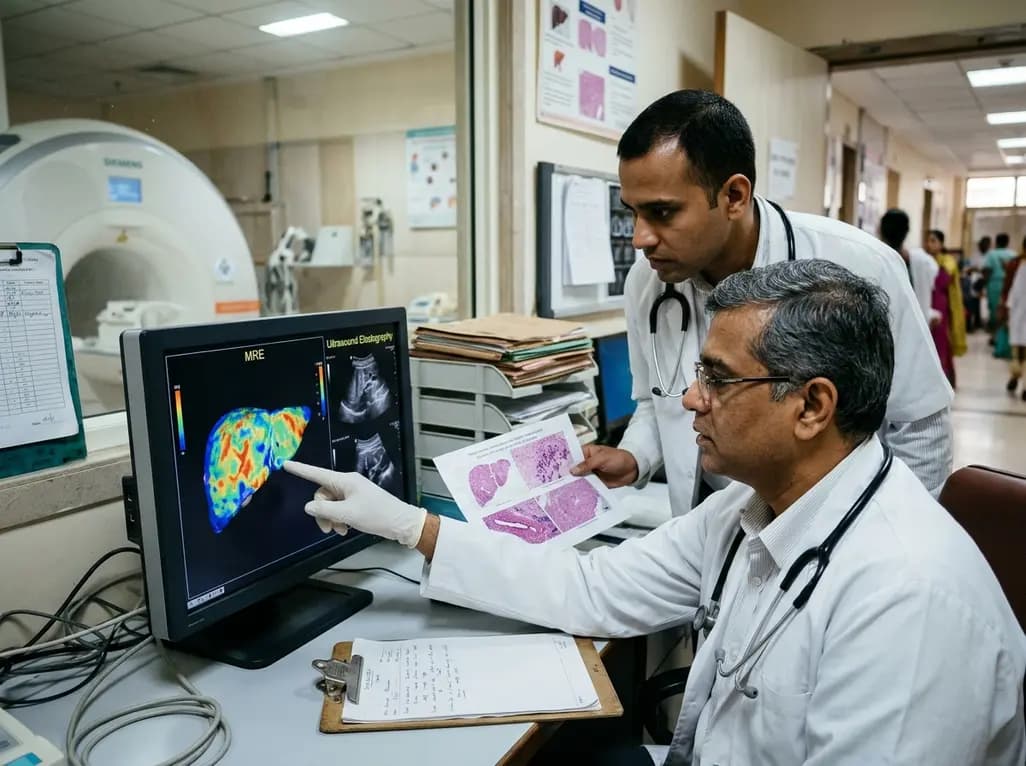
A comparative study reveals magnetic resonance elastography outperforms ultrasound elastography in assessing liver fibrosis in Fontan-associated liver disease, demonstrating stronger histological agreement and superior discrimination.
Today
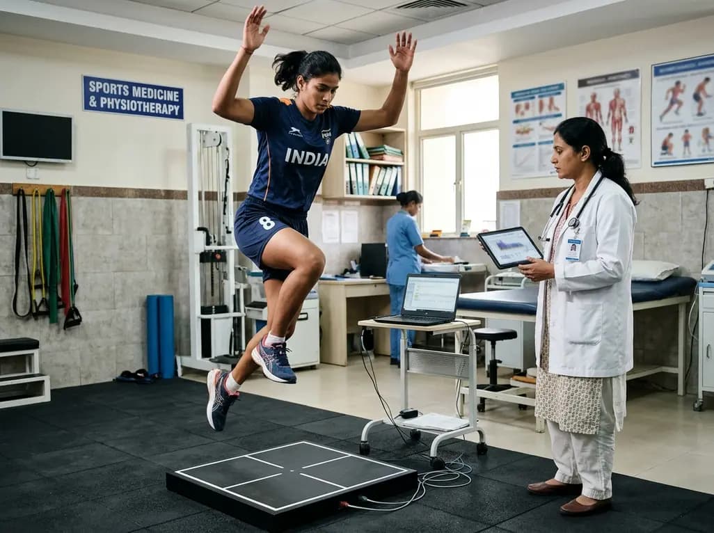
A systematic review and meta-analysis highlights the substantial impact of plyometric jump training on jump performance, reactive strength, and landing kinematics in female team-sport athletes, providing evidence-based programming guidelines for sports medicine and orthopedics.
Today

A comprehensive review of the role of growth hormone in diabetic kidney disease and podocytopathy. Learn how elevated GH activates ZEB2, Notch, and TGF-beta1, driving podocyte loss and glomerulosclerosis, and explore novel targeted therapeutic approaches.
Today
