.webp&w=384&q=75)

Loading, please wait...
Loading, please wait...
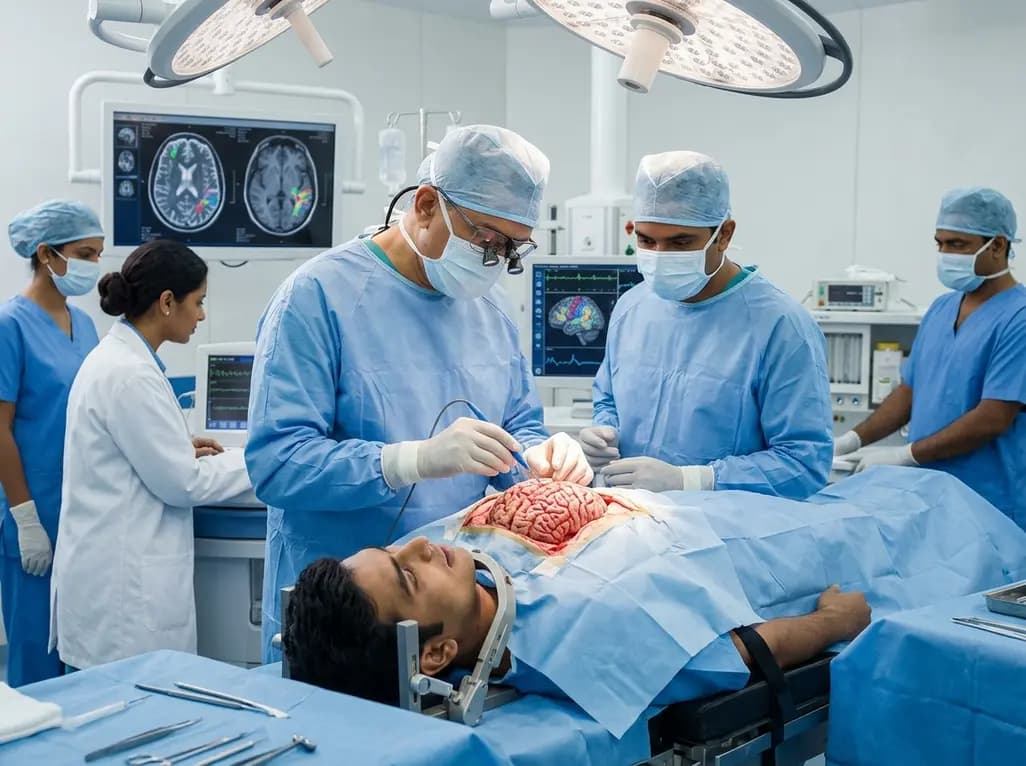
Awake craniotomy has revolutionized the field of neuro-oncology by allowing surgeons to maximize tumor resection while meticulously preserving eloquent brain functions. A cornerstone of this precision is Negative Motor Area mapping. This technique identifies cortical regions where electrical stimulation does not produce muscle contraction but instead inhibits ongoing motor activity. In clinical practice, identifying these areas is vital for avoiding permanent motor deficits. Historically, the localization of the Negative Motor Area (NMA) has been difficult due to its functional overlap with other regions. Specifically, both the NMA and the frontal language area (FLA) cause speech arrest when stimulated. This diagnostic ambiguity poses a challenge for neurosurgeons in India and across the globe who must differentiate between motor inhibition and language processing. Consequently, recent research has focused on refining our anatomical understanding of these zones. By using advanced intraoperative tools, clinicians can better navigate the complex topography of the lateral frontal cortex. Understanding these functional boundaries is essential for achieving the gold standard of maximal safe resection in glioma surgery. Moreover, preserving the patient\'s quality of life depends on the surgeon\'s ability to map these inhibitory gateways accurately during the intraoperative phase.
Developing a reliable anatomical blueprint for the NMA is a primary goal for modern neurosurgery. A recent study involving 18 awake surgery cases utilized postoperative 3D modeling to project cortical mapping results. This approach allowed researchers to pinpoint the NMA with greater accuracy than previous investigations. Notably, in every case where the NMA and FLA occupied different gyri, the Negative Motor Area mapping localized the NMA inferior to the precentral gyrus. This consistency suggests a more stable anatomical landmark than previously suspected. Furthermore, the use of 3D projection helps visualize how tumor growth might shift these eloquent areas. In Indian clinical settings, where high-grade gliomas often present with significant mass effect, this localization data is invaluable. Surgeons can now anticipate the position of the NMA relative to the precentral gyrus during preoperative planning. Additionally, this anatomical knowledge reduces the time required for intraoperative mapping. Instead of exploring broad cortical regions, the surgical team can focus stimulation on the inferior aspect of the precentral gyrus. Consequently, this targeted approach minimizes the risk of accidental injury to the primary motor cortex. Therefore, establishing these spatial relationships enhances the safety profile of complex resections involving the lateral frontal lobe.
The primary hurdle in awake mapping is the phenomenon of speech arrest, which is shared by multiple cortical regions. When stimulation of the FLA occurs, speech arrest results from a disruption in language processing or word retrieval. In contrast, stimulation of the NMA leads to speech arrest via motor inhibition of the vocal apparatus. Distinguishing between these two can be difficult because the clinical presentation during surgery often looks identical. However, precise mapping techniques allow for a more nuanced differentiation. For instance, the FLA is typically located within the inferior frontal gyrus, whereas the NMA often resides on the precentral gyrus itself. Furthermore, surgeons can use tasks beyond simple counting to test these regions. Specifically, asking the patient to perform finger tapping while speaking can reveal if the inhibition is global or language-specific. Moreover, the integration of real-time monitoring by speech-language pathologists has become a standard in multidisciplinary teams. These experts help identify subtle cues that differentiate motor-based apraxia from aphasic errors. Consequently, this collaborative approach ensures that the surgeon does not misidentify the NMA as Broca\'s area. Ultimately, the ability to separate these functions allows for more aggressive resection in the frontal lobe without compromising the patient\'s communicative abilities.
Effective Negative Motor Area mapping requires a standardized protocol for electrical stimulation. Surgeons generally utilize bipolar electrodes to deliver low-current pulses directly to the cortex while the patient is fully conscious. During this process, the patient performs repetitive tasks such as naming objects or moving their limbs. If stimulation causes an immediate cessation of the activity without causing a seizure, the area is marked as a functional zone. Notably, the study conducted between 2000 and 2013 emphasized the importance of projecting these intraoperative marks onto 3D models. This allows for a longitudinal analysis of how cortical functions relate to the underlying anatomy. In addition, the use of neuronavigation systems in India has made it easier to coregister these functional maps with preoperative MRI scans. Furthermore, clinicians must maintain a high level of vigilance for "positive" motor responses, which indicate the primary motor cortex. By mapping both the positive and negative motor zones, the surgeon can define the boundaries of the precentral gyrus with high confidence. Subsequently, this information guides the path of resection, ensuring that the scalpel avoids critical nodes of the motor network. This systematic methodology remains the most reliable way to navigate the functional variability of the human brain.
A significant concern for any neurosurgeon is the consequence of resecting an eloquent area that has been invaded by a tumor. In the analyzed study, four cases required the removal of the NMA due to direct tumor infiltration. In these instances, the patients experienced immediate apraxia of speech during the procedure. This symptom persisted into the early postoperative period, causing temporary distress for the patients and their families. However, the long-term data provides a very encouraging prognosis. Specifically, the speech apraxia improved significantly within just a few months of the surgery. Most importantly, all four patients eventually achieved full social rehabilitation and were able to return to their professional work. This suggests that the NMA, while functionally important, may possess a higher degree of plasticity or redundancy than the primary motor cortex. Furthermore, the brain seems capable of compensating for the loss of this inhibitory gateway through other nodes in the motor network. Therefore, if a tumor necessitates the resection of the NMA, surgeons can proceed with the knowledge that serious, long-term complications are unlikely. This finding offers reassurance to both clinicians and patients when navigating the difficult trade-offs between oncological control and functional preservation.
The findings regarding the localization of the NMA have broad implications for the future of neurosurgery in India. As awake craniotomy becomes more accessible across different healthcare tiers, standardized mapping data helps maintain high surgical standards. Furthermore, the discovery that the NMA is commonly localized inferior to the precentral gyrus provides a reliable starting point for future probabilistic brain maps. These maps could eventually be integrated into augmented reality systems, allowing for real-time functional overlays during surgery. In addition, the successful social rehabilitation of patients following NMA resection highlights the need for robust postoperative rehabilitation programs. While the deficit is temporary, intensive speech therapy can accelerate the recovery process. Moreover, further research is needed to determine if the NMA\'s location varies significantly in patients with long-standing epilepsy or pediatric cases. Consequently, the neurosurgical community must continue to share mapping data to refine our collective understanding. Ultimately, the goal remains the same: to push the boundaries of tumor resection while ensuring that every patient retains the ability to speak, work, and interact with the world. This study serves as a vital step toward that objective by clarifying the role and resilience of the negative motor area.
The Negative Motor Area (NMA) acts as a functional gateway that inhibits ongoing motor activity when stimulated. Unlike the primary motor cortex, which causes muscle contraction, the NMA causes the cessation of movement. This is particularly evident during speech, where NMA stimulation can lead to temporary speech arrest or motor apraxia.
Surgeons differentiate these areas during awake mapping by analyzing the nature of speech arrest and anatomical position. The NMA is typically located on the precentral gyrus, while the FLA is in the inferior frontal gyrus. Furthermore, specialized tasks and intraoperative monitoring help identify if the arrest is due to motor inhibition or language dysfunction.
No, the loss of function following NMA resection is generally temporary. While patients may experience immediate apraxia of speech during and after the surgery, symptoms typically resolve within a few months. Most patients are able to achieve full functional recovery and return to their normal social and professional activities shortly after.
Disclaimer: This content is for informational and educational purposes only and does not constitute medical advice. It is not intended to be a substitute for professional medical judgment, diagnosis, or treatment. Always seek the advice of your physician or other qualified health provider with any questions you may have regarding a medical condition. Refer to the latest local and national guidelines for clinical practice.
References
Tsuzuki S et al. Localization and symptoms associated with removal of negative motor area during awake surgery. Br J Neurosurg. 2025 Aug. doi: 10.1080/02688697.2023.2271082. PMID: 37855108.
Duffau H. The Huge Plasticity of the Brain in Glioma Patients: The "Paradox" of Eloquent Areas. Neurosurgery. 2020.
Schucht P et al. Anatomy of the negative motor area: A cortical stimulation study. Journal of Neurosurgery. 2021.
"
Read summarized clinical updates, watch expert medical content, and earn CME certifications right from your smartphone.
Open your smartphone camera to scan and install.





A recent study investigates the anatomical localization of the Negative Motor Area (NMA) during awake surgery. Findings show the NMA is typically inferior to the precentral gyrus, and while its resection causes temporary speech apraxia, long-term functional recovery and social rehabilitation remain excellent.
2 weeks back
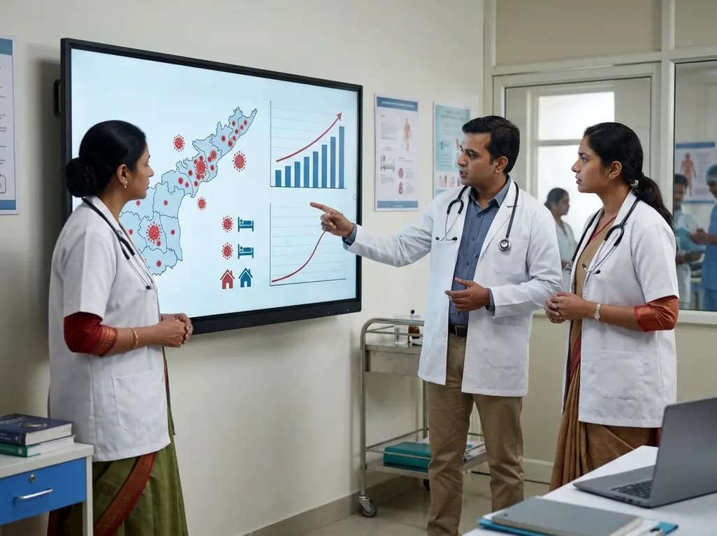
Andhra Pradesh reported 10 new Covid-19 cases, taking the state tally to 49 while deaths remain at four. With 24 patients hospitalized and 16 under home isolation, the Health Department has intensified monitoring. Medical professionals should review regional distribution, diagnostic protocols, and management plans.
Today

An 11-year Swedish registry study of 618 uterine sarcoma patients found that minimally invasive surgery yielded survival comparable to open surgery in early stages. However, adjuvant chemotherapy conferred no survival benefit in localized or advanced disease, highlighting stage and histology as key outcomes.
3 days back

A cross-sectional study evaluates post-intensive care syndrome in cardiac patients 2-4 weeks post-ICU discharge, highlighting cognitive, psychological, and functional impairments and the need for structured multidisciplinary rehabilitation.
3 days back

Anterior cruciate ligament reconstruction failure lacks uniform definition. A narrative review proposes an integrative framework incorporating objective and subjective instability, persistent pain, restricted motion, graft rupture, and secondary meniscal injury to standardize clinical reporting.
3 days back
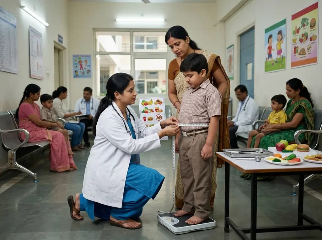
With World Obesity Atlas data warning that over 41 million Indian children are overweight or obese, ICMR and NIN have unveiled a 10-point policy roadmap. The initiative calls for mandatory front-of-pack labeling, HFSS taxes, strict marketing bans, and healthier school environments to curb non-communicable diseases.
Today
