.webp&w=384&q=75)

Loading, please wait...
Loading, please wait...
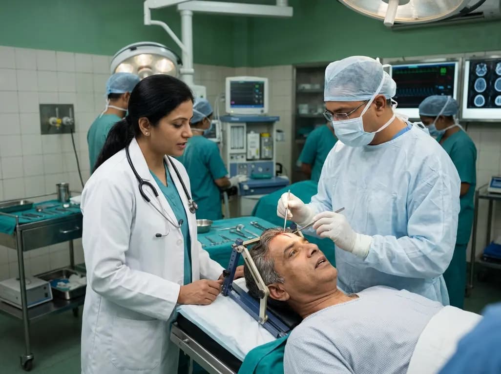
In the evolving landscape of neuro-oncology, achieving the maximal safe resection of gliomas remains a primary objective for surgeons worldwide. Specifically, awake surgery has emerged as a gold-standard technique for navigating the eloquent regions of the brain. During these procedures, clinicians utilize Negative Motor Area mapping to identify regions that, when stimulated, cause a cessation of ongoing motor activity. These areas, known as Negative Motor Areas (NMA), represent a unique functional network. Unlike the primary motor cortex, which produces positive motor responses like muscle twitching, the NMA acts as an inhibitory gateway. For surgeons in India managing complex brain tumors, understanding the precise localization of these areas is vital to preserving quality of life while maximizing the extent of resection. Consequently, recent clinical investigations have sought to refine our anatomical understanding of the NMA and its relationship with language centers.
Functional brain mapping during awake craniotomy provides real-time feedback that traditional imaging cannot match. Furthermore, the dynamic nature of the human brain often means that tumors can shift eloquent areas from their expected anatomical positions. Therefore, direct cortical stimulation (DCS) is essential for identifying these inhibitory zones. However, identifying the NMA presents a significant diagnostic hurdle. Because stimulation of the NMA can lead to speech arrest, it is frequently confused with the frontal language area (FLA), or Broca’s area. This confusion can lead to overly cautious surgical margins, potentially leaving behind viable tumor tissue. By establishing a more precise anatomical blueprint of the NMA, surgeons can better differentiate between motor inhibition and language processing deficits during the intraoperative phase.
The primary challenge in Negative Motor Area mapping lies in the symptom of speech arrest. When a surgeon applies an electrical current to the cortex, the patient may suddenly stop speaking. This phenomenon occurs regardless of whether the probe is on the NMA or the FLA. Specifically, the NMA inhibits the physical movement of the vocal apparatus, while the FLA is responsible for the cognitive and linguistic assembly of speech. Consequently, the clinical presentation is identical to the naked eye. To overcome this, researchers have analyzed cases where these two functional zones reside in different brain gyri. By isolating these instances, it becomes possible to map the specific coordinates of the NMA without the confounding influence of the language cortex.
Moreover, the function of the NMA is not limited to speech. It also governs the inhibition of limb movements. During awake mapping, patients are often asked to perform repetitive tasks, such as tapping their fingers or opening and closing their hands. If stimulation causes these movements to halt without a corresponding muscle contraction, the surgeon has likely identified an NMA. This distinction is critical because the long-term consequences of resecting an NMA differ significantly from resecting a primary language area. While the loss of Broca’s area can lead to permanent expressive aphasia, the loss of an NMA may only result in transient motor or speech apraxia. Therefore, the ability to accurately distinguish these regions allows for more aggressive surgical planning in the context of invasive gliomas.
Recent studies involving 18 cases of awake surgery have provided significant clarity regarding the anatomical landmarks of the NMA. In these cases, clinicians utilized 3D modeling to project preoperative mapping results and compare them with postoperative outcomes. Notably, the researchers found that the NMA was localized inferior to the precentral gyrus in all successful mapping attempts. This consistent localization suggests that while individual variability exists, there is a predictable anatomical trend that surgeons can rely upon. In contrast, the frontal language area typically occupies the posterior portion of the inferior frontal gyrus. Consequently, using these landmarks as a guide, surgeons can better anticipate the functional identity of a speech-arrest site.
Additionally, the relationship between the NMA and the precentral gyrus is fundamental to understanding cortical organization. The precentral gyrus is the home of the primary motor cortex (M1). The fact that the NMA sits just inferior to this area suggests a close functional integration between the excitatory and inhibitory motor networks. Furthermore, by using advanced intraoperative navigation and 3D modeling, neurosurgeons can now visualize these boundaries with higher precision than ever before. This level of anatomical detail is particularly relevant in the Indian clinical setting, where the prevalence of high-grade gliomas requires a delicate balance between radical resection and functional preservation. Ultimately, these localized findings offer a roadmap for safer surgical corridors through the frontal lobe.
One of the most significant findings in the study of the NMA is the patient's recovery profile following resection. When a tumor invades the same gyrus as the NMA, surgeons are often faced with a difficult choice: leave the tumor or remove the functional tissue. In cases where the NMA was resected alongside the tumor, patients immediately experienced apraxia of speech. Apraxia of speech is characterized by a difficulty in planning and coordinating the muscle movements necessary for phonation. Unlike aphasia, the patient's cognitive language ability remains intact; they know what they want to say, but the physical execution is impaired. This distinction is vital for patient counseling and postoperative expectations.
However, the prognosis for recovery from NMA-related apraxia is remarkably positive. In the cases studied, while the symptoms were present during and immediately after surgery, they were not permanent. Specifically, the apraxia of speech improved significantly within a few months. This suggests a high degree of neuroplasticity in the motor inhibitory networks. Furthermore, the brain appears capable of reorganizing these inhibitory functions, likely through the recruitment of the supplementary motor area (SMA) or the contralateral hemisphere. For neurosurgeons, this means that the NMA—while eloquent—may be considered "resectable" when compared to the primary motor or language cortices. This flexibility allows for a more comprehensive removal of tumor cells, which is directly correlated with improved survival rates in glioma patients.
The ultimate goal of any neurosurgical intervention is the patient’s return to their previous social and professional life. Regarding NMA resection, the data on social rehabilitation is encouraging. Patients who experienced transient apraxia of speech were generally able to return to work within two to three months. This timeline is consistent with the recovery observed in other transient syndromes, such as Supplementary Motor Area syndrome. Consequently, the temporary nature of these deficits justifies the surgical decision to resect the NMA when it is necessary for oncological control. In the context of the Indian workforce, where many patients are the primary breadwinners for their families, this high rate of social rehabilitation is a critical metric of surgical success.
Moreover, the recovery process is often facilitated by targeted speech and language therapy. Because the deficit is motor-planning based rather than linguistic, therapy focuses on repetitive muscle coordination exercises. Additionally, the psychological impact of temporary speech loss should not be underestimated. Clinicians must provide clear preoperative education to patients and their families about the possibility of transient apraxia. When patients understand that their inability to speak clearly is a temporary phase of the healing process, their compliance with rehabilitation increases. This comprehensive approach to care ensures that the benefits of an aggressive surgical resection are not overshadowed by the stress of temporary neurological impairment.
As we look toward the future of neurosurgery, the integration of Negative Motor Area mapping with other advanced modalities like functional MRI (fMRI) and navigated transcranial magnetic stimulation (nTMS) will be paramount. While direct cortical stimulation remains the gold standard, preoperative mapping can help surgeons plan their approach and identify potential NMA and FLA overlap zones. Furthermore, the use of white matter tractography, such as Diffusion Tensor Imaging (DTI), allows clinicians to see the underlying connections that support these motor inhibitory networks. For example, the frontal aslant tract (FAT) is believed to play a role in connecting the SMA and the NMA with the language centers. Understanding these deep connections will further refine our ability to preserve function.
In conclusion, the localization of the NMA inferior to the precentral gyrus provides a stable landmark for surgeons performing awake craniotomies. While the resection of this area triggers immediate and sometimes alarming symptoms like apraxia of speech, the long-term outcome is favorable. The findings underscore that the NMA, unlike the FLA, does not harbor indispensable, permanent functions. This insight empowers neurosurgeons in India and across the globe to pursue more radical tumor resections when the NMA is involved. By prioritizing both oncological efficacy and functional recovery, we can continue to push the boundaries of what is possible in the treatment of malignant brain tumors.
The Negative Motor Area (NMA) is a specific region in the frontal lobe of the brain. When stimulated during surgery, it causes the immediate cessation of motor activity or speech without causing muscle contractions. It acts as an inhibitory control center for physical movements and vocalization during the planning phase of action.
Distinguishing these areas is difficult because both cause speech arrest when stimulated. However, the NMA is typically located inferior to the precentral gyrus, whereas the Frontal Language Area (FLA) usually resides in the inferior frontal gyrus. Surgeons use anatomical landmarks, repetitive motor tasks, and 3D mapping to identify the specific functional role of each site.
Yes, NMA resection is generally considered safe if required for tumor removal. While it often leads to temporary apraxia of speech, these symptoms typically resolve within a few months. Most patients achieve full functional recovery and are able to return to their normal social and professional activities without long-term complications.
Disclaimer: This content is for informational and educational purposes only. It does not constitute medical advice or establish a doctor-patient relationship. Refer to the latest local and national guidelines for clinical practice.
References
Tsuzuki S et al. Localization and symptoms associated with removal of negative motor area during awake surgery. Br J Neurosurg. 2025 Aug. doi: 10.1080/02688697.2023.2271082. PMID: 37855108.
Mikuni N. Awake craniotomy for glioma. Neurologia medico-chirurgica. 2015; 55(4): 300-308.
Tate MC et al. Probabilistic map of critical functional regions of the human cerebral cortex: anatomical variations and supramodal nodes. Brain. 2014; 137(10): 2773-2782.

Read summarized clinical updates, watch expert medical content, and earn CME certifications right from your smartphone.
Open your smartphone camera to scan and install.





New research clarifies the localization of the Negative Motor Area (NMA) during awake surgery. Findings show that NMA typically resides inferior to the precentral gyrus. While NMA resection may cause temporary apraxia of speech, most patients recover fully within months, allowing for aggressive tumor removal.
3 months ago

A breakthrough study evaluated H5N1 transmission efficiency across mammalian isolates. Researchers discovered that human-derived D1.1 harboring the PB2 D701N mutation spread efficiently in ferrets, unlike strains carrying PB2 E627K. This finding highlights critical genetic markers for zoonotic surveillance.
Today

A comparative cohort study evaluates vNOTES vs TLH hysterectomy for benign conditions, demonstrating shorter operative duration, faster return to daily activities, and equivalent female sexual function scores without increased dyspareunia.
Today

Review how tailored exercise programs benefit frail older patients with heart failure. Explore pathophysiological mechanisms, individualized training prescriptions, and strategies to overcome adherence barriers in clinical practice.
Yesterday

This comparative study evaluates posterior labral tears with and without glenohumeral instability, highlighting distinct demographic patterns, MRI underestimation in stable shoulders, and successful arthroscopic repair outcomes.
Yesterday

A clinical study demonstrates that early intervention with tocilizumab, either as monotherapy or bridged with corticosteroids, achieves significant proptosis reduction and resolves active inflammation in treatment-naive thyroid eye disease.
Today
