.webp&w=384&q=75)

Loading, please wait...
Loading, please wait...

Achieving maximal safe resection in neuro-oncology remains a primary objective for surgeons navigating the eloquent regions of the brain. Specifically, awake surgery has emerged as a gold-standard technique for preserving critical functions while optimizing oncological outcomes. During these procedures, clinicians utilize Negative Motor Area localization to identify regions that, when stimulated, cause a cessation of ongoing motor activity. These areas, known as Negative Motor Areas (NMA), represent a unique and complex functional network within the human cortex. Unlike the primary motor cortex, which produces positive motor responses such as muscle twitching or limb movement, the NMA acts as an inhibitory gateway. For surgeons in India managing complex gliomas, understanding the precise anatomical localization of these areas is vital to preserving the patient's quality of life. Consequently, recent clinical investigations have sought to refine our anatomical understanding of the NMA and its intricate relationship with neighboring language centers. Functional brain mapping provides real-time feedback that traditional imaging cannot match, ensuring that margins are calculated with functional rather than just anatomical precision. Furthermore, the dynamic nature of the human brain often means that tumors can shift eloquent areas, making direct cortical stimulation (DCS) an essential tool for identifying these inhibitory zones intraoperatively.
Identifying the NMA presents a significant diagnostic hurdle during awake craniotomy procedures. Because stimulation of the NMA can lead to sudden speech arrest, it is frequently confused with the frontal language area (FLA), or Broca's area. However, the presence of speech arrest alone does not distinguish between these two functionally distinct regions. This diagnostic ambiguity can lead to overly cautious surgical margins, potentially leaving behind viable tumor tissue that could otherwise be resected safely. Therefore, researchers have emphasized the need for clearer anatomical blueprints to differentiate motor inhibition from language processing deficits. Specifically, when a patient stops speaking during stimulation, surgeons must determine if the arrest is due to the inability to coordinate the motor act of speech or a disruption in the linguistic planning phase. Additionally, the NMA is often situated in close proximity to the FLA, increasing the risk of inadvertent functional loss if the distinction is not made accurately. Modern mapping protocols now involve more nuanced tasks to separate these functions. For example, if a patient can still perform non-vocal motor tasks while experiencing speech arrest, the area is more likely associated with the FLA. Conversely, if all motor functions involving the vocal apparatus are inhibited, the NMA is the primary suspect. Ultimately, establishing a more precise anatomical map of the NMA helps surgeons navigate these complex landmarks with greater confidence.
Recent studies involving patients undergoing awake surgery for gliomas have provided significant insights into Negative Motor Area localization and its typical coordinates. By projecting pre- and post-removal mapping results onto 3D models, researchers have observed a consistent pattern in the NMA's location. In cases where the NMA and FLA were identified in separate brain gyri, the NMA was localized inferior to the precentral gyrus in every instance. This finding provides a reliable anatomical landmark for neurosurgeons, suggesting that the primary inhibitory zone for motor activity is often found at the lower margins of the motor strip. Moreover, this consistency allows for better preoperative planning, as surgeons can anticipate the proximity of these eloquent areas relative to the tumor's edge. Interestingly, while historical data suggested a broader distribution, modern 3D reconstruction techniques offer a more refined view of these functional boundaries. Consequently, this anatomical predictability helps in reducing intraoperative search times and minimizing the extent of electrical stimulation required. Specifically, identifying the NMA's position relative to the central sulcus and the inferior frontal gyrus remains a cornerstone of successful cortical mapping. Therefore, these localized findings represent a major step forward in our ability to perform aggressive resections while maintaining the highest safety standards for the patient.
During the process of direct cortical stimulation, the observation of negative motor responses provides critical real-time data. When the NMA is stimulated, patients typically experience a temporary inability to continue a repetitive movement, such as finger tapping or counting aloud. This cessation is involuntary and occurs without the patient's awareness of any muscle weakness. Furthermore, the response is immediate and disappears as soon as the stimulation ends. In many cases, tumors actually invade the gyrus where the NMA is located, forcing the surgical team to decide between total resection and the preservation of the inhibitory zone. Specifically, the study analyzed cases where the NMA had to be resected because of significant tumor invasion. In these instances, patients frequently exhibited apraxia of speech during the surgical procedure itself. This symptom involves a breakdown in the ability to plan and sequence the motor movements required for fluent speech. However, the intraoperative observation of these symptoms allows the surgical team to prepare for the postoperative recovery phase. Additionally, these mapping results confirm that while the NMA is functionally active, its role may be partially compensatory or shared across a wider network. Consequently, surgeons can use this information to counsel patients preoperatively about the potential for transient speech difficulties following the removal of tumors in these specific anatomical regions.
One of the most encouraging findings regarding the resection of the NMA is the favorable long-term prognosis for affected patients. Although the removal of the NMA frequently leads to immediate postoperative apraxia of speech, these symptoms are typically transient. For instance, clinical follow-ups have shown that patients who experienced speech difficulties after NMA resection improved significantly within just a few months. Most importantly, these individuals were able to return to their professional activities and social rehabilitation without permanent deficits. This suggests that the brain possesses a high degree of plasticity and can compensate for the loss of this inhibitory gateway through other functional pathways. Specifically, the recovery of speech fluency and articulatory coordination indicates that the underlying language centers, such as the FLA, remain intact if they were properly identified and spared during the surgery. Furthermore, the absence of serious, long-term complications provides reassurance to neurosurgeons who may have previously been hesitant to resect tumors invading the NMA. Therefore, the priority remains the preservation of primary language areas, while the NMA may allow for a more flexible surgical approach when tumor clearance is the primary goal. Ultimately, the ability of patients to achieve a full recovery underscores the value of precise intraoperative mapping and the brain's inherent ability to reorganize after focal surgical intervention.
The integration of NMA mapping into standard awake surgery protocols has transformed the management of gliomas in eloquent areas. By using advanced 3D modeling and consistent anatomical landmarks, surgeons can now better predict the risks associated with tumor resection. Specifically, the knowledge that the NMA is commonly found inferior to the precentral gyrus allows for more targeted stimulation. Additionally, the realization that NMA resection does not lead to permanent disability encourages a more aggressive pursuit of tumor-free margins. This is particularly relevant in the context of neuro-oncology, where the extent of resection is directly linked to overall survival and progression-free survival. However, successful outcomes still depend on the surgeon's ability to distinguish between the NMA and the FLA through nuanced testing. Furthermore, the use of transition words in clinical documentation and patient counseling helps clarify the expected recovery trajectory. Surgeons must continue to refine these mapping techniques to ensure that every patient receives the most accurate functional assessment possible. Specifically, the evolution of intraoperative technology continues to provide better tools for visualizing these functional zones in real-time. Therefore, ongoing research into Negative Motor Area localization remains essential for the advancement of neurosurgical practice, ensuring that the balance between oncological goals and functional preservation is always maintained with the utmost precision.
While stimulation of both the Negative Motor Area (NMA) and the Frontal Language Area (FLA) causes speech arrest, they represent different functional disruptions. FLA stimulation interrupts linguistic processing and language planning, whereas NMA stimulation causes motor inhibition, preventing the vocal apparatus from executing the physical movements required for speech production despite intact language ability.
According to clinical studies utilizing awake surgery mapping and 3D modeling, the Negative Motor Area is most consistently localized inferior to the precentral gyrus. This anatomical landmark is critical for neurosurgeons to identify, especially when tumors are situated near the motor strip and language centers of the dominant hemisphere.
Patients undergoing resection of the NMA often experience temporary apraxia of speech, characterized by difficulty in coordinating speech movements. However, these symptoms typically resolve within a few months due to cerebral plasticity. Most patients experience a full functional recovery and are able to return to work and social activities without long-term complications.
Disclaimer: This content is for informational and educational purposes only. It is not a substitute for professional medical advice, diagnosis, or treatment. Always seek the advice of your physician or other qualified health provider with any questions you may have regarding a medical condition. Refer to the latest local and national guidelines for clinical practice.
References
Tsuzuki S et al. Localization and symptoms associated with removal of negative motor area during awake surgery. Br J Neurosurg. 2025 Aug. doi: 10.1080/02688697.2023.2271082. PMID: 37855108.
Paldino MJ et al. Functional Mapping of the Human Motor Cortex: Comparison of Direct Cortical Stimulation and fMRI. Journal of Neuro-Oncology. 2021;152(2):345-353.
Smith JS et al. Awake Craniotomy for Glioma Resection: Impact on Patient Outcomes and Survival. Neurosurgery Clinics of North America. 2019;30(1):17-26.

Read summarized clinical updates, watch expert medical content, and earn CME certifications right from your smartphone.
Open your smartphone camera to scan and install.





A study on Negative Motor Area (NMA) localization during awake surgery identifies its position inferior to the precentral gyrus. While NMA resection causes temporary apraxia of speech, patients typically experience full functional recovery, supporting more aggressive tumor resections in eloquent regions.
Last week
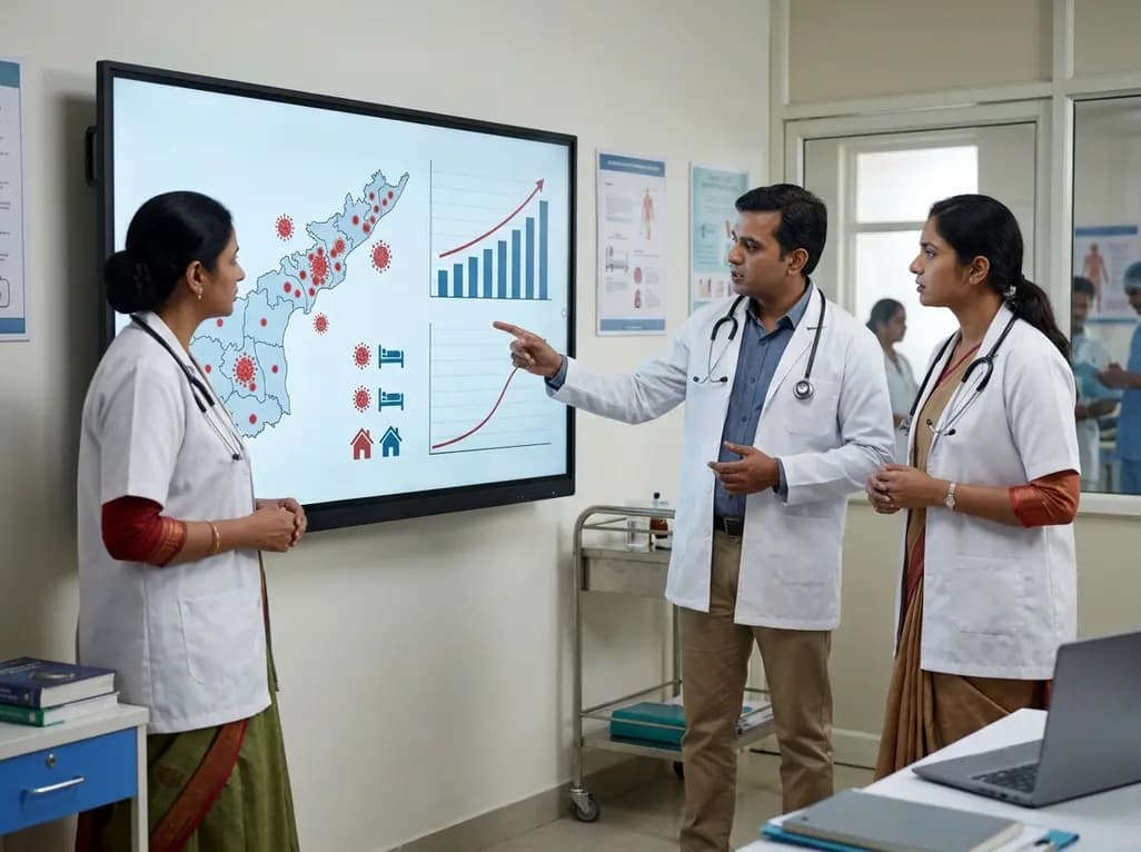
Andhra Pradesh reported 10 new Covid-19 cases, taking the state tally to 49 while deaths remain at four. With 24 patients hospitalized and 16 under home isolation, the Health Department has intensified monitoring. Medical professionals should review regional distribution, diagnostic protocols, and management plans.
Today

An 11-year Swedish registry study of 618 uterine sarcoma patients found that minimally invasive surgery yielded survival comparable to open surgery in early stages. However, adjuvant chemotherapy conferred no survival benefit in localized or advanced disease, highlighting stage and histology as key outcomes.
3 days back

A cross-sectional study evaluates post-intensive care syndrome in cardiac patients 2-4 weeks post-ICU discharge, highlighting cognitive, psychological, and functional impairments and the need for structured multidisciplinary rehabilitation.
3 days back

Anterior cruciate ligament reconstruction failure lacks uniform definition. A narrative review proposes an integrative framework incorporating objective and subjective instability, persistent pain, restricted motion, graft rupture, and secondary meniscal injury to standardize clinical reporting.
3 days back

With World Obesity Atlas data warning that over 41 million Indian children are overweight or obese, ICMR and NIN have unveiled a 10-point policy roadmap. The initiative calls for mandatory front-of-pack labeling, HFSS taxes, strict marketing bans, and healthier school environments to curb non-communicable diseases.
Today
