

Loading, please wait...
Loading, please wait...

Biliary atresia represents a critical neonatal cholangiopathy requiring prompt diagnosis and timely surgical intervention. Although Kasai portoenterostomy effectively restores biliary drainage in many affected infants, progressive hepatic fibrosis remains a persistent clinical concern. Consequently, pediatric hepatologists require dependable prognostic indicators to track post-surgical recovery and anticipate severe long-term complications. Accurate risk stratification helps medical teams optimize surveillance protocols and prepare families for potential pediatric liver transplantation. In this clinical setting, predicting native liver survival serves as an essential objective benchmark for evaluating surgical success and monitoring disease progression after portoenterostomy.
Surgical reconstruction via Kasai portoenterostomy offers the primary therapeutic option for infants with biliary atresia. Nevertheless, post-surgical trajectories vary substantially across pediatric cohorts due to variable intrahepatic inflammation. While certain infants clear jaundice completely, others progress relentlessly toward biliary cirrhosis. Therefore, early identification of high-risk infants allows clinicians to deliver aggressive, proactive care. Historically, pediatric specialists relied on total bilirubin levels measured three months post-surgery to estimate clinical prognosis. However, bilirubin clearance alone cannot depict the true severity of parenchymal tissue damage. Furthermore, hepatic fibrogenesis frequently advances despite adequate macroscopical biliary flow. Because repeated surveillance liver biopsies carry major procedural risks, noninvasive scoring indices provide an appealing alternative for continuous monitoring. Recent clinical studies indicate that objective laboratory markers calculated at hospital discharge accurately forecast native liver survival. Consequently, integrating standardized noninvasive scores into post-surgical workflows enhances early risk stratification. This strategic approach empowers multidisciplinary teams to personalize outpatient visits, guide nutritional support, and determine timely transplant referrals before hepatic decompensation ensues.
Noninvasive fibrosis indices offer profound insight into hepatic parenchymal architecture following surgical drainage. Specifically, recent clinical investigations evaluated the aspartate aminotransferase to platelet ratio index alongside the infantile biliary atresia liver fibrosis score. Healthcare providers calculate both indices using standard blood panels collected routinely at hospital discharge after portoenterostomy. In a notable observational cohort study, elevated discharge values of both biomarkers strongly correlated with reduced native liver survival. Specifically, higher numerical scores directly reflected advanced histological fibrosis and ongoing hepatocyte injury. Moreover, multivariable regression models confirmed that higher discharge scores conferred a significantly elevated hazard for early liver transplantation or mortality. Because these parameters rely solely on routine liver biochemistries and automated platelet counts, they require no expensive laboratory equipment. Therefore, secondary community centers and specialized tertiary referral hospitals can easily deploy them. Additionally, serial biomarker calculations provide objective longitudinal tracking during critical post-surgical recovery months. Thus, discharge noninvasive scoring equips clinicians with actionable prognostic information without exposing delicate infants to invasive procedures.
Despite the growing utility of circulating biomarkers, intraoperative wedge liver biopsy continues to provide an essential prognostic baseline. During portoenterostomy, surgeons routinely harvest liver tissue specimens for detailed histopathological staging using the validated Ishak fibrosis score. Notably, clinical evidence demonstrates that higher Ishak scores correlate strongly with poorer native liver survival. Marked structural alterations, bridging fibrosis, and extensive nodular formation signify severe prenatal or perinatal hepatic insult. Furthermore, infants displaying advanced baseline fibrosis typically experience delayed jaundice resolution and accelerated parenchymal decline. However, relying exclusively on histopathology presents clear operational limitations during routine longitudinal follow-up. Biopsies capture only a single static cross-sectional snapshot, whereas hepatic fibrogenesis represents a highly dynamic cellular phenomenon. Additionally, tissue sampling variations between different liver lobes can alter staging precision. Consequently, pairing baseline Ishak fibrosis staging with dynamic discharge biochemical scores overcomes these limitations. This combined strategy provides pediatric hepatologists with a comprehensive evaluation of both existing architectural scarring and ongoing hepatocellular damage, greatly refining individualized post-surgical prognostication.
Portal hypertension represents an exceptionally dangerous sequela following Kasai portoenterostomy in pediatric patients. Progressive intrahepatic vascular remodeling and sinusoidal resistance frequently induce splenomegaly, hypersplenism, and sudden gastrointestinal variceal bleeding. Fortunately, recent observational investigations demonstrate that discharge biomarker values effectively predict subsequent portal hypertension onset. Both the aspartate aminotransferase to platelet ratio index and the infantile biliary atresia liver fibrosis score demonstrated robust predictive utility. Pathophysiologically, worsening parenchymal stiffness elevates intrahepatic vascular resistance and promotes progressive portosystemic collateral formation. Concurrent splenic platelet pooling accelerates thrombocytopenia, directly increasing both calculable biomarker scores. Therefore, sharp elevations in these noninvasive ratios alert clinicians to impending vascular decompensation well before overt physical symptoms emerge. In daily practice, early risk identification prompts pediatric gastroenterologists to schedule surveillance endoscopies and detect vulnerable esophageal varices promptly. Furthermore, proactive monitoring enables timely medical prophylaxis or endoscopic band ligation, significantly mitigating catastrophic hemorrhage risks. Thus, utilizing simple discharge biomarkers preserves vascular stability and protects pediatric patients against severe clinical crises.
Translating these prognostic insights into clinical workflows requires structured post-surgical follow-up protocols. Following portoenterostomy, surgical teams should systematically calculate noninvasive fibrosis scores from discharge blood samples. When infants demonstrate elevated discharge scores alongside advanced baseline Ishak fibrosis, clinicians should assign them to high-risk surveillance pathways. Consequently, these fragile patients require shorter follow-up intervals, frequent liver enzyme tracking, and routine Doppler ultrasonography to monitor portal flow dynamics. Additionally, aggressive nutritional therapy remains critical for this population, as chronic cholestasis severely impedes fat-soluble vitamin absorption. Pediatric gastroenterologists must prescribe specialized formulas containing medium-chain triglycerides and fat-soluble vitamin formulations to support optimal growth. Moreover, objective biomarker stratification fosters transparent, empathetic communication with anxious families regarding expected clinical milestones. Parents gain clear expectations regarding long-term disease trajectories and the potential requirement for pediatric liver transplantation. Accordingly, referral centers can initiate pre-transplant workups proactively before acute decompensation strikes. Ultimately, embedding noninvasive discharge scores into standard protocols optimizes clinical resources and enhances long-term patient outcomes.
The aspartate aminotransferase to platelet ratio index and infantile biliary atresia liver fibrosis score reflect active hepatocyte cytolysis and progressive hypersplenism. When infants display elevated discharge biomarker scores, they possess more extensive hepatic fibrogenesis and ongoing biliary epithelial injury. Consequently, these children experience accelerated parenchymal decline, which substantially diminishes their native liver survival probabilities and necessitates timely consideration for future liver transplantation.
Portal hypertension represents a life-threatening complication after portoenterostomy, predisposing children to severe variceal bleeding, ascites, and recurrent cholangitis. Early identification through discharge noninvasive biomarkers enables gastroenterologists to initiate preventive endoscopic screening before catastrophic hemorrhage occurs. Furthermore, pediatric teams can promptly start medical prophylaxis and schedule nutritional interventions. This proactive monitoring approach significantly lowers acute morbidity, prevents emergency hospital admissions, and stabilizes children awaiting transplant evaluation.
Noninvasive biomarkers cannot entirely replace baseline liver biopsy during portoenterostomy. Baseline histopathology using Ishak scoring provides indispensable diagnostic confirmation of biliary atresia, identifies structural ductal injury, and establishes the initial degree of hepatic fibrosis. However, noninvasive panels like discharge APRi and iBALF effectively complement biopsy results. They offer simple, repeatable, and non-traumatic surveillance tools that track post-surgical disease evolution without subjecting fragile pediatric patients to the severe hazards of repeat surgical biopsies.
Disclaimer: This content is for informational and educational purposes only... Refer to the latest local and national guidelines for clinical practice.
References

Read summarized clinical updates, watch expert medical content, and earn CME certifications right from your smartphone.
Open your smartphone camera to scan and install.





Discharge APRi, iBALF, and Ishak fibrosis scores provide essential prognostic information regarding native liver survival and portal hypertension risk in infants undergoing Kasai portoenterostomy for biliary atresia, guiding targeted post-surgical surveillance and clinical care.
Today

A breakthrough study demonstrates that fullerol-primed extracellular vesicles generated in a 3D SECCS bioreactor attenuate ferroptosis and restore myogenic regeneration in diabetes via the FOXO3 pathway, offering a cell-free therapeutic strategy for diabetic musculoskeletal degeneration.
Today
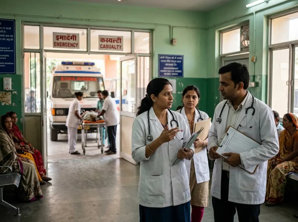
A recent maternal mortality case in Kota has drawn national scrutiny toward operational deficits in emergency transport networks. With low ambulance utilization among high-risk pregnant women, delayed inter-facility transfers, and preventable clinical complications, urgent systemic reforms are vital for patient safety.
Today
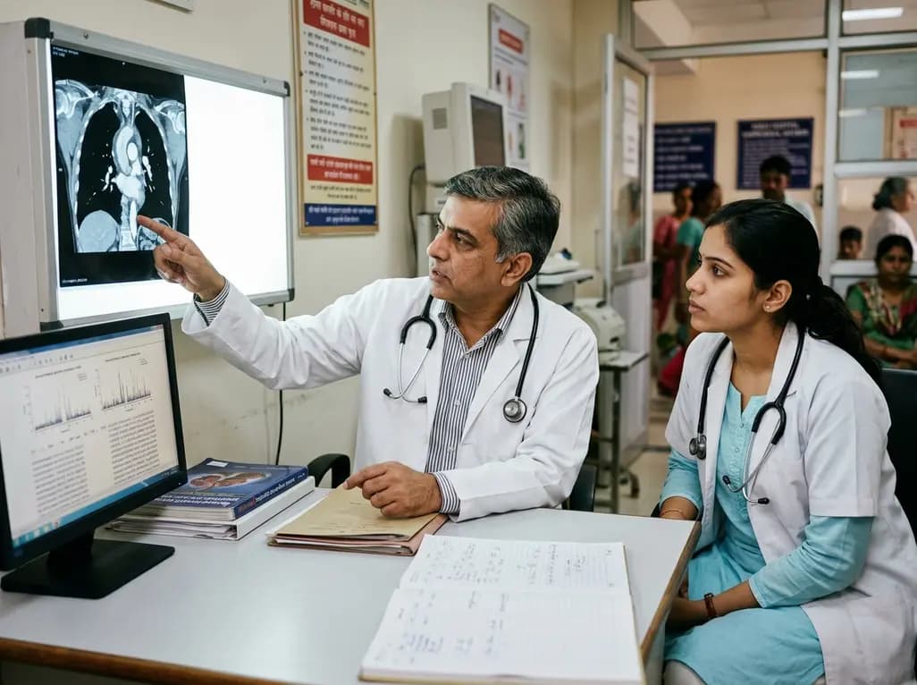
A study reveals that the MYLK c.4079G>A, p.(Gly1360Asp) variant is linked to high-penetrance heritable thoracic aortic disease and early-onset aortic dissection, showing a founder effect in Finnish cohorts. Findings emphasize early cascade screening and vigilant surveillance.
Yesterday
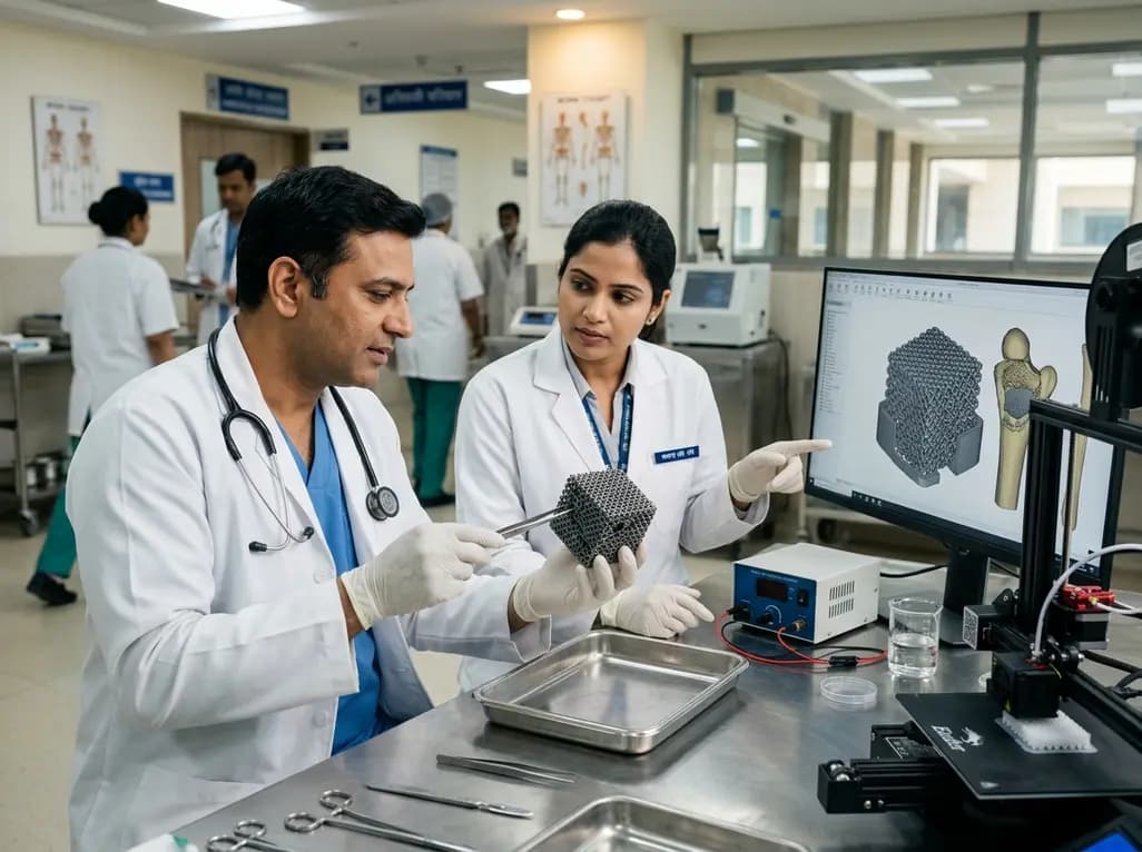
A novel sacrificial-template technique combines FDM polymer 3D printing and micro-arc oxidation to fabricate orientation-controlled porous titanium scaffolds. These bioactive constructs offer tailored stiffness, exceptional multidirectional load resistance, and robust osseointegration for bone defect repair.
Today
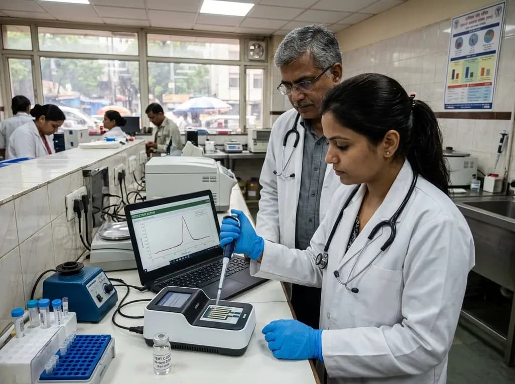
A novel label-free electrochemical biosensor utilizing magnetic nanocomposites enables ultrasensitive TERT C228T mutation detection (LOD: 0.67 pM) for rapid risk stratification in papillary thyroid cancer, offering promising point-of-care utility for endocrine oncology.
Today
