

Loading, please wait...
Loading, please wait...

Diagnostic precision represents a cornerstone in managing acute and chronic parenchymal renal disorders. Performing a native kidney biopsy provides critical insight into tissue morphology, glomerular integrity, and tubulointerstitial changes. While non-invasive laboratory evaluations offer preliminary clues, histological confirmation remains the definitive gold standard. A retrospective ten-year analysis from the Szeged Nephrology and Hypertension Center offers valuable real-world evidence on diagnostic concordance. Specifically, the study illuminates how often preliminary clinical impressions align with confirmed tissue pathology. Consequently, nephrologists must re-examine the relationship between bedside clinical suspicion and microscopic reality to enhance patient outcomes.
Clinicians frequently encounter complex renal presentations where serum biomarkers and urinalysis provide ambiguous findings. Therefore, conducting a native kidney biopsy serves as an indispensable tool for establishing precise diagnoses and determining targeted treatments. Tissue examination allows physicians to distinguish between aggressive inflammatory processes and slowly progressive sclerotic damage. Furthermore, histological grading provides accurate prognostic markers that guide the intensity of immunosuppressive therapies. In clinical practice, avoiding unnecessary toxic treatments is just as critical as initiating prompt disease-modifying therapy.
Histopathology frequently uncovers unsuspected co-existing pathologies, such as acute interstitial nephritis or subclinical immune-complex deposition. Additionally, tissue sampling helps clinicians monitor treatment responses and assess chronicity indices. Many systemic diseases present with isolated renal manifestations, making tissue analysis essential for comprehensive systemic evaluation. Consequently, nephrologists rely on percutaneous renal sampling to resolve diagnostic dilemmas and reduce long-term morbidity. Ultimately, the biopsy procedure bridges the gap between syndromic clinical classification and targeted molecular pathobiology.
Understanding the prevailing patterns of renal pathology helps clinicians prioritize diagnostic workups effectively. In the ten-year Szeged registry, nephrotic syndrome emerged as the primary indication for renal intervention, accounting for 38.8% of all procedures. Other common clinical triggers included unexplained acute kidney injury, persistent sub-nephrotic proteinuria, and progressive chronic kidney disease. Histological evaluation identified membranous glomerulonephritis as the single most frequent diagnosis, representing 16.7% of all biopsy specimens. IgA nephropathy followed closely at 11.5%, while focal segmental glomerulosclerosis accounted for 10.9% of confirmed cases.
Moreover, secondary glomerulopathies, including lupus nephritis and systemic vasculitis, represented another substantial subset of cases. These distribution patterns mirror epidemiological trends observed across diverse adult populations worldwide. However, local demographic factors and regional referral practices invariably influence exact disease frequencies. For example, metabolic comorbidities frequently modify clinical presentations, masking underlying immune glomerulonephritis. Therefore, recognizing these common histopathological entities enables physicians to design tailored diagnostic algorithms and implement timely therapeutic strategies.
The correlation between pre-biopsy impressions and histological findings reveals the clinical precision of nephrologists. In this ten-year retrospective cohort, clinical hypotheses aligned with histological results in 71.9% of patients with documented pre-biopsy diagnoses. Thus, experienced clinicians accurately predicted the underlying disease process in nearly three out of every four patients. Nevertheless, a full 28.1% of cases showed complete diagnostic discordance. This significant discrepancy demonstrates that clinical syndromic assessments cannot reliably substitute for direct tissue evaluation.
Indeed, atypical clinical presentations frequently mislead even seasoned practitioners. For instance, primary podocytopathies may present with subtle proteinuria rather than full-blown nephrotic syndrome. Similarly, proliferative glomerulonephritis occasionally mimics benign non-inflammatory nephropathies. Histopathology clarifies these ambiguous cases by revealing specific architectural disruptions and immune deposition patterns. Consequently, direct histological confirmation prevents misdiagnosis and averts inappropriate drug regimens. These findings reinforce the principle that histology remains irreplaceable for achieving true diagnostic accuracy.
A deeper examination of diagnostic mismatches reveals striking patterns across specific clinical entities. Clinicians suspected focal segmental glomerulosclerosis far more often than pathologists confirmed it, with clinical suspicion reaching 35.3% versus a histological rate of 11.8%. This marked divergence suggests that practitioners frequently over-presume focal segmental glomerulosclerosis in therapy-resistant nephrotic states. In reality, membranous nephropathy or minimal change disease often accounts for these presentation patterns.
Likewise, clinicians suspected diabetic nephropathy in 12.2% of cases, yet histology confirmed it in only 7.5%. Consequently, many diabetic patients presenting with renal decline actually harbor non-diabetic renal diseases. These non-diabetic entities include IgA nephropathy, membranous glomerulonephritis, and pauci-immune vasculitis. Therefore, attributing renal deterioration solely to longstanding diabetes without biopsy verification creates significant clinical risk. Biopsy results in such patients frequently lead to immediate therapeutic changes, including the prompt introduction of immunosuppressive agents. Ultimately, these findings urge clinicians to maintain a low threshold for tissue evaluation in atypical diabetic presentations.
While percutaneous native kidney sampling provides vital diagnostic clarity, clinicians must carefully weigh its procedural safety. In the analyzed ten-year cohort, minor complications occurred in 21.7% of patients, consisting primarily of transient macroscopic hematuria and small, self-limiting perinephric hematomas. Major complications, however, remained rare at only 2.9%, requiring interventions such as blood transfusions or angiographic embolization. These safety outcomes align with established global benchmarks for real-time ultrasound-guided percutaneous procedures.
To minimize adverse events, clinicians must strictly normalize pre-procedural coagulation parameters, blood pressure, and antiplatelet therapy. Furthermore, post-biopsy monitoring protocols remain essential for the early detection and management of bleeding. Beyond procedural mechanics, seamless interdisciplinary collaboration plays a pivotal role in optimizing patient outcomes. Nephrologists must supply comprehensive clinical histories, including duration of symptoms and serological markers, to the renal pathologist. In return, pathologists contextualize microscopic findings alongside clinical data. Therefore, regular clinicopathological case conferences enhance diagnostic interpretation, refine pre-biopsy reasoning, and foster superior clinical decisions.
Clinical syndromic presentations often overlap significantly across diverse renal pathologies. For instance, conditions like membranous glomerulonephritis, focal segmental glomerulosclerosis, and minimal change disease present identically as nephrotic syndrome. A native kidney biopsy provides definitive tissue architecture, identifying underlying immune deposits and chronicity indices. Consequently, histological evaluation prevents misdiagnosis, eliminates inappropriate immunosuppressive treatment, and establishes an accurate long-term prognosis that routine laboratory testing cannot provide alone.
Diagnostic mismatch arises primarily because clinical features reflect organ dysfunction rather than precise cellular mechanisms. For example, clinicians often suspect diabetic nephropathy in diabetic individuals with heavy proteinuria, yet biopsy frequently reveals superimposed non-diabetic glomerulonephritis. Additionally, sampling variability and overlapping biomarker patterns can obscure primary podocytopathies. Therefore, histological analysis provides essential clarification, exposing unexpected immune-complex deposition, microvascular injuries, or interstitial inflammation that bedside clinical assessments cannot reliably detect.
Close clinicopathological collaboration ensures accurate tissue interpretation within the appropriate clinical context. Specifically, nephrologists should provide comprehensive clinical data, including disease onset, serological profiles, and response to previous therapies. In response, pathologists integrate light microscopy, immunofluorescence, and electron microscopy findings with these clinical details. Furthermore, regular multidisciplinary clinical conferences resolve ambiguous borderline cases, refine diagnostic reasoning, and directly guide individualized patient management to optimize therapeutic outcomes.
Disclaimer: This content is for informational and educational purposes only. It is not intended to substitute for professional medical advice, diagnosis, or treatment. Always seek the advice of your physician or other qualified health provider with any questions you may have regarding a medical condition. Refer to the latest local and national guidelines for clinical practice.
References

Read summarized clinical updates, watch expert medical content, and earn CME certifications right from your smartphone.
Open your smartphone camera to scan and install.





A 10-year review of 451 native kidney biopsies demonstrates that clinical suspicion matches histology in 71.9% of cases, while revealing notable discrepancies in conditions like FSGS and diabetic nephropathy. The study highlights procedural safety and the necessity of close clinicopathological collaboration.
Today

An intracranial EWSR1::CREM fusion sarcoma in a 31-year-old female manifested as a paraneoplastic inflammatory syndrome with anemia and secondary infertility. The dural mass mimicked meningioma, but gross-total resection resolved her systemic inflammation and restored natural fertility within one month.
Today
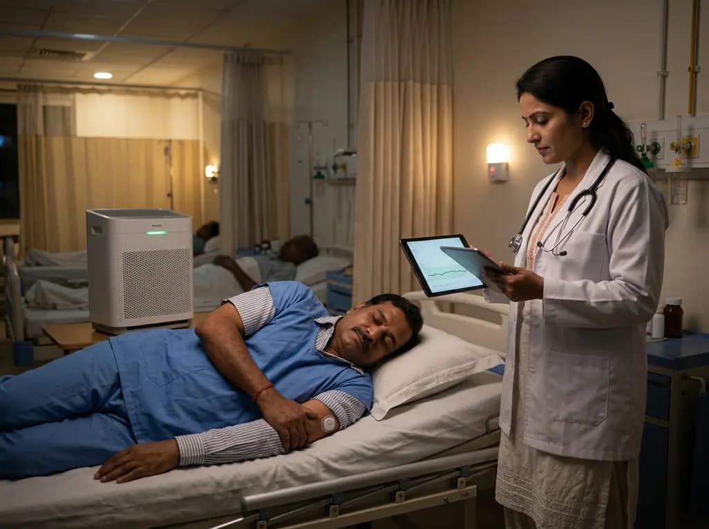
A recent clinical trial demonstrates that using bedroom HEPA air filtration during sleep significantly reduces nocturnal particulate exposure, curbs glycemic variability, and mitigates postprandial glucose surges in prediabetic patients, underscoring indoor air quality as an actionable cardiometabolic risk factor.
Today
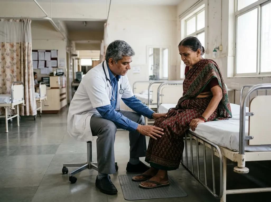
An age-matched cross-sectional study reveals that lower muscle quality and impaired lower-extremity function strongly correlate with an increased fear of falling in older women, highlighting the necessity of targeted resistance training and balance interventions in geriatric care.
Today

The DOMINO study protocol presents a multidisciplinary implementation mapping approach to overcome provider, patient, and payer barriers to antiobesity medications, offering vital insights for Indian clinicians managing the growing diabesity burden.
Today
