.webp&w=384&q=75)

Loading, please wait...
Loading, please wait...
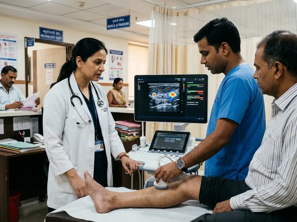
Neuromuscular diseases comprise over 600 distinct pathologies that affect the peripheral nervous system and skeletal musculature. Consequently, affected individuals frequently endure significant physical disability and progressive morbidity. Establishing a definitive diagnosis often requires invasive, costly, and lengthy evaluations. In recent years, high-resolution sonography has emerged as a non-invasive, radiation-free diagnostic modality. However, manual image interpretation remains labor-intensive and subjective. To overcome these diagnostic bottlenecks, investigators have developed innovative muscle ultrasound deep learning frameworks. These automated systems accelerate clinical evaluation while maintaining exceptional accuracy across diverse clinical settings.
Diagnosing neuromuscular disorders requires significant clinical expertise because presenting symptoms often overlap considerably. Clinicians regularly evaluate patients with diffuse weakness, rapid fatigue, or progressive muscle atrophy. Traditional diagnostic pathways rely heavily on needle electromyography, biochemical testing, and genetic panels. Furthermore, invasive muscle biopsies often become necessary to confirm structural pathology. While these diagnostic tools remain valuable, they can cause substantial patient discomfort. Consequently, patients frequently face a protracted diagnostic journey before receiving targeted therapies.
Musculoskeletal sonography presents an attractive diagnostic alternative for bedside evaluation. Real-time ultrasound reliably visualizes architectural disruption, fatty replacement, and fibrous tissue proliferation within diseased muscles. Moreover, clinicians can rapidly examine multiple muscle groups during a single patient visit. However, traditional visual grading requires substantial operator experience and specialized training. Variations in equipment settings and subjective scoring further complicate routine clinical adoption. Therefore, objective automated screening methods are vital to streamline neuromuscular referrals.
In standard neuromuscular ultrasound, physicians frequently apply the visual Heckmatt grading scale. This ordinal four-point scoring system assesses structural muscle integrity by evaluating echogenicity and deep bone echo reflection. Under normal physiological conditions, healthy muscle tissue appears relatively hypoechoic with distinct fascial boundaries and strong bone reflections. In contrast, diseased muscle tissue demonstrates increased echogenicity due to widespread fibrofatty infiltration. Severe pathological changes progressively attenuate the acoustic ultrasound beam, obscuring underlying bone reflections entirely.
Although the Heckmatt scale provides valuable qualitative information, inter-observer variability presents a significant practical challenge. Different sonographers may assign disparate scores to identical acoustic scans. Additionally, manual quantification of grayscale values demands significant computational time, limiting rapid clinical throughput. Researchers sought to resolve these limitations by training artificial intelligence models on standardized Heckmatt parameters. Consequently, automated visual grading standardizes image interpretation and enables rapid diagnostic screening across different healthcare facilities.
Recent breakthroughs utilize multimodal neural networks that integrate structural ultrasound data with patient demographics. Specifically, engineers designed an intermediate data fusion model that evaluates sonographic images from six key anatomical muscles. This architecture processes standard scans from the biceps brachii, forearm flexors, rectus femoris, and lower leg compartments. Furthermore, investigators enriched the network with demographic variables, specifically patient age and body mass index.
In a comprehensive test cohort of 320 patients, this multimodal network achieved an area under the precision-recall curve of 0.87. Notably, SHAP analyses revealed that acoustic image features drove model predictions, while age and BMI exerted negligible influence. Thus, the deep learning model robustly captures intrinsic neuromuscular alterations rather than superficial demographic differences. By automating the screening pipeline, this algorithm rapidly distinguishes genuine neuromuscular disease from non-neuromuscular complaints, drastically accelerating the clinical diagnostic workflow.
Integrating artificial intelligence into point-of-care sonography provides major practical benefits for modern clinical practice. First, automated image analysis significantly reduces the time required for comprehensive neuromuscular assessments. Neurologists and radiologists can quickly obtain objective assessments without calculating grayscale histograms manually. Therefore, clinics can increase diagnostic throughput while preserving high diagnostic fidelity.
Second, this screening framework guides subsequent diagnostic investigations with superior precision. Patients identified with probable neuromuscular disease can proceed immediately to confirmatory molecular genetic testing or targeted electromyography. Conversely, patients with negative screening scans avoid painful, low-yield invasive procedures. Moreover, automated quality assessment facilitates standardized evaluations in community health centers lacking subspecialized neuromuscular experts. Consequently, this technology helps democratize specialized diagnostic care across diverse healthcare settings.
Widespread adoption of deep learning tools requires extensive validation across multiple external centers. Different ultrasound machines, transducer frequencies, and institutional scanning protocols introduce acoustic variability that algorithms must reliably handle. Therefore, ongoing research focuses on cross-vendor generalization and federated learning pipelines. These collaborative initiatives ensure that algorithmic performance remains robust across diverse patient populations and imaging platforms.
Additionally, researchers are expanding algorithms to classify specific disease subcategories, such as inflammatory myopathies and motor neuron disorders. Combining real-time video ultrasound analysis with static image grading will further enhance clinical accuracy. As software architectures become lighter, developers can embed these deep learning models directly into portable point-of-care ultrasound devices. Ultimately, automated muscle ultrasound will serve as an indispensable clinical decision-support tool, improving diagnostic speed and patient outcomes worldwide.
Muscle ultrasound provides a rapid, non-invasive, and radiation-free method to evaluate structural muscle changes. It allows clinicians to visualize muscle atrophy, fibrous proliferation, and intramuscular fat substitution in real time. Because sonography causes minimal discomfort, physicians frequently use it to screen patients before recommending painful electromyography or invasive muscle biopsies.
The Heckmatt score is an ordinal four-point visual grading system based on tissue echogenicity and bone reflection clarity. Normal muscles exhibit low echogenicity with bright bone reflections. In progressive neuromuscular pathology, muscle tissue becomes increasingly hyperechoic due to fibrofatty infiltration, which progressively attenuates sound waves and obliterates deep bone reflections.
Deep learning algorithms automate image segmentation and feature extraction, which eliminates human inter-observer variability. By rapidly processing multi-muscle ultrasound scans through neural networks, artificial intelligence delivers an objective prediction within seconds. This automated approach improves diagnostic consistency and enables non-specialist centers to perform accurate screening.
Disclaimer: This content is for informational and educational purposes only. It is not intended to provide medical advice, diagnosis, or treatment. Refer to the latest local and national guidelines for clinical practice.
References
1. Kok P et al. A Combined Deep Learning Approach to Screen Patients for Neuromuscular Pathology. Ultrasound Med Biol. 2026 Aug 15. doi: undefined. PMID: 42603762.
2. Marzola F, van Alfen N, Doorduin J, Meiburger KM. Machine learning-driven Heckmatt grading in facioscapulohumeral muscular dystrophy: A novel pathway for musculoskeletal ultrasound analysis. Clin Neurophysiol. 2025;172:61-69.
3. Wijntjes J, van der Hoeven J, Saris CGJ, et al. Visual versus quantitative analysis of muscle ultrasound in neuromuscular disease. Muscle Nerve. 2026;73(2):189-198.

Read summarized clinical updates, watch expert medical content, and earn CME certifications right from your smartphone.
Open your smartphone camera to scan and install.





A multimodal deep learning framework utilizing muscle ultrasound images and clinical parameters achieves high precision in screening neuromuscular diseases, streamlining diagnostic pathways and reducing operator dependency.
last month
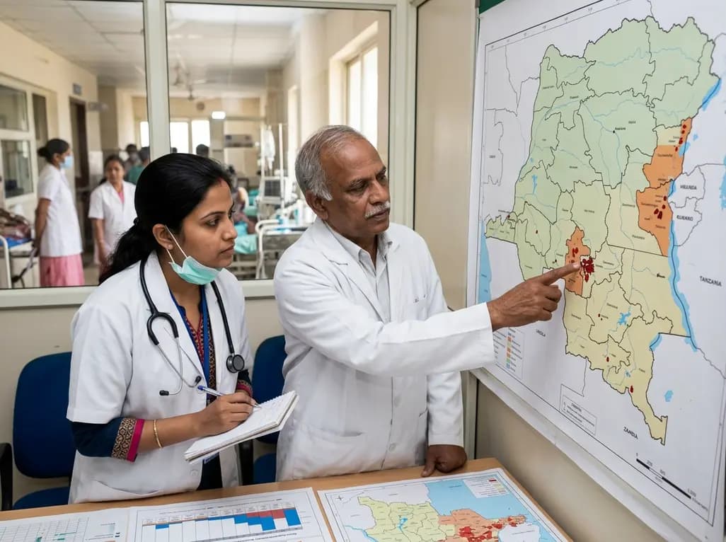
Epidemiologists and health leaders warn that active Ebola virus transmission in the Democratic Republic of Congo remains entrenched within communities. With fewer than ten percent of expected contacts monitored, public health officials urge rapid surveillance scale-up and reinforced healthcare precautions.
Today

A scoping review evaluates 116 studies on AI in women's reproductive health, finding that 83.6% carry high risk of bias. While ensemble models show promise, disparities and lack of validation hinder LMIC deployment.
Today

Differential oxygenation during peripheral TA-NRP can cause undetected coronary hypoxemia due to arch vessel clamping. A novel circuit with continuous aortic root monitoring detects desaturation early, facilitating left ventricular unloading and preventing allograft dysfunction and unnecessary heart discard.
Today
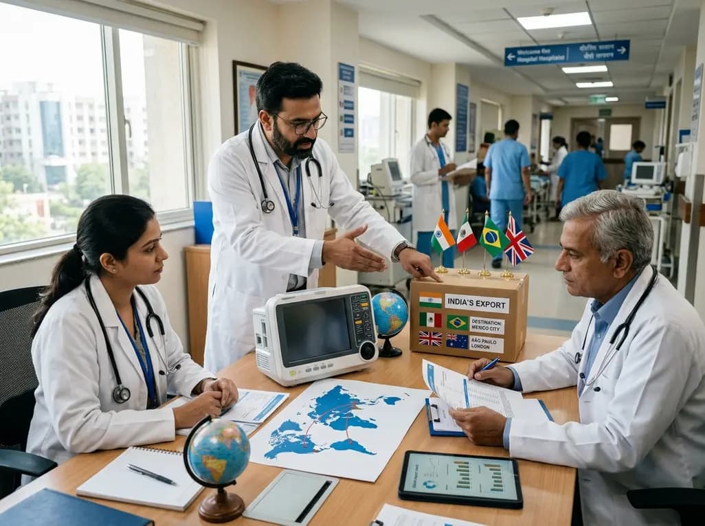
India faces significant opportunities to expand medical device exports to high-growth markets like Mexico, Brazil, and the UK. By addressing its trade deficit, advancing domestic component manufacturing, and aligning with global regulatory standards, the country aims to become an international healthcare hub.
Today

A post hoc analysis of the PARM-T2D study shows pemafibrate exerts baseline-dependent effects on LDL-C and particle size in type 2 diabetes and hypertriglyceridemia. It lowers triglycerides and remnants while favorably remodeling atherogenic LDL particles based on initial lipid control status.
Today
